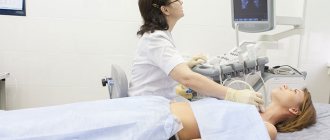
What is fluoroscopy indicated for?
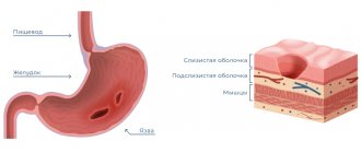
During an X-ray of the stomach, the specialist receives a detailed image of the internal structure of the organ. This allows us to draw conclusions about the shape and size of the stomach and the condition of the mucous membrane.
Definitions of stomach shape
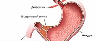
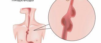
The shape of the stomach is clearly visible on the x-ray. Thanks to the image, the specialist can diagnose local narrowing of the lumen. Pathology may indicate the presence of a neoplasm in the wall of the stomach or in neighboring organs. If fluoroscopy shows widening of the lumen, there may be a bulge in the stomach wall (diverticulum).
Condition of the mucous membrane
In some cases, when performing an X-ray of the stomach, the use of a contrast agent is required; the procedure is good because it shows changes in the structure of the mucous membrane. The study is prescribed if an ulcer, chronic atrophic gastritis or other anomalies is suspected.
Organ size
An increase in the size of the stomach occurs due to overeating, stress or inflammation, as a result of complications after surgery or for other reasons. The condition is characterized by pain, nausea, bloating, tachycardia and low blood pressure. To diagnose the pathology, a fluoroscopy of the stomach is done with contrast.
Relief
To determine the relief of the mucous membrane, an x-ray of the stomach is performed with double contrast. In normal condition, the mucous membrane has a relief in the form of twisting grooves. If fluoroscopy of the stomach reveals a change in the direction of the grooves, their increase or decrease, pathologies are possible.
Structural structure
Changes in tissue structure and disruption of the integrity of the stomach walls indicate a serious illness. The pathology will be visible immediately after specialists take an X-ray of the stomach.
Carrying out the procedure
To properly prepare for a stomach x-ray, you need to understand what it is and how the examination is carried out. Depending on the purpose of the procedure, the technique may vary. Therefore, preparing a patient for an X-ray of the stomach, in addition to general requirements, may include specific steps that the doctor warns about.
In most cases, the specialist prescribes an x-ray of the stomach without contrast. In this case, the patient takes a horizontal position on the couch, after which a series of photographs is taken.
If the examination is carried out with contrast, preparation for an x-ray of the stomach includes the use of a barium-based drug. The substance does not cause harm to health and is eliminated naturally. X-ray data of the stomach with contrast are more complete than with a conventional examination.
In cases where a detailed examination of the walls of the stomach is required, their natural folds must be straightened. Then the specialist prescribes a fluoroscopy of the stomach with double contrast.
After taking the contrast agent, air is pumped into the stomach through a tube. Under slight pressure, the folds of the walls are straightened, which allows you to obtain more detailed images of the surface.
How to prepare for a stomach x-ray?
Before undergoing the examination, the patient needs to prepare for an x-ray of the stomach. It is advisable to start preparing a couple of days before the procedure, eliminating foods that contribute to increased gas formation from the diet. This is one of the main requirements on how to prepare for an x-ray of the stomach, because increased gas formation will interfere with obtaining clear images and the examination will be in vain.
The last meal is allowed 8 hours before the examination, since the patient needs to completely cleanse the organ in order to prepare for an x-ray of the stomach. Dinner on the eve of the procedure should consist of easily digestible foods. You should not have breakfast before a stomach x-ray.
There are other restrictions that must be complied with. Otherwise, the survey results will be inaccurate. For example, when preparing a patient for an X-ray of the stomach, experts do not recommend chewing gum or smoking. Increased salivation is a factor that contributes to the secretion of gastric juice, which negatively affects the condition of the mucous membrane and can cause spasms.
Decoding the results
When deciphering the results of an X-ray of the stomach, the specialist evaluates a whole range of data: from the shape and size of the organ, the condition of the tissues and the relief of the inner surface of its walls to the speed of movement of the contrast agent along the digestive tract. Deviations from the norm can indicate many pathologies.
For example, if fluoroscopy of the stomach reveals slow progress of contrast through the esophagus, stomach and intestines, motor disturbances are possible. Abnormalities in the mucous membrane may indicate a peptic ulcer, and changes in the lumen of the stomach may indicate a neoplasm.
Indications for X-ray
Due to the fact that the data provided by an x-ray of the stomach is irreplaceable in making a diagnosis for many diseases, the procedure is often prescribed by specialists. However, the examination is based on the use of ionizing radiation. Therefore, fluoroscopy of the stomach is prescribed only when the doctor suspects a dangerous disease.
The occurrence of heartburn
When a patient suffers from heartburn for a long time, this may indicate atrophic gastritis or peptic ulcer disease. To make an accurate diagnosis and begin effective treatment, a specialist prescribes an x-ray examination of the stomach.
Nausea before or after eating
Vomiting before and after meals may indicate the development of cancer. To detect a tumor, a specialist usually prescribes a fluoroscopy of the stomach with contrast.
Indigestion
Indigestion is characterized by heartburn, pain in the upper abdomen and a feeling of nausea. It may not be associated with a particular pathology. However, if indigestion occurs along with other symptoms, a specialist may suspect a serious illness. To make a correct diagnosis, a fluoroscopy of the stomach is prescribed.
Feeling of stomach stopping
Gastric atony occurs as a result of loss of muscle tone in the organ. The sensation of stopping occurs due to damage to nerve cells. There can be many reasons for atony: from stress to infection. Often, fluoroscopy of the stomach is required to determine the cause of the disease.
Intestinal disorder
18% of adults worldwide suffer from intestinal disorders from time to time. The disease can have many causes: from simple stress to dangerous gastrointestinal infections and allergies. A specialist prescribes an X-ray of the stomach in cases where it is not possible to establish the cause of an intestinal disorder by other methods.
X-ray of the stomach, esophagus and duodenum
Duration
60 minutes
We make an appointment for an hour.
The study itself lasts 30-40 minutes. Within half an hour after the fluoroscopy, you will receive the images on disk or by email. Soreness
No pain
X-ray of the stomach is a non-invasive technique.
Anesthesia
No anesthesia required
The examination is a non-invasive, painless procedure and no anesthesia is required.
Additional factors
Use of contrast agent
To perform an objective x-ray examination, additional contrast with an aqueous solution of barium sulfate in an amount of 250-300 ml is used.
Preparation
Preparation required
Preparation for fluoroscopy of the stomach is simple.
The study is carried out on an empty stomach. It is advisable to make an appointment for the first half of the day. For fluoroscopy of the esophagus (when the esophagus is examined separately), almost no preparation is required. Rehabilitation
Not required
After your exploration, you will be offered sweet tea or coffee.
You will feel good. Work ability
Saved
You can safely go to work after the procedure.
Sports activities
No restrictions
The procedure is non-invasive.
You can exercise even on the day of the procedure. X-ray of the stomach is a diagnostic study, which is a non-invasive technique that makes it possible to examine the shape, size, position of the esophagus, stomach and duodenum, as well as to identify their anatomical features and pathological changes.
The study helps gastroenterologists identify ulcers, gastritis and other diseases and pathologies. Don’t worry and remember: the main thing is to start treatment at an early stage!
X-ray examination of the esophagus, stomach and duodenum allows us to detect and differentiate the following pathological processes:
- impaired movement of the contrast mass through the esophagus, stomach and duodenum (impaired gastrointestinal motility);
- inflammatory changes in the esophagus, stomach and duodenum: esophagitis, gastritis, duodenitis;
- peptic ulcer of the stomach and duodenum;
- diseases of the esophagus: achalasia of the esophagus, varicose veins of the esophagus, hiatal hernia, diverticula of the esophagus and others;
- deformation of the duodenum and its parts due to scarring, stenosis, external compression;
- neoplasms in the walls of the stomach and duodenum, etc.
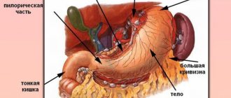
About stomach examination
The stomach is an abdominal organ with a hollow structure. To perform an objective x-ray examination, additional contrast is used with an aqueous solution of barium sulfate in an amount of 250-300 ml, which is harmless to the patient’s body, is not absorbed in the gastrointestinal tract and is excreted unchanged.
Sometimes, if it is necessary to examine the inner surface of the stomach and antrum, double contrast is used.
When conducting a study that lasts 30-40 minutes, the doctor must evaluate the passage of the contrast mass through the esophagus, stomach and duodenum, note the changes and take x-rays. They are processed at the end of the study and enable the radiologist to correctly assess the condition of the organs being examined and make a diagnosis.
The examination is usually carried out in an upright position. We also perform fluoroscopy in the Trendelenburg position. This is the name for the position of the patient on the operating table when the pelvic organs are slightly above the head.
Indications for the study:
- difficulty swallowing, pain in the chest when swallowing;
- acute or chronic pain in the abdominal cavity;
- vomit;
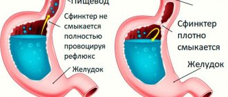
- heartburn and reflux (backflow of partially digested food or digestive juices);
- diarrhea or constipation;
- sudden weight loss;
- pathological change in the color and consistency of stool, blood in the stool.
Preparing for the study is simple. The main thing is that fluoroscopy of the stomach is performed on an empty stomach. You can read more in this section.
X-ray of the esophagus + radiography (as a separate study):
To correctly make a diagnosis, taking into account the patient’s complaints, the radiologist conducts an independent examination of the esophagus. To do this, the patient is given several sips of barium suspension and a series of polypositional (in different positions: sideways, obliquely to the right and left, lying and standing) images are taken under the scope of an RG-scope. The shape, position of the esophagus in the chest cavity, the speed of passage of the barium suspension, the location of the folds of the mucous membrane, additional narrowings or protrusions in the wall of the esophagus (diverticula) are assessed. When examining the esophagus, the doctor can detect a hiatal hernia, assess its size and position, which will tell cardiologists and gastroenterologists a plan for further treatment of the patient.
With diseases of the esophagus, patients often complain:
- for swallowing disorder - dysphagia;
- to “lump in throat”;
- for heartburn, chest pain;
- for heart rhythm disturbances;
- for pain in the heart.
preparation is required for fluoroscopy of the esophagus. It is only important that the patient comes to the clinic on an empty stomach.
Attention! After gastroscopy, X-ray examination is not performed.
Contraindications for carrying out
X-ray is a safe examination that has no absolute contraindications and allows you to quickly obtain clear images of internal organs.
However, pregnant women (especially in the first trimester) and children under 14 years of age are not recommended to undergo x-rays. Examinations using a contrast agent are contraindicated in those who have recently had an intestinal biopsy.
At MedikCity, fluoroscopy is performed using a digital X-ray machine GMM Opera T30csx (ITALRAY CLINODIGIT COMPACT). One of its important advantages is high image detail: the examination area can be enlarged on the screen without loss of quality. This allows you to see even small details.
Fluoroscopy results are not the only basis for diagnosis. Anamnesis, laboratory tests and other instrumental diagnostic methods are also taken into account.
Our X-ray room is open for you from 9.00 to 21.00. You can find out the cost of x-ray examination in a special section.
You can learn more about preparing for fluoroscopy from your attending physician or call center operators.
The material was prepared with the participation of a specialist:
Kostacheva Galina Aleksandrovna
Radiologist
Memo on preparing patients for x-ray examination
Preparing patients and conducting x-ray examinations of the stomach and small intestine.
How are patients prepared for x-ray examination of the stomach and small intestine?
Patients with normal bowel function do not require any special preparation for an X-ray examination of the stomach
.
In case of pathology of the stomach and intestines, 2-3 days before the study, foods that contribute to gas formation (brown bread, vegetables, fruits, legumes, milk, etc.) are excluded from the diet of the test subject. 14 hours before the examination, the patient stops eating, takes 30 ml of castor oil in the evening, and after 2-3 hours he is given a cleansing enema with 1-1.5 liters of warm water, chamomile infusion or soap solution (5 g of baby soap). 2-3 hours before the study, a repeat cleansing enema at room temperature is given. On the day of the study, the patient should not drink or smoke.
If there is a large amount of liquid, mucus, or food debris in the patient’s stomach (for example, with an organic narrowing of the gastric outlet), the stomach should be rinsed 2-3 hours before the test.
For severe flatulence and persistent constipation, a cleansing enema is recommended 1.5-2 hours before the test.
How is an X-ray examination of the stomach and duodenum performed?
As a contrast agent for x-ray examination of the stomach and duodenum
use a suspension of barium sulfate, which is prepared at the rate of 100 g of powder per 80 ml of water.
Preparing patients and conducting x-ray examinations of the gallbladder and biliary tract.
How are patients prepared for x-ray examination of the gallbladder and biliary tract?
For X-ray examination of the gallbladder and biliary tract
Two main methods are most often used:
cholecystography
(x-ray examination of the gallbladder with preliminary oral administration of a contrast agent) and
cholegraphy
(x-ray examination of the bile ducts with intravenous administration of a contrast agent). Before cholecystography and cholegraphy, the patient must follow a diet for 3 days to prevent flatulence (excluding raw cabbage, black bread, milk, etc.). Accumulations of gas in the intestines, giving rounded foci of clearing on an x-ray image, can overlap the shadow of the gallbladder, complicating the correct interpretation of the data obtained. Special mandatory cleansing enemas, as well as so-called “fat breakfasts” on the eve of the study are not required. A cleansing enema is given only in cases of severe flatulence.
How is cholecystography performed?
During cholecystography, the patient on the eve of the study takes a radiopaque iodine-containing drug (cholevid, yopagaost, etc.) at the rate of 1 g per 20 kg of the patient’s body weight, washed down with sweet tea, but 0.5 g every 5 minutes for half an hour. The contrast agent, entering the liver, is excreted in the bile and accumulates in the gallbladder. In this case, the maximum concentration of the drug in the gallbladder is observed 15-17 hours after administration; therefore, if cholecystography is scheduled for 9-10 a.m., then the drug should be taken the night before at 5-7 p.m. It is necessary to warn patients about the possibility of nausea and loose stools after taking these radiocontrast drugs.
The next day, X-rays (x-rays) of the gallbladder are taken. How is gallbladder x-ray analyzed?
When analyzing radiographs, the intensity of the shadow of the gallbladder, its shape, size, position, the presence or absence of deformation, concretions (stones), etc. are assessed.
How is gallbladder motor function tested?
To clarify the motor function of the gallbladder, the patient is given a so-called choleretic breakfast (2 raw egg yolks or 20 g of sorbitol in 100-150 ml of water), after which after 30-45 minutes (preferably serially, every 15 minutes), repeated photographs are taken and determined contractility of the gallbladder.
How is cholegraphy performed?
When conducting cholegraphy, a contrast agent (bilignost, bilitrast, etc.), which is also secreted by the liver and contrasts the bile ducts, is administered intravenously. Taking into account the possibility of allergic reactions, a test dose (1-2 ml) of a 50% solution of bilignost or biligrafin, warmed to body temperature, is first administered intravenously. If there are no allergic reactions (itching, chills) after 5-10 minutes, the main part of the drug is slowly administered. More intense filling of the ducts occurs after additional administration of 0.5 ml of a 1% morphine solution to the patient. Subsequent images are taken 20, 30-40 and 45-60 minutes after administration of the contrast agent.
How is bile duct radiographs analyzed?
On radiographs, the size, contours, lumen of the intra- and extrahepatic bile ducts, the presence or absence of stones in them are assessed, and the concentration and contractile functions of the gallbladder are clarified. To more accurately determine the condition of the common bile duct, intravenous cholegraphy is often supplemented with an X-ray examination of the duodenum ( duodenography
).
What are the contraindications for cholecystography and cholegraphy?
Cholecystography is not performed in case of severe liver damage, hypersensitivity to iodine, but cholegraphy. in addition, in acute inflammatory diseases of the bile ducts that occur with an increase in temperature (cholangitis), severe hyperfunction of the thyroid gland.
Preparing patients and conducting x-ray examination of the large intestine.
What is the purpose of an x-ray examination of the colon?
X-ray examination of the colon (irrigoscopy)
carried out using a contrast enema. The use of irrigoscopy makes it possible to determine the shape, position, condition of the mucous membrane, tone and peristalsis of certain parts of the colon and plays an important role in recognizing its various diseases - tumors, polyps, diverticula, intestinal obstruction.
How is preparation for irrigoscopy carried out?
To prepare a patient for irrigoscopy, food that promotes flatulence is excluded from his diet for 3 days, and porridge, jelly, omelettes, boiled meat and fish products are prescribed. Three times a day, chamomile infusion is given internally, a gas tube is inserted;
On the eve of the study, the patient is given 30 g of castor oil before lunch, and in the evening they give a cleansing enema, preferably twice with an interval of 1 hour. The patient does not eat dinner. In the morning, the patient is given a light breakfast and again given 2 cleansing enemas.
How is an X-ray examination of the colon (irrigoscopy) performed?
A suspension of barium sulfate is used as a contrast agent (at the rate of 400 g of powder per 1600 ml of water), which is best prepared in an electric mixer. The suspension, warmed to body temperature, is administered using an enema.
Preparing patients and conducting x-ray examination of the urinary system.
How do you prepare for an x-ray examination of the urinary system (urography)?
Before a survey of the kidneys, gas-forming foods (brown bread, potatoes, sauerkraut, legumes, sweet fruits, whole milk, etc.) are excluded from the patient’s food for 2-3 days, and saline laxatives are not prescribed. The night before, a cleansing enema of warm water with chamomile infusion is given. In the morning, 3 hours before the study, a cleansing enema is given again. On the day of the procedure, the patient should not eat or drink.
During an X-ray examination with contrast agents containing iodine, a sensitivity test is performed the day before the procedure. In case of an allergic reaction, the study is contraindicated.
urography procedure performed?
?
30 minutes before the study, the patient empties the bladder and X-rays check for the presence of gases in the intestines. If there is a large amount of gas, the enema is repeated and after 45 minutes a survey of the kidneys is taken.
In retrograde urography, a contrast agent is injected through a catheter into the bladder ( cystography
) or through special catheters into the renal pelvis. X-rays are then taken.
Preparing patients for x-ray examination of the bronchi, trachea, and chest.
How is a patient prepared for bronchography?
Bronchography
- This is an X-ray examination of the bronchi and trachea using contrast agents. During preparation, the patient’s sensitivity to iodine drugs is checked, postural drainage of the bronchi is carried out, expectorants, bronchodilators, and antibiotics are prescribed. Before the procedure, atropine is administered subcutaneously, and, if necessary, pipolfen and seduxen.
What are the features of the procedure?
Bronchography is performed under general anesthesia or local anesthesia.
After the procedure, the patient is not allowed to eat for 3 hours.
Catheters for administering contrast agents are sterilized by boiling.
How is a chest x-ray performed?
Examination of the chest (x-ray and x-ray) is carried out without special preparation of the patient. The method of photographing an X-ray image on 7x7 cm or 10x10 cm film is called fluorography.