The computed tomography method is often used to diagnose respiratory pathologies. This is a safe X-ray examination that takes 15-30 minutes and allows you to assess the condition of the lungs. The images are formed from 1000 layer-by-layer images of tissues. Any deviations from the norm are visible on them. Based on the images, the doctor can accurately diagnose and prescribe effective treatment. The method is used to clarify pneumonia, tuberculosis, chronic diseases, neoplasms, and assess lung function.
What is CT?
A CT scanner is a scanner with X-ray sensors. The radiation dose is safe for the body. The scanner contains a table on which a person sits. During the study, the patient lies still and follows the doctor's instructions. Over the course of half an hour, the scanner rotates around the table and takes a series of pictures.
The tomograph has precise settings for layer-by-layer scanning of tissues. If necessary, the device takes pictures every 0.5-1 mm. With this resolution, it is impossible to miss even minimal changes in tissues.
At the end of the study, computer equipment creates a digital 3D model of the respiratory system from a series of images. The attending physician can examine the lungs in any projection and examine problem areas as closely as possible.
What to take with you to the examination
If a CT scan is performed for the first time, the patient needs to take with him a referral, passport and compulsory medical insurance policy. But if a person has previously undergone lung diagnostics, then it is necessary to show the doctor the results of past studies. It can be:
- X-ray;
- fluorogram;
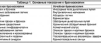
- bronchoscopy results;
- discs with MRI or CT recordings.
Any available data can help diagnose the patient's condition. Therefore, even if only extracts from the medical record remain from the last examination, it is better to take them with you and show them to the doctor.
What diseases are diagnosed by this method?
A pulmonologist, surgeon, oncologist or internist can refer you for a CT scan of the lungs. The list of diagnoses is extensive:
- pneumonia;
- tuberculosis;
- chronic obstructive pulmonary disease;
- damage to the lymphatic system;
- benign and malignant neoplasms;
- metastases in the lungs;
- parasitic, fungal infections;
- sarcoidosis;
- silicosis;
- anthracosis;
- pulmonary embolism;
- traumatic damage to the respiratory system;
- fluid in the pleural cavities;
- fibrosis of lung tissue;
- interstitial lung diseases (fibrosing alveolitis, vasculitis).
Advantages of the method:
- maximum information content;
- 3D image of tissues;
- you can accurately determine the location, size, nature of the pathological focus;
- high detail of images;
- safety for the body (low dose of radiation);
- assessment of all lung segments without blind spots;
- detection of tumors up to 5 mm in size at the initial stages.
What diseases will it detect?
CT scanning of the lungs and bronchi will allow you to quickly and accurately identify all types of respiratory diseases, including tuberculosis, pneumonia and cancer.
Tuberculosis
Tuberculosis is an infectious disease caused by Koch bacilli. Most often it affects the lungs. The classic symptom of tuberculosis is a prolonged cough with sputum, which in later stages develops into hemoptysis. Requires complex, long-term treatment. It is worth noting that in the modern world, tuberculosis in the early stages of development is treatable, so at the slightest sign of this disease you should consult a doctor.
Pneumonia
Pneumonia, or pneumonia, is an infectious inflammation of the lung tissue. Pneumonia usually affects children, adults over 65 years of age, and people with weakened immune systems. There are now many effective treatments for pneumonia. However, the mortality rate from this disease still remains high due to late seeking of medical help.
Lungs' cancer
Lung cancer is a malignant neoplasm of the epithelial tissue of the bronchus, which appears under the influence of carcinogens contained in cigarettes. Often ends with removal of the lung. High percentage of deaths. At the same time, at an early stage, treatment of lung cancer is possible and successful.
When does the doctor refer for a CT scan?
Symptoms indicating diseases of the respiratory system are shortness of breath, prolonged dry or wet cough, which is accompanied by fever. Today, computed tomography is widely used to detect lung lesions due to coronavirus infection.
Often the reason for undergoing an examination is pain in the chest, which is accompanied by cold sweat, pallor or cyanosis of the skin. Such symptoms are a consequence of arterial pulmonary thromboembolism.
Other diseases can also cause damage to the respiratory system: cirrhosis, hepatitis, HIV, sarcoidosis. Tomography will allow the doctor to get a complete picture of the disease.
MRI and CT with and without contrast - what is the difference?
Check the possibility of treatment in this area at the moment
Magnetic resonance and computed tomography are considered the most informative, safe and fast diagnostic methods in modern medicine. With their help, pathologies of various organs and systems are identified, the condition of the body is monitored during the rehabilitation period, and congenital anomalies are found. At CDC-24 you can undergo examinations using new generation tomographs, including MRI and CT with and without contrast
. The difference lies in the need to use medications that are administered intravenously or orally. Enhanced examinations are safe, but require certain preparation and are done exclusively as prescribed by a specialist: cardiologist, neurologist, gastroenterologist, oncologist, traumatologist, etc.
Why is contrast used in MRI?
In most cases, diagnostics using radio waves and magnetic fields is sufficient to detect abnormalities in internal organs, joints, brain structures and the spine. But sometimes there is a need to obtain the most accurate and detailed image, and then the doctor recommends a contrast method. The injected substance influences the properties of water molecules in the area under study, thereby improving visualization and making it possible to find the smallest anomalies due to the amplified signal.
Some people think that the main difference between an MRI with and without contrast is
is that enhanced diagnostics are harmful, but conventional magnetic resonance imaging is not. In fact, both procedures are safe, and the substances used do not affect the body in any way; their purpose is to “illuminate” the picture. They are quickly eliminated from the body and do not cause a hypersensitivity reaction (with rare exceptions). As for adverse reactions, they occur infrequently and disappear within 1-2 hours - as a rule, they are a slight headache, rashes and dizziness.
Depending on the area under study, enhancer substances may differ in composition (with iron oxide, manganese compounds, gadolinium, etc.) and method of use. Most drugs are administered by intravenous injection, but when studying the state of the gastrointestinal tract, oral varieties are used.
Does MRI without contrast show tumors?
It is possible to detect neoplasms without introducing special drugs into the patient’s body. However, in some cases, for example, with a small tumor or its complex location, amplification is required in order to most accurately determine the type and stage of the pathology. The study is also prescribed to determine malignancy and when deciding on surgical intervention.
Indications and contraindications
Enhanced MRI is performed on patients with suspected cancer, aneurysms, pituitary gland pathologies, as well as nerve damage and disorders of the cardiovascular system. Diagnostics allows us to identify necrotic lesions of soft tissues of any location, injuries of the musculo-ligamentous apparatus, diseases of the reproductive organs, lungs, liver, gallbladder and spleen, as well as degenerative changes in the central nervous system.
Indications for the examination include:
- convulsions, migraines, frequent fainting;
- decrease or loss of hearing, vision;
- palpable neoplasms in the neck area;
- arthrosis of joints, hernias, protrusions;
- multiple sclerosis;
- joint pain;
- muscle atrophy, paralysis, paresis of the lower and upper extremities;
- infectious lesions of the brain or spinal cord;
- neoplasms in the chest, pelvic organs and gastrointestinal tract;
- infertility in men and women.
There are no restrictions on the frequency of the procedure - it is prescribed as many times as required for effective treatment of the patient. Before conducting diagnostics at CDC-24, it is mandatory to check for contraindications, which include:
- pregnancy, regardless of trimester;
- individual intolerance to contrast agent;
- chronic heart failure;
- serious diseases of the kidneys and other organs of the urinary system;
- the presence of metal objects and devices in the body;
- fear of closed spaces.
MRI with and without contrast: which is better?
Both methods are effective and safe, and their differences are due to the use of drugs that enhance the signal. Compared to conventional magnetic resonance imaging, enhanced MRI:
- Lasts longer - the patient will have to stay 30-40 minutes longer inside the tomograph.
- Allows you to get more detailed pictures.
- May cause minor allergic reactions.
- Provides additional information about the state of the structures under study.
- Requires large financial costs.
- Assumes compliance with additional training recommendations.
PET CT, CT with and without contrast: what studies show, what are their differences
If we are talking about injuries and diseases of dense structures, then patients are prescribed computed tomography and PET CT, and CT can also be performed with the introduction of a contrast agent. In the first case, the radiologist studies the anatomical features of organs and tissues, using signal-enhancing drugs if necessary, and in the second, he determines deviations in dynamics by examining functional activity. Positron emission tomography is considered the most advanced method and provides more information than CT with contrast. The disadvantages of the examination include its duration (and the patient must remain motionless during the entire diagnosis) and high cost.
Conclusion
To summarize, we can say that the difference between MRI, CT with and without contrast
lies in the quality of visualization. Enhanced examination makes it possible to detect pathologies at the initial stage and is indispensable in differential diagnosis. Most often, the procedure is performed on patients with cardiac, neurological symptoms, as well as malignant and benign neoplasms. When contrast is administered, the examination takes longer and in some cases may be accompanied by side effects.
Who is the procedure contraindicated for?
- Restrictions apply to patients weighing more than 145 kg. Many devices are simply not designed to carry such a large weight.
- CT scanning is not recommended for pregnant women.
- The study cannot be carried out if the patient has severe diseases of the nervous system.
- CT angiopulmonography is not performed for people with renal failure, severe diabetes mellitus, thyroid pathologies and an allergy to iodine, which is contained in the X-ray contrast agent.
Preparing for a CT scan of the chest
A thoracic CT scan can be performed immediately on the day of your visit.
Preliminary preparation includes:
- Questioning the patient to identify possible contraindications;
- Making entries in the outpatient card, signing voluntary informed consent for the study;
- Mandatory removal by the patient of all metal objects that may affect the quality of the study;
- Explaining to the patient the basic rules of diagnostics, as well as explanations of actions in the event of a sudden deterioration of the condition.
Carrying out a CT scan of the lungs using contrast does not cause negative reactions in most patients, with the exception of individual intolerance to the components of the substance.
A CT scan takes about 2 minutes, and up to 10 minutes when using contrast. During the entire procedure, the radiologist monitors the progress of the procedure and the patient’s condition.
How is a computed tomography scan of the lungs performed?
The patient is positioned on the tomograph table with his hands behind his head. The machine begins to rotate, taking a series of X-ray images. Many patients fear that they will feel uncomfortable in a confined space. But there is no need to worry about this - in modern tomographs there is no closed space.
The doctor operating the machine may ask the patient to hold his breath for 10-15 seconds. An important condition is that you need to lie still while the tomograph is operating, otherwise the images will be blurry.
When scanning is prohibited
The main danger of the technology is the presence of an X-ray field during the examination. It is minimized to low-dose levels, which is considered safe for relatively healthy people. However, this type of diagnosis is not allowed:
- young children;
- women at any stage of pregnancy;
- cancer patients who have recently undergone radiation therapy;
- patients living or working in places with increased background radiation, often requiring radiography.
In some cases, limitations may be associated with the use of contrast. To enhance the clarity of the images, an iodine-based staining agent is injected intravenously. For more information about contrast enhancement, please follow the link: Lung CT with Contrast.
Where can I get a CT scan?
Sign up for a computed tomography scan at the MRI Center by phone or through the feedback form on the website. Doctors of the highest category, polite staff, and the latest generation tomographs - Philips, Siemens, Toshiba - are waiting for you. You will receive high-quality images, an expert opinion and a chance for a speedy recovery.
There are no queues at our diagnostic centers - studies are carried out by appointment for a specific time. The air in the rooms is disinfected after each patient, all surfaces are thoroughly treated with antiseptic solutions.
Do I need to prepare for the procedure?
There is no need to prepare specifically or for a long time for a tomography session, but when going for a scan, it is important to consider the following points:
- Interaction with the device occurs in a lying position, so it is better to wear loose, comfortable clothes that do not restrict movement when lying on a high couch.
- All foreign objects, metal jewelry, and small change must be removed from the body and pockets, as distortions and artifacts may appear in the photographs.
- Tell the specialist about chronic pathologies, possible pregnancy, the presence of built-in implants, electronic stimulators.
- To register or conclude an agreement for the service, bring your passport, as well as the results of other studies and a referral from the treating doctor.
What to do if you need to undergo a test urgently
To urgently undergo the study, call: +7 (812) 385-77-56. Our operators see a summary table of available places in most diagnostic clinics in St. Petersburg, so they can make an appointment in the near future.
Sources used:
- Gedymin L.E., Filippov V.P., Evgushchenko G.V., et al. The role of biopsy in the diagnosis of pulmonary diseases at the prehospital level of observation // Clinical Medicine. - 2009. - No. 4. — P.41-44.
- Ivanova E.V., Bilichenko T.N., Chuchalin A.G. Morbidity and mortality of the working age population in Russia due to respiratory diseases in 2010 -2012. // Pulmonology. - 2015. - T. 25. - No. 3. - P.291-297.
- Karashchuk N.P., Kiseleva M.V. Lung cancer and tuberculosis // Scientific Medical Bulletin of Ugra. -2014. — No. 1-2(5-6). — P.71-73.
- Kartashov M.V., Kartashova O.M., Kotlyarov P.M. First experience of using diffusion-weighted magnetic resonance imaging for small cell lung cancer // Medical visualization. – 2011. – No. 4. – P.28-33.
- Kotlyarov P.M. Post-processing of multislice computed tomography data in the refined diagnosis of pathological changes in diffuse lung diseases // Pulmonology. - 2021. - No. 4. — P.472-477.
- Kotlyarov P.M., Sergeev N.I. Radiation research methods in the differential diagnosis of parasitic and tumor lesions of the lungs // Siberian Journal of Oncology. – 2021. – No. 15(4). – P.33-39.
- Ternovoy S.K., Nasnikova I.Yu., Morozov S.P. Modern computed tomography in clinical medicine // Kremlin Medicine. Clinical Bulletin. - 2008. - No. 2. — P.9-13.
- Tyurin I.E. Differential diagnosis of single lesions in the lungs // Polyclinic. - 2014. - No. 3-1. — P.28-32.
- Yudin A.L., Afanasyeva N.I., Abovich Yu.A., et al. High-resolution computed tomography in the diagnosis of interstitial pneumonia // Medical visualization. -2002. - No. 4. — P.40-48.
Text approved by therapist Laeva Alina Vadimovna
CT lungs Admiralty CT lungs Bucharest CT lungs Vasileostrovskaya CT lungs Volkovskaya CT lungs Gorky CT lungs Gostiny Dvor CT lungs Dostoevskaya CT lungs Elizarovskaya CT lungs Zvenigorodskaya CT lungs Zvezdny CT lungs Komendantsky Prospekt CT lungs Krestovsky Island CT lungs Kupchino CT lungs Ladozhskaya CT lungs Ligovsky Prospect CT lungs Lomonosov CT lungs Mayakovskaya CT lungs International CT lungs Moscow CT lungs Moscow Gate CT lungs Nevsky Prospekt CT lungs Novocherkasskaya CT lungs Bypass channel CT lungs Obukhovo CT lungs Pobeda Park CT lungs Alexander Nevsky Square CT lungs Primorskaya CT lungs Proletarskaya CT lungs Bolshevikov Avenue CT lungs Rybatskoe CT lungs Sadovaya CT lungs Sennaya Square CT lungs Spasskaya CT lungs Sports CT lungs Staraya Derevnya CT lungs Technological Institute CT lungs Dybenko Street CT lungs Frunzenskaya CT lungs Chkalovskaya CT lungs Elektrosila show all metro show more
Indications and contraindications
Before sending for an x-ray examination, the doctor takes into account the risk-benefit ratio of the diagnosis
Indications for CT of the chest are complaints from the lungs and organs and structures of the mediastinum, which cannot be interpreted unambiguously after assessing the results of primary studies: radiography, ultrasound, etc. CT of the chest can be done as an additional diagnosis if there is:
- hemoptysis, shortness of breath, decreased respiratory excursions of the chest, cough, wheezing;
- limitation of mobility and pain in the joints of the shoulder girdle, thoracic spine, palpable formations and post-traumatic pain in the chest area, crepitus of the ribs (characteristic crunching sound during fractures), etc.;
- difficulty swallowing, if esophageal stenosis is suspected;
- chest pain, aggravated by movement, feeling of lack of air;
- enlargement of the cervical, supraclavicular and subclavian lymph nodes.
CT scan of the lungs after coronavirus
A CT scan of the lungs for coronavirus is done not only to assess lung damage, but also to monitor the recovery process as part of therapy. The first is done three days after the start of treatment, if it does not produce results and the patient does not recover. The next tomography can be repeated after a week if the patient’s condition does not improve.
With favorable treatment during the rehabilitation period, a CT scan of the lungs can be performed twice (interval - 2-3 weeks) to monitor the dynamics of lung recovery after coronavirus. In total, it is recommended to do no more than 5 CT scans per year.
Lung tissue is elastic and capable of regeneration. If the pathology is detected in time and treatment measures are taken, the patient’s body can cope with the infection within 1 month, and after rehabilitation, the functionality of the lungs will be completely restored.
If a patient was admitted to a medical facility with more than 50% lung damage, suffered severe pneumonia or acute respiratory distress syndrome, then fibrosis may develop. The consequences of pulmonary fibrosis resemble scars, and such pathological changes may be irreversible. However, if small areas are affected, then from a functional point of view they are easily compensated by healthy ones and are not felt throughout life. The feasibility and number of repeated computed tomography scans during the rehabilitation period are determined by the doctor.