Throat bone (fish bone)
Among cases of foreign bodies of the upper respiratory tract in the practice of an otorhinolaryngologist, fish bones are the most common. The peak demand for removal of fish bones occurs in the summer months, when the diet contains a lot of freshly caught river fish. Samara is no exception, as it is located on the Volga River. Removing and pushing through fish bones is done at home with a crust of bread. Most often, small, thin bones—ribs—get stuck. The bone becomes lodged in the upper respiratory and digestive tracts at the time of ingestion. The most favorite places for bone fixation in the pharynx are the palatine tonsils, lingual tonsil, lateral ridges, posterior palatine arches, and pyriform sinuses. The palatine tonsils become a target for fish bones, since they actively accompany the bolus of food at the moment of swallowing. The lingual tonsil suffers for the same reasons. The tissue of the palatine and lingual tonsils is represented by lymphadenoid tissue, which is very loose and easily threaded onto a thin fish bone. Concomitant pathology in the form of chronic tonsillitis with hypertrophy of the tonsils increases the risk of bone entering the tissue. In the case where the bone is stuck in the upper parts of the pharynx and is in the line of sight, removing the fish bone in such a situation is not difficult. The situation with bone fixation in the lower parts of the pharynx requires the participation of a specialist. It is extremely difficult to remove such a bone without the help of an otolaryngologist. Complications of pharyngeal trauma caused by fish bones are rare. This form of sore throat is classified as traumatic; if the bone remains in the tonsil tissue for a long time, paratonsillitis can develop, which will end in a peritonsillar abscess. Acute pharyngitis, lateropharyngeal abscess, mediastinitis, phlegmon of the pharynx, neck, sepsis, laryngeal stenosis as a complication are quite rare. Removal of fish bones in Samara is performed by ENT doctors at Outpatient Center No. 1. First medical aid. During pharyngoscopy, you should carefully examine the palatine tonsils, moving away the palatine arches; with indirect laryngoscopy, you should carefully examine the root of the tongue, the vallecula of the tongue, and pear-shaped pouches. Finger examination is allowed. The foreign body is removed with a forceps or tweezers under visual control, after which it is recommended to rinse the oropharynx with an antiseptic solution and adhere to a gentle diet. If the foreign bodies are located in a different location in the pharynx, the patient should be urgently hospitalized in the otorhinolaryngology department. Specialized assistance. Foreign bodies of the lingual tonsil, vallecula of the tongue root and pyriform pouches are removed during indirect laryngoscopy in adults and direct hypopharyngoscopy in children using a laryngeal forceps or forceps. Anti-inflammatory therapy is prescribed. If a foreign body is not detected in the pharynx, and the pain syndrome persists, it is necessary to exclude a foreign body of the esophagus. For this purpose, fibrohypopharyngoscopy and esophagoscopy are performed.
Foreign bodies of the pharynx
Causes.
They are usually localized in the oropharynx and laryngopharynx, where they enter with food, sometimes during manipulations in the mouth (pin, needle, toothpick). The most common foreign body in the pharynx is a fish bone, which penetrates into the loose tissue of the palatine, lingual tonsils, and into the vallecula of the root of the tongue. Less often, foreign bodies (coin, meat bone) are fixed in pear-shaped pockets. Foreign bodies enter the nasopharynx from the nasal cavity (needle), or from the lower parts of the pharynx during vomiting. This occurs more often in children and the elderly.
Symptoms.
Pain in the throat when swallowing with irradiation into the ear (stabbing with a fish bone), discomfort in the projection of a foreign body, sometimes hypersalivation, vomiting, difficulty swallowing.
Complications.
Bleeding, acute pharyngitis, lateropharyngeal abscess, mediastinitis, phlegmon of the pharynx, neck, sepsis, laryngeal stenosis.
First medical aid.
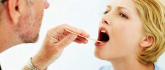
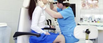
During pharyngoscopy, you should carefully examine the palatine tonsils, moving away the palatine arches; with indirect laryngoscopy, you should carefully examine the root of the tongue, the vallecula of the tongue, and pear-shaped pouches. Finger examination is allowed.
The foreign body is removed with a forceps or tweezers under visual control, after which it is recommended to rinse the oropharynx with an antiseptic solution and adhere to a gentle diet. If the foreign bodies are located in a different location in the pharynx, the patient should be urgently hospitalized in the otorhinolaryngology department.
Specialized assistance.
Foreign bodies of the lingual tonsil, vallecula of the tongue root and pyriform pouches are removed during indirect laryngoscopy in adults and direct hypopharyngoscopy in children using a laryngeal forceps or forceps. Anti-inflammatory therapy is prescribed. If a foreign body is not detected in the pharynx, and the pain syndrome persists, it is necessary to exclude a foreign body of the esophagus. For this purpose, fibrohypopharyngoscopy and esophagoscopy are performed.
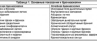
4.Treatment
The method of choice is the use of minimally invasive bronchoscopy, since modern models of endoscopes are very maneuverable in space, provide sufficient image quality and allow active use of manipulating forceps. However, in some situations, a foreign body is wedged in and fixed so firmly, and the walls of the trachea or bronchi become so thin, tense, inflamed or necrotic (not to mention cases of traumatic perforation and massive bleeding) that there is no alternative to surgical intervention with open access. In any case, after the intervention, intensive antibiotic and anti-inflammatory therapy is carried out, measures are taken to stop complications that have developed and prevent their further aggravation.
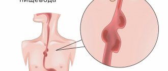
Foreign bodies of the esophagus
Causes.
Hasty eating, missing teeth, inadequate dentures, decreased pharyngeal reflex, alcohol intoxication, cicatricial narrowing of the esophagus. Foreign bodies usually get stuck in the area of physiological narrowings, most often at the level of the first thoracic vertebra.
Symptoms.
The onset of the disease is sudden and associated with food intake. Characterized by pain in the throat or behind the sternum with irradiation to the back, interscapular area, dysphagia, aphagia, drooling, general weakness, malaise, pain on palpation of the neck (on the left), aggravated by tapping on the spine, possibly forced positioning of the head.
When a foreign body is localized in the area of the first physiological narrowing of the esophagus, the head is tilted forward, down, the patient holds it motionless, and turns the whole body. When a foreign body is localized in the thoracic esophagus, the patient is in a semi-bent position (“carrying posture”).
Indirect laryngoscopy reveals swelling, hyperemia of the mucous membrane in the area of the aryepiglottic folds, arytenoid cartilages, and accumulation of saliva in the pyriform pouch (usually the left one). Vomiting and coughing are possible. A large foreign body can cause difficulty breathing through the larynx.
Complications.
Perforation of the esophagus, periesophagitis, mediastinitis, bleeding from the great vessels.
First medical aid.
. Immediate evacuation to hospital. Attempts to push a foreign body by swallowing bread crusts or using bougies are prohibited.
Specialized assistance
provided by otorhinolaryngologists together with endoscopists. To do this, indirect laryngoscopy and x-ray examination of the cervical spine in two projections (according to G.M. Zemtsov) are performed, which makes it possible to detect the shadow of a foreign body, indirect signs of a non-contrast foreign body of the esophagus or damage to its walls.
These symptoms are:
- straightening of the cervical spine due to tension in the scalene muscles;
- expansion of the prevertebral space;
- the presence of a symptom of an air “arrow” - an accumulation of air released from the stomach below the level of a foreign body, the pointed end of the “arrow” indicating the location of the foreign body;
- striped clearings in the prevertebral space are a sign of air penetration into the retroesophageal tissue or the development of putrefactive inflammation with the formation of gas.
Fibroesophagoscopy is also performed for diagnostic and therapeutic purposes. If it is impossible to remove a strangulated foreign body of the esophagus during esophagoscopy, an esophagotomy is performed. Anti-inflammatory therapy is prescribed.
Foreign bodies in the respiratory tract
Causes.
Aspiration of liquid or obstruction by particles of food or soil during a sudden deep breath, falling, crying, fright, talking, laughing. This is facilitated by the distraction of the victim’s attention while eating, the habit of holding foreign objects in the mouth, a decrease in the laryngeal-pharyngeal reflex, wearing removable dentures, alcohol intoxication, lack of consciousness due to traumatic brain injury, or poisoning.
Foreign bodies of the bronchi (88%) are more common, less common are the trachea (8.8%) and larynx (3.2%). The clinical picture depends on the nature, shape and level of presence of the foreign body in the respiratory tract.
Foreign bodies of the larynx
Symptoms.
When foreign bodies cover more than half of the glottis (live fish, a piece of food, adenoid tissue), fulminant stenosis develops: suffocation occurs, the voice disappears, and consciousness is lost.
Sharp and thin metal foreign bodies (pins, sewing needles, fish bones) initially do not cause severe breathing problems. A convulsive cough occurs, accompanied by sudden difficulty breathing, voice disorder, vomiting, and pain in the larynx are possible. During laryngoscopy, a foreign body can be detected that has stuck into the area of the arytenoid cartilage and aryepiglottic fold. The addition of mucosal edema causes an increase in inspiratory dyspnea.
Complications.
A foreign body obstructing the lumen of the larynx causes fulminant stenosis, and without proper assistance in the next few minutes leads to death. If the larynx is not completely obstructed by a foreign object, acute laryngeal stenosis develops in the coming hours.
First medical aid.
At the IV (terminal) stage of laryngeal stenosis, a conicotomy or cricoconicotomy is performed; at the III (decompensated) stage of stenosis, an urgent tracheostomy is performed. Dehydrating, diuretic, antihistamine, and corticosteroid drugs are administered. The victim is immediately evacuated to the ENT department.
Specialized assistance
consists of immediate removal of the foreign body during indirect (in children) or direct fibrolaryngoscopy with the participation of an endoscopist and an anesthesiologist, carrying out anti-edematous, anti-inflammatory, symptomatic therapy.
Emergency assistance if coin-shaped bodies enter the respiratory tract
If you hit objects that look like a coin, it is advisable to use a technique called the “piggy bank effect.” If a person swallows a coin, you need to force the foreign object to change its location. With strong blows to the chest area, there is a possibility that the foreign object will turn in the other direction and free up the air passage or move to the bronchi (when a coin or button ends up in one bronchus, the victim will be able to breathe and have time to get to the ambulance).
The most common way to shake the chest is to tap the back with your palm. The “American police method” is also considered an effective procedure. Technique of execution: you should stand behind the victim and take him by the shoulders, then move him away from you to outstretched arms and only then sharply hit his back against his own chest. This manipulation can be carried out 3-4 times. The technique is effective if the rescuer has a flat male chest.
Tracheal foreign bodies
Symptoms.
Foreign bodies (nuts, beans, peas, watermelon seeds), carried away by the inhaled air, can pass through the glottis and become fixed on the mucous membrane of the trachea. This leads to paroxysmal convulsive dry cough, difficulty breathing, chest pain, vomiting during coughing attacks. Symptoms of “balloting” or “popping” of a foreign object in the trachea are detected. The addition of mucosal edema leads to inspiratory dyspnea.
Complications.
Foreign bodies capable of swelling (bean seeds), in combination with reactive edema of the tracheal mucosa, can lead to its stenosis, especially in young children, and the development of tracheitis.
First medical aid.
Prescribe sedatives, dehydrating, antihistamines, corticosteroids, antibiotics, oxygen inhalations. For decompensated stenosis, tracheostomy is performed.
Specialized assistance
consists of urgent removal of a foreign body during upper tracheoscopy under anesthesia using muscle relaxants. If it is impossible to remove a swollen foreign body through the glottis, a tracheostomy is performed on a bronchoscopic tube and removed through an incision in the trachea. Prescribe anti-inflammatory, decongestant, symptomatic therapy.
Performing a cricothyroidotomy
Only medical professionals have the right to perform cricothyroidotomy, since this is a serious procedure that requires training and mastery of the technique. During the manipulation, an incision is made between the cricoid and thyroid cartilages (the hole will be located above the trachea and the victim will be able to breathe freely).
People around you can help carry out emergency manipulation; for this you need to: fix the head of the choking person (it is advisable to hold it between both knees so that the medical worker makes an accurate cut), press the victim’s hands to the floor or ground, ask people for an object in the form of a tube (it will help let air in and out of the lungs).