Renal angiomyolipoma (also known as hamartoma) is a benign tumor of the kidney. Women get sick 4 times more often than men, manifestations occur in middle and older age. According to statistics, this is one of the most common kidney tumors.
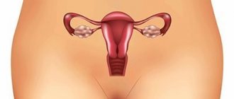
The tumor is formed by adipose tissue, smooth muscle, epithelium and blood vessels. Hamartomas also occur in other organs, including the hypothalamus, lungs, and skin. Angiomyolipoma is found in the kidneys, in which muscle tissue and blood vessels predominate. The danger of kidney angiomyolipoma is that the tissues grow unevenly, can degenerate, and the vessels can form aneurysms that are prone to rupture.
At CELT you can get a consultation with a urologist.
- Initial consultation – 3,000
- Repeated consultation – 2,000
Make an appointment
Causes and forms of renal angiomyolipoma
Kidney ultrasound
- Cost: 2,700 rub.
More details
The exact causes of angiolipoma are unknown. There is ongoing debate as to whether isolated angiomyolipoma is congenital or acquired during life. An autosomal dominant type of inheritance of such tumors has been proven, when the mutant gene is transmitted through the male line.
An acquired tumor may be associated with hormonal changes during pregnancy and the development of other types of tumors (especially vascular and connective tissue). Angiomyolipoma can also develop with various kidney lesions - from injuries to chronic inflammation.
Two forms of the disease are known:
- Sporadic or isolated, which develops independently, without connection with other pathologies. This is a single tumor enclosed in a capsule, developing in one kidney in the medulla or cortex. A common form, found in 9 out of 10 cases.
- Bourneville-Pringle syndrome or a congenital form that develops against the background of tuberous sclerosis. In this form, multiple angiomyolipomas are found in both kidneys.
The structure of angiomyolipoma can be typical and atypical: with typical, all types of tissues are present (adipose, muscle tissue, epithelium, blood vessels), and with atypical, there is no fat. This can only be determined by histological examination of the punctate or the specimen removed during surgery.
Pathogenesis
The pathogenesis of AML is practically unknown. The tumor arises from perivascular epithelioid cells, which are located around the vessels and can be characterized as large smooth muscle polygonal cells with signs of melanocytic differentiation. These cells are characterized by a relatively high rate of proliferation and growth (on average 1.5 mm per year), which occurs under the influence of precisely unknown factors. It is assumed that hormonal factors play a leading role in the development of AML, as evidenced by the presence of specific estrogen / progesterone .
There is information about characteristic gene mutations both in sporadic cases and in cases associated with tuberous sclerosis of the kidneys (loss of heterozygosity, mutations of the TSC2/TSC1 gene locus localized on chromosome 16p13). Histologically, the tumor is represented by thick-walled blood vessels, smooth muscle fibers and mature adipose tissue in different quantitative ratios. Structural variants of AML can vary significantly and depend on the maturity of the smooth muscle tissue in the smooth muscle component of the tumor.
Symptoms of angiolipoma
Kidney CT
More details
Manifestations of the disease depend on the size: up to 4 cm in diameter, the tumor behaves asymptomatically. However, even with enlargement, angiomyolipoma of the kidney may not manifest itself for a long time. Thus, in 80% of those examined, formations measuring 5 cm are found, and in 18% - 10 cm, found by chance during a kidney examination for another reason.
A size of 4-5 cm is considered extremely safe, since the vast majority of people do not have any symptoms. In the future, the tumor requires more oxygen. Muscle tissue forms faster than blood vessels, which cannot keep up with muscle growth.
As a result, the vessels stretch, the load on them increases significantly. Areas of thinning and aneurysms form in the walls of blood vessels, which easily rupture. In addition, the structure of the vascular wall in a tumor itself is rarely normal. Hemorrhages are the most common complications of such tumors.
Angiomyolipoma of the kidney can manifest itself with the following symptoms:
- dull pain or discomfort in the lower back and abdomen on the side of the tumor;
- fatigue, weakness;
- an enlarged kidney or a clearly palpable round elastic lump in the abdomen;
- blood in the urine;
- surges in blood pressure.
When a vessel ruptures and bleeds, a picture of hemorrhagic shock develops, acute pain in the lower back appears, visible blood in the urine, and an increasing compaction in the kidney area is felt. If blood has spilled into the abdominal cavity, a picture of an “acute abdomen” is formed.
Complications of the disease also include compression of neighboring organs, tumor tissue necrosis, vascular thrombosis and cancerous degeneration. Sometimes the tumor remains benign, but small nodules form in neighboring organs, often in the liver.
List of sources
- Ivanov D.D. (2017a) Nephrology “under the microscope”.
- Angiomyolipoma of the kidney: independent and associated disease. Ukr. honey. Chasopis, 4(120): 78–79.
- Kuchinsky G.A., Matveev V.B., Mironova G.T., Lukyanenko A.B. Some clinical issues and diagnostics of renal angiomyolipoma // Urol. nephrol. – 1995. – No. 2. – P.41-45.
- Nechiporenko N.A., Nechiporenko A.N. Benign kidney tumors: diagnosis and treatment // Materials of the III Polish-Belarusian Symposium of Urologists. – Augustov, 2003. – P.21-23
- Nechiporenko N.A., Galkin L.P., Balla A.A. Features of the echoscopic structure of kidney parenchyma tumors // Healthcare of Belarus. – 1993. – No. 8. – P.52-54.
- Chistyakova O.V., Sokolova I.N., Bogatyrev V.N., Sorokin K.V. Cytological method in the preoperative diagnosis of angiomyolipomas. Abstracts of the III Congress of Oncologists and Radiologists of the CIS, Minsk May 25-28, 2004, Part 1, pp. 323-324
Diagnosis of angiolipoma
At the initial stage of formation, angiomyolipoma is discovered by chance on an ultrasound or X-ray examination. For lower back pain of varying intensity, instrumental diagnostics are performed, which quickly establishes the pathology. Laboratory testing of urine and blood is required. Micro- or macrohematuria is detected in the urine.
Most often, a tumor is detected on ultrasound as a rounded isolated area of reduced echogenicity. The typical location, round shape and uniformity suggest that this is an angiomyolipoma. Small isolated tumors are most often found in the right kidney. Damage to the left kidney is less common.
The second most informative examination method is multislice computed tomography (MSCT) with contrast. This is a multi-slice study that allows you to study the structure of the kidney in real time. With MSCT, you can clearly assess the blood supply to the kidney and blood flow in the tumor.
MRI is also used for diagnosis, which better visualizes the medulla and cortex of the kidney. These methods complement each other. In addition, MRI does not use x-rays, which is important for some categories of patients.
Ultrasound angiography (duplex scanning of the renal arteries) is used to visualize the vessels. If the study detects a tumor in the form of a tangle of blood vessels, then changes in the vascular wall, expansion, narrowing and other formations are clearly visible on the monitor.
If angiomyolipoma is suspected, a biopsy of the tumor tissue can be performed, which is performed under ultrasound guidance or during endoscopic surgery. Histological examination allows us to clarify the diagnosis.
Research methods are selected by the attending physician depending on the characteristics of a particular case.
Postoperative period
After laparoscopic surgery, recovery is much faster and the patient feels almost no pain. Small incisions are left on the skin, which heal quickly. You will be able to get up and eat after the first day, this will speed up your recovery. And you will be discharged from the hospital within 2-3 days. Rehabilitation takes 1-2 months.
After open surgery, this will not be easy due to cut muscles or even removed ribs. On average, it takes from 1 week to 1 month before you are allowed to go home. But rehabilitation will last about 1.5 years. You will be prescribed painkillers, recommended moderate exercise to restore blood circulation, and prescribed a special diet. After the operation, you will need to regularly visit a urologist for monitoring purposes, and also undergo a CT examination on average every six months.
How long you live after surgery for kidney cancer depends on the type and stage of cancer, as well as how you undergo the surgery.
Immunotherapy
Kidney cancer after surgery can go into remission or metastasize. For such patients, immunotherapy is relevant - maintaining the body's resources in order to encourage them to fight the remaining disease. For treatment, tumor cells from a removed tumor are used: a vaccine is made from it, which stimulates the immune system to fight cancer and recognize cancer cells as hostile. This technique reduces the likelihood of relapse. It is important that such treatment is chosen by a very experienced doctor, so as not to aggravate the condition or encourage unwanted cells to grow.
Our doctors
Mukhin Vitaly Borisovich
Urologist, Head of the Department of Urology, Candidate of Medical Sciences
34 years of experience
Make an appointment
Perepechay Dmitry Leonidovich
Urologist, Candidate of Medical Sciences, doctor of the highest category
40 years of experience
Make an appointment
Khromov Danil Vladimirovich
Urologist, Candidate of Medical Sciences, doctor of the highest category
35 years of experience
Make an appointment
Kochetov Sergey Anatolievich
Urologist, Candidate of Medical Sciences, doctor of the highest category
34 years of experience
Make an appointment
Treatment of angliopoma
Treatment of this tumor depends on many factors: its size, the age and gender of the patient, the presence of concomitant diseases, the general level of health and condition of the body.
For small tumors, treatment is not required; the condition is monitored and the examination is repeated periodically. The frequency of the control ultrasound examination is prescribed by the attending physician, at first it is usually once a quarter. If there is no tumor growth for a long time, monitoring is carried out less frequently.
If the tumor is large, the only treatment is surgical. Urgent surgery is needed if bleeding develops from the tumor vessels.
Types of surgical treatment for renal angiolipoma:
- tumor enucleation;
- kidney resection;
- kidney removal (nephrectomy);
- superselective embolization of arteries feeding the tumor;
- cryoablation.
Enucleation is the removal of the tumor along with the capsule. The method is gentle, the surrounding tissues are not harmed, but is applicable only if the process is undoubtedly benign. It is performed endoscopically (through punctures).
Kidney resection is the removal of the tumor along with part of the kidney. It is used if the capsule is not tight, or the tumor has grown into neighboring tissues, but the rest of the kidney tissue is healthy and functioning normally. Resection can be performed through a classical approach (tissue dissection) or laparoscopic - through punctures of the anterior abdominal wall. In CELT, laparoscopic partial nephrectomy is used in most cases.
Nephrectomy, or removal of the entire kidney, is used when saving the organ is not possible. Nephrectomy is used in cases where many tumors are found in the kidney, and their isolated removal is futile.
Embolization is the administration of drugs that cause the walls of blood vessels feeding the tumor to stick together. Without receiving nutrition, the tumor gradually decreases in size.
Cryoablation is the effect of liquid nitrogen on tumor tissue, causing the pathological tissue to die. This method is possible with a single tumor, a generally good level of health, and high body defenses.
CELT specialists have been treating kidneys for more than 25 years, during which time they have accumulated invaluable practical experience. Our doctors use those treatment methods that will bring recovery to a particular patient.
Forecast
The prognosis in most cases is favorable; a tumor up to 5 cm does not require planned removal; regular ultrasound monitoring and monitoring of kidney function are sufficient. Patients with AML are considered to have chronic kidney disease. In this case, the prognosis is determined by the absence/level of albuminuria , preservation of glomerular filtration rate and control of possible growth/risk of bleeding. Below is a schematic diagram of monitoring a patient with AML.
Monitoring scheme for a patient with AML
Our services
The administration of CELT JSC regularly updates the price list posted on the clinic’s website. However, in order to avoid possible misunderstandings, we ask you to clarify the cost of services by phone: +7
| Service name | Price in rubles |
| Ultrasound of the kidneys and adrenal glands | 2 700 |
| MRI of the retroperitoneal abdominal organs (kidneys and adrenal glands) with intravenous contrast | 14 000 |
All services
Make an appointment through the application or by calling +7 +7 We work every day:
- Monday—Friday: 8.00—20.00
- Saturday: 8.00–18.00
- Sunday is a day off
The nearest metro and MCC stations to the clinic:
- Highway of Enthusiasts or Perovo
- Partisan
- Enthusiast Highway
Driving directions