Signs of bone sarcomas
Diagnosing bone sarcomas at the initial stage of development is extremely difficult. Specialists and patients often mistake dull pain in the joints for inflammatory processes or the consequences of injuries, and are in no hurry to conduct a thorough diagnosis. As the tumor grows, the tissue above it becomes swollen, with a pronounced vascular network.
The joint becomes inactive, contracture develops and a pronounced limitation of physical activity occurs; patients often take a forced position of the body. The tumor itself, when palpated, can be painful and inactive. The local temperature of the pathological focus is often elevated. The pain tends to get worse at night.
Among young patients, the following are common: osteoscarcoma, Ewing's sarcoma, and malignant fibrous histiocytoma. At a more mature age, the following types of bone sarcomas are diagnosed: chondrosarcoma, malignant fibrous histiocytoma, fibrosarcoma. As a rule, such neoplasms metastasize to the lungs, bones and liver.
Methods for diagnosing bone sarcomas:
- X-ray examination;
- tumor puncture with morphological examination of tissues;
- computer, magnetic resonance imaging;
- positron emission tomography scintigraphy.
Diagnosis of osteosarcoma
It must be remembered that the earlier the diagnosis of osteosarcoma is made, the better the prognosis (outcome) of the disease.
Pain in the affected bone is the most common complaint of the patient. The pain is not constant at first and usually gets worse at night. In case of damage to the lower limb, physical activity leads to increased pain and lameness. Swelling in the area of pain may occur many weeks later.
Although osteosarcoma makes the bone less strong, fractures are rare. Telangiectatic osteosarcomas are a rare type of tumor, but in 30% of cases they lead to bone fracture.
Normally, at a young age, pain and swelling are common, so in many cases the diagnosis of osteosarcoma is made late.
Bone x-rays can establish the diagnosis of osteosarcoma, but microscopic confirmation with a tumor biopsy is necessary.
Computed tomography (CT) , along with intravenous contrast, allows you to obtain a detailed bone structure and determine the volume of the lesion (nearby muscles, fatty tissue, etc.) and select the site of bone biopsy.
CT also makes it possible to detect metastases in the lungs.
Magnetic resonance imaging (MRI) scans the soft tissue, tumor, and bone marrow in more detail. In some cases, it is possible to detect small foci of osteosarcoma, which is important for treatment planning. This method is especially valuable when examining the brain and spinal cord.
Radionuclide bone scans are performed using technetium-99. As a result of this study, damage to other bones and lung tissue can be detected.
Features of treatment of bone sarcomas
With the development of modern medicine, significant breakthroughs have been achieved in the treatment of sarcomas. Today, many patients have access to organ-preserving surgical treatment methods, as well as combined methods, including radiation therapy and chemotherapy drugs (cytostatics). Many bone tumors are sensitive to cytostatics and are treated quite successfully with their use. If you seek medical help in a timely manner and carry out effective treatment, the prognosis is generally favorable.
Psychological aspects in the treatment of osteosarcoma
Most cases of osteosarcoma occur during adolescence, the most sensitive period of a person's life.
All this leaves a deep imprint on the patient’s life, including the ability to attend school, engage in sports, work and other activities. The first year of treatment is a particularly difficult period.
Help from family members, friends, medical personnel and psychologists can have a positive impact on a person with osteosarcoma.
Conversations with patients who have already undergone treatment for the same disease may also be useful.
Osteosarcoma
Osteosarcoma is a malignant neoplasm that affects the human skeletal system and rapidly develops with the formation of multiple metastases. The tumor occurs mainly among young people. Sarcoma originates mainly in the metaphysis - the section of long tubular bones adjacent to the epiphyseal plate. The tendency to early metastasis makes this disease extremely malignant and dangerous.
The majority of patients with osteosarcoma are young men under 25-30 years of age. A surge in the disease is observed during puberty and accelerated skeletal growth. The tubular bones of the lower extremities are affected several times more often than the upper ones. As a rule, the pathological focus covers the femur, humerus or tibia.
Signs of progression
Like all malignant bone tumors, osteosarcoma is diagnosed quite late. Many people (and doctors too) mistake dull, sudden pain in the joints for signs of an inflammatory process (myositis) or echoes of an injury received several months ago. In fact, it is osteosarcoma that progresses, taking over more and more healthy bone tissue.
As the disease progresses, more noticeable signs of pathology begin to appear: - the appearance of a tumor and characteristic effusion in the joint; - increased pain; - pain of the pathological focus upon palpation; - decreased activity, lameness, restrictions in movement (joint contracture); - tumor growth into muscles; - formation of metastases, spread of malignant cells through the bloodstream to the lungs and brain; - swelling, redness of the tumor, the appearance of a pronounced vascular network on its surface.
If you seek medical help at the first pathological signs, undergo a routine x-ray examination, and be vigilant, then subsequent treatment will be more effective. Often, osteosarcoma is diagnosed in the later stages of development, when metastases are detected in many organs and tissues, but even in this case, well-chosen treatment can save a person’s life and health.
What is osteosarcoma?
Bones contain several types of cells. Osteoblasts are responsible for the formation of the foundation of bones, namely connective tissue and inorganic substances (minerals) that provide bone strength.
Osteoclasts help regulate the level of inorganic substances in the blood, influencing the deposition and removal of these substances from the bones, which allows the bones to maintain the necessary shape. Bone marrow, found in some bones, contains fat and, most importantly, hematopoietic cells that produce various blood cells.
There are flat and long tubular bones. Flat bones help protect the brain and organs of the chest, abdomen, and pelvis from injury. For example, the bones of the skull and sternum are flat bones, while the bones of the upper and lower extremities are long tubular bones.
Osteosarcoma is the most common bone tumor.
Like other malignant tumors, osteosarcoma can spread to nearby tissue (muscle, tendons, fatty tissue), as well as through the bloodstream to other bones, lungs and other internal organs. This process is called metastasis.
Most often, osteosarcoma occurs at the ends of long bones, especially in the area of the knee joints. 80% of osteosarcomas in children and adolescents develop in these areas. The second most common location for osteosarcomas is the upper humerus. However, osteosarcoma can occur in any bone, including the pelvis and jaw.
Not all malignant bone tumors are osteosarcomas. Ewing's sarcoma is the second most common malignant tumor in children and adolescents. Other tumors are usually found in adults and very rarely in children. These include chondrosarcoma, which arises from cartilage, and malignant fibrous histiocytoma.
Benign bone tumors include osteomas, chondromas, osteochondromas, eosinophilic granulomas, fibromas, xanthomas, giant cell tumors, and lymphangiomas.
There are several subtypes of osteosarcoma with different prognosis of the disease.
Osteosarcomas are classified into low, intermediate and high grades of malignancy.
Among high-grade osteosarcomas, osteoblastic, chondroblastic, fibroblastic, mixed, small cell and telangiectatic variants are found.
The periosteal variant refers to osteosarcomas of intermediate malignancy, while the paraosteal and intramedullary variants refer to osteosarcomas of low malignancy.
The degree of malignancy of osteosarcomas determines the prognosis of the disease.
Thus, after complete removal of low-grade osteosarcoma, chemotherapy is usually not required and the outcome of the disease is favorable.
For high-grade osteosarcomas, both surgery and chemotherapy are necessary. The outcome of the disease in patients with intermediate-grade osteosarcomas is variable.
Most osteosarcomas in children are high-grade tumors.
Diagnostic methods
The initial stage of osteosarcoma has practically no symptoms. The patient may experience minor aching pain in the joint, but there are almost never external signs of the disease (swelling, redness, small dilated veins). The absence of pronounced symptoms misleads both patients and doctors themselves.
Often, when visiting hospitals and clinics, people with osteosarcomas begin to undergo anti-inflammatory therapy. Some of the prescribed techniques may not only be ineffective, but also dangerous. For example, warming physiotherapeutic procedures can accelerate the growth of tumor cells. The patient should be interested in the prescribed treatment and insist on a high-quality and more detailed diagnosis. Especially in cases where the doctor, due to lack of time or for some other reason, does not strive to get to the bottom of the truth and find the true cause of the patient’s poor health.
Common diagnostic methods used for osteosarcoma include the following:
- radiography: the most accessible diagnostic method for all malignant bone tumors;
- tissue biopsy, tumor histology;
- laboratory examination of blood parameters: in patients with osteosarcoma, some indicators are elevated (lactate dehydrogenase, alkaline phosphatase);
- computed tomography: allows you to detect metastases, but, as a rule, the smallest of them are never detected;
- skeletal scintigraphy;
- arteriography.
If questionable results of the study are obtained, it is recommended to carry out a number of additional diagnostic procedures. A thorough examination is necessary to select the correct treatment tactics and predict the outcome of the disease.
Features of treatment of patients with osteosarcomas
High-quality treatment of osteosarcoma should be carried out in a special oncology department, in a clinic specializing in providing medical care to patients with bone tumors. Treatment tactics are selected strictly on an individual basis, taking into account the stage of the disease, tumor size, patient’s age and other characteristics.
As a rule, the choice of doctors when treating patients with sarcomas is complex treatment, including surgical removal of the tumor, radiation and drug therapy. A course of chemotherapy is usually given before surgery. The use of modern chemotherapy drugs makes it possible to reduce the size of osteosarcoma and destroy micrometastases that were not detected during diagnosis.
During the operation, the entire pathological focus is removed, including healthy tissue. Until recently, patients with osteosarcomas underwent amputation. Today, the possibilities of surgery make it possible to eliminate tumors while preserving a limb, replacing part of the bone with an endoprosthesis made of metal, plastic, or the patient’s own bone tissue. Currently, amputations are carried out only for special indications (for large tumor sizes, often relapsing course of the disease). In addition to resection of the tumor focus, operations are performed to remove distant metastases from the lungs and resection of lymphatic vessels affected by the oncological process.
After surgery, chemotherapy is given. A repeated course of using cytostatics allows you to completely destroy all the smallest metastases in the body. Thanks to the active use of new generation combination drugs, it is possible to avoid many complications that usually occur after chemotherapy treatment. In rare cases, radiation therapy is also used.
Classification
According to the WHO classification, there is a localized (locally advanced) version of osteosarcoma - 80% and a generalized (metastatic) - 20% of cases. Based on the examination of the material obtained through biopsy, the pathologist gives a histological assessment of the tumor. Tumors are divided into central (high and low grade of malignancy), superficial, intracortical, gnotic, extraskeletal and secondary - associated with Paget's disease, radio-induced, as a rule, this is a variant of high grade of malignancy.
In addition to histological classification, the TNM system is used, which evaluates the size of the tumor, the extent of damage to regional lymph nodes, the presence or absence of distant metastases, and the degree of differentiation of tumor tissue.
Based on the data from instrumental studies and histological findings, the disease is staged.
Prognosis for treatment for osteosarcoma
The prognosis for osteosarcoma can vary depending on the size of the tumor, the stage at which surgery was performed, the results of chemotherapy, and the age of the patient. With properly organized treatment and timely assistance, the survival rate is quite high - 70%.
The success of treatment depends on the quality of tumor resection and all detected metastases, as well as on the results of chemotherapy. In many clinics in Europe, care for patients with sarcomas is provided at a very high level, and similar conditions are organized in large oncology centers in Russia. A special role is given to primary diagnosis, since time is almost priceless for people with sarcomas.
Treatment of osteosarcoma
Over the past 30 years, significant advances have been made in the treatment of osteosarcoma. In the 60s, the only treatment method was amputation of the limb. Depending on the stage of the disease, patient survival for 2 years ranged from 5 to 20%. Since the introduction of effective chemotherapy, it has become possible to use it before and after surgery, and in some cases it was possible to avoid amputation of a limb.
Currently, the 5-year survival rate of patients with localized osteosarcoma exceeds 70%. The survival rate of patients with chemotherapy-sensitive tumors varies between 80-90%.
After examining the patient and clarifying the stage of osteosarcoma, a treatment plan is developed. It should be borne in mind that due to the rarity of osteosarcomas, treatment should be carried out only in specialized departments with sufficient experience.
Patients with localized but intractable osteosarcoma after chemotherapy generally have a poor prognosis. This category includes patients with osteosarcomas of the base of the skull, spine and pelvic bones.
Surgical treatment can consist of either removing the tumor and preserving the limb, or amputating the limb. Limb-sparing surgery can be performed in 50-80% of cases, although in some cases amputation must be resorted to later.
For some patients, amputation is the only type of possible surgery.
This applies to patients with extensive spread of the tumor into surrounding tissues, germination of blood vessels and nerves. After prosthetics, such patients are able to walk within 3-6 months after amputation.
Surgical removal of metastases is used in patients with damage to lung tissue. This takes into account the number, size and location of metastases, as well as the effectiveness of chemotherapy.
Chemotherapy refers to systemic treatments.
An antitumor drug introduced into the body enters the bloodstream and reaches tumor cells, destroying them. In most cases, osteosarcoma patients receive chemotherapy before or after surgery.
The following drugs are used: methotrexate in high doses in combination with leucovorin, which neutralizes the drug and prevents side effects, doxorubicin (Adriamycin), cisplatin, carboplatin, etoposide, ifosfamide, cyclophosphamide.
Chemotherapy leads to the destruction of tumor cells.
However, this also damages normal cells. The severity of side effects depends on the type of drug, dose and duration of use.
Temporary side effects include nausea, vomiting, deterioration or loss of appetite, hair loss, mouth ulcers, menstrual irregularities, hemorrhagic cystitis, impaired renal, liver and heart function. Damage to bone marrow cells may be accompanied by infections and bleeding.
In rare cases, chemotherapy increases the risk of developing second tumors, such as leukemia, but it should be kept in mind that the value of chemotherapy for osteosarcoma outweighs the risk of second tumors.
Radiation therapy
To treat patients with osteosarcoma, external irradiation using high-energy rays or particles is used.
However, this type of therapy has a limited role in osteogenic sarcoma. It can be effective in patients after incomplete tumor removal. In addition, radiation therapy is used to control pain and in patients if the tumor recurs (comes back) after surgery.
Ewing's sarcoma and PNET
Ewing sarcoma and peripheral primitive neuroectodermal tumors (PNET) belong to a large group of closely related tumors that grow in bones and soft tissues and are characterized by an unfavorable course.
This group of malignant neoplasms can affect patients of working age. According to long-term observations, the average age of patients does not exceed 25-30 years. Tumors of this type are most often diagnosed in childhood and adolescence, mainly in males. In 1921, Ewing's sarcoma was discovered by James Ewing, an American oncologist and pathologist.
Over many years of studying these oncological tumors, scientists have come to the conclusion that they are neurogenic in nature and must belong to the same family. Ewing's sarcoma and primitive neuroectodermal tumors have similar molecular genetic features. Neoplasms grow very quickly, are difficult to diagnose early, and almost always give metastases during the period of their detection. Because of this aggressive spread, they are considered the most dangerous tumors affecting the skeletal system.
But modern medicine has made a huge breakthrough in improving methods for diagnosing and treating cancer of this type. Patients with malignant bone tumors, if they seek professional help in a timely manner, today receive highly effective complex treatment that eliminates the pathological focus, metastases and prevents relapses of the disease.
Causes of development of bone PNET and Ewing's sarcoma
Despite the persistent research work carried out by scientists regarding a family of tumors similar to Ewing's sarcoma, the exact cause of the appearance of these tumors could not be determined. But today there are a number of predisposing factors to the development of these diseases:
- hereditary factor: it has been noted that people with gene mutations or cases of sarcomas in the family are more likely than others to suffer from PNET and Ewing's sarcoma;
- race: according to statistics, malignant bone tumors most often affect people of the Caucasian race, mainly males (boys suffer from Ewing's sarcoma twice as often as girls);
- benign bone tumors;
- young age (5-30 years);
- traumatic factor: a connection between bone injuries and the development of sarcomas can be traced, but it has not been confirmed;
- X-ray examination: X-ray diagnostics is the main method for detecting pathological foci in bone tissue;
- computed tomography, magnetic resonance imaging: allow you to determine the stage of development of sarcoma, the exact location of the tumor, the degree of damage to tissues and blood vessels;
- tumor histology: this method cannot be considered as the main one, since the results of a biopsy of other tumors may coincide with the results of a histological examination of Ewing’s sarcoma;
- laboratory tests: in the presence of sarcomas, pronounced leukocytosis, increased ESR and lactate dehydrogenase levels are observed;
- bone marrow biopsy;
- scintigraphy: the use of radiopharmaceuticals to detect bone pathologies;
- ultrasound diagnostics;
- molecular genetic diagnostics.
Risk factors for developing osteosarcoma
Age and height. The highest risk of osteosarcoma occurs during the period of rapid growth of the child, namely, in adolescence. Children diagnosed with a tumor are usually taller than their peers. This indicates a relationship between rapid bone growth and the risk of osteosarcoma.
Irradiation of bones. People who have had radiation therapy for another tumor have an increased risk of developing post-radiation osteosarcoma. Exposure to radiation at a young age, as well as high doses of radiation therapy (over 60 gray), increases the risk of osteosarcoma.
X-rays used for diagnostic purposes have virtually no effect on the risk of developing osteosarcoma.
Some bone diseases. Children and adolescents with certain bone diseases have an increased risk of developing osteosarcoma when they reach adulthood.
Paget's disease is a benign but precancerous disease that affects one or more bones. The disease usually occurs over the age of 50 years. In 5-10% of cases with severe Paget's disease, bone sarcomas develop, and, as a rule, osteosarcomas.
Osteochondromas are benign tumors of bones and cartilage. In the presence of multiple hereditary osteochondromas, the risk of developing osteosarcoma is increased. The more osteochondromas a patient has, the higher the risk of osteosarcoma.
Hereditary tumor syndromes. Children with some hereditary tumor syndromes have an increased risk of developing osteosarcoma.
People with Li-Fraumeni syndrome have an increased risk of breast cancer, adrenal cancer, brain tumors, osteosarcoma, and other tumors.
Retinoblastoma is a rare eye tumor in children. In 40% of cases, retinoblastoma is hereditary. Children with these tumors have an increased risk of osteosarcoma. In the case of radiation therapy to the eye area, the risk of developing osteosarcoma of the skull bones increases.
Methods for diagnosing Ewing's sarcoma and PNET
- FISH (fluorescent in situ hybridization) method: determination of translocation between certain chromosomes with identification of gene changes and synthesis of the EWS/FLI1 protein, which occurs only in patients with Ewing sarcoma;
— polymerase chain reaction (PCR): the method allows you to repeatedly copy a certain section of DNA, detecting the slightest changes in genes.
- angiography.
Diagnosing sarcoma can be quite difficult for a primary care doctor. In city clinics and provincial hospitals, this disease is often mistaken for a common inflammatory process, bruise or sprain, without prescribing an initial X-ray diagnosis. As a result of this, the treatment regimen chosen is completely incorrect, and some methods of therapy can even aggravate the patient’s condition and contribute to the progression of the disease.
Staging of osteosarcoma
Staging means determining the extent of the process.
In principle, osteosarcomas are divided into localized and metastatic .
With localized osteosarcoma, only the bone and adjacent tissues, including muscles, tendons, etc., are affected.
In the case of metastatic osteosarcoma, there is multiple damage to the bones and lungs (85%). In addition, the brain and other internal organs may be involved.
Patients with metastases have a worse prognosis than patients with localized disease. However, in the case of surgical removal of metastases and chemotherapy, the prognosis improves.
Clinical picture
Ewing's sarcoma and primitive neuroectodermal tumors can develop in any bone, affecting the surrounding soft tissue. Almost half of Ewing's sarcomas affect the lower extremities and pelvic region. Often the bones of the upper extremities are also involved in the pathological process. PNETs most often affect the bones and soft tissues of the chest, lungs, mediastinal organs and pleura.
The main sign of pathology is pain. At first, the pain is mild and may spontaneously weaken or even disappear for a while. But as the cancer process develops, the pain progresses and becomes more noticeable. All patients with bone sarcoma note swelling in the area of development of the tumor process. The swelling gradually affects the surrounding articular surfaces, causing impaired mobility of the joint and the limb as a whole.
During palpation, the area of edema is painful, characterized by severe hyperemia, an increase in the vascular pattern and pastosity, and is hot to the touch. There is a moderate increase in lymph nodes. If a tumor develops in the pelvic area, then dysfunction of the internal organs of this area may occur (fecal incontinence, urinary incontinence, paralysis). The pain tends to intensify at night, and often cannot be relieved by traditional methods. As tumor cells increase, the load on the bones increases, which can result in fractures.
Some patients at the time of diagnosis of the disease note signs of intoxication: patients complain of weakness, weight loss, and a slight increase in body temperature. Tumors of this family tend to metastasize very quickly to the lungs, bone marrow, and bones.
Many patients do not seek medical help when pain and swelling occur, as they attribute these symptoms to minor inflammation that develops after an injury or illness. Due to late diagnosis of the tumor, before effective treatment measures are taken, severe metastasis already develops, as a result of which treatment of Ewing sarcoma and PNET is often ineffective. It is extremely important to promptly contact doctors as soon as early signs of pathology appear.
Sarcomas should be treated only in specialized clinics that have modern diagnostic equipment and experience in managing patients with similar diseases. Modern medicine, alas, is imperfect, many doctors are incompetent, and delay in this case is simply unacceptable. Patients with sarcomas need treatment from real professionals.
The most important thing is diagnosis and timely treatment. Insist on being examined, even if the doctor does not see the need for it. The patient is an active participant in the treatment process. Many specialists have long recognized this fact and are successfully implementing it into their modern practice.
Treatment of osteogenic sarcoma in children
Treatment methods for osteosarcoma have remained virtually unchanged over the past 20 years. To date, the main methods of treatment remain polychemotherapy and surgery.
There are two options for surgical intervention:
- organ-preserving,
- organ-carrying (crippling).
In the absence of contraindications, in most cases organ-preserving treatment is performed to help the child preserve not only the functional abilities of the limb, but also to minimize cosmetic defects. For this purpose, all the achievements of modern medicine in bone grafting and reconstructive surgery are used.
Endoprosthetics
Endoprosthesis replacement is an option for organ-preserving intervention. This is an operation to replace a joint with implants that have the anatomical shape of a healthy joint and allow full range of motion. Total endoprosthesis replacement is more often used due to the impossibility of preserving the joint (Fig. 5).
Rice. 5. Total knee replacement consists of three components: femoral, tibial and patella
Arthrodesis
Other options for organ-preserving intervention include arthrodesis. This is a surgical procedure that fixes the joint in a permanent position. The bones are rigidly connected for complete immobilization and fastened with metal clamps (special screws, knitting needles, pins). This joint provides support and motor function.
Rice. 6. Arthrodesis
Exoprosthetics
In cases of large spread of the tumor to surrounding tissues, involvement of the neurovascular bundle in the process, or growth of the tumor mass during treatment, amputation/disarticulation is performed, followed by prosthetics with an exoprosthesis (Fig. 7, 8, 9). Considering the quality of modern exoprostheses and the development of medical rehabilitation, it is possible to fully preserve functionality even with such organ-removing treatment. Rehabilitation aims not only to restore functional abilities, but also to help the child cope with the social and emotional consequences of limb loss.
Fig.7 Exoprosthesis of the upper limbs
Rice. 8 Exoprosthesis of the lower extremities
Fig.9 Exoprosthetics of the lower limb
Rotational plastic
Rotation plasty is a treatment method that is the method of choice in relation to mutilating amputation/disarticulation operations. The principle is resection of the affected part of the femur and rotation of the tibia by 180°. In this case, the ankle joint takes on the function of the “knee joint” (Fig. 10). The indication for this type of intervention is a tumor in the distal part of the femur with a pronounced soft tissue component, when the surrounding soft tissue is involved in the tumor process and organ-preserving surgery is impossible. In this case, a necessary condition for the operation is the preservation of the sciatic nerve.
Fig. 10 Rotational plastic
Methods for effective treatment of Ewing's sarcoma and neuroectodermal bone tumors
In recent years, methods of treating oncological lesions of the skeletal system have significantly improved, which has a positive effect on the life expectancy of patients and the prognosis of the course of these pathologies. Treatment should be comprehensive, affecting both the tumor itself and the entire body - with the goal of destroying all areas with metastases.
Treatment methods include the following:
- Surgical removal of the pathological focus. During the operation, the femur, pelvic bones, and ribs may be partially removed. The scale of resection depends on the extent of the pathological process. Endoprostheses can be placed in place of the removed bone areas. Today, all surgeons in the world try not to carry out radical interventions that end in limb amputation and disability.
- Radiation therapy. Radiation exposure is carried out repeatedly: before surgery and after surgery. High-quality radiation therapy can effectively combat metastases and prevent relapses of the disease. The radiation dose is selected individually. One course of radiation treatment usually lasts 4-6 weeks.
- Chemotherapy. The introduction of certain chemotherapy drugs into the body (cyclophosphamide, adriamycin, vincristine) helps destroy the smallest metastases, which are very common in sarcoma. Diagnosing them can be difficult, and chemotherapy affects all human organs and tissues, making it possible to fight micrometastasis and slow down tumor growth. Cytostatics are prescribed in courses with breaks of 2-3 weeks between them. If metastases are detected in the bone marrow, the use of large doses of chemotherapy drugs with a stem cell transplant is indicated.
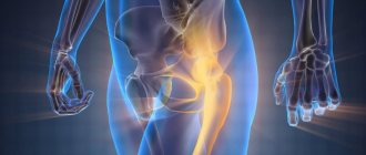
Hip tumor
Or, in other words, chondrosarcoma is a malignant tumor growing from cartilage tissue.
Chondrosarcoma of bones is distinguished by the following features:
- a slight swelling of the skin is detected;
- swelling appears on the patient’s thigh;
- there is a change in the functionality of organs, they are compressed or displaced under the influence of the tumor;
- walking causes severe pain.
An oncologist can identify and diagnose sarcoma of the pelvic bone. For examination, he prescribes the following types of diagnostics:
- X-ray examination (determines the location of the tumor);
- biopsy (sampling of cancer cells to determine the stage of the disease and the nature of the tumor);
- osteoscintigraphy, represented by three types of examination:
- Computed tomography, which determines the exact size and area of spread of the cancer;
- PET CT, assessing the state of oncology;
- Angiography, which evaluates the condition of blood vessels at the site of tumor spread.
It is worth saying that the sooner the disease is detected, it is fully examined and treatment is prescribed, the more positive the outcome of the disease will be.
Thanks to the modern methods of Israeli clinics, highly professional specialists with extensive experience in this field, and the latest medical technology, more and more people have recovered.
For example, statistics on the treatment of patients by a doctor of the highest category, Igor Kazansky, gives the following indicators: 70% of cases with localized sarcoma and 80–90% of cases when the tumor stopped growing under the influence of chemotherapy.
Prices for hip replacement in Israel
| Medical services | Cost USD |
| Advanced blood test, including tumor markers | 580 |
| Revision of histological preparations - slides and paraffin blocks (on a first-come, first-served basis - results within 14 days) | 650 |
| OR Revision of histological preparations - slides and paraffin blocks (urgently - primary result within 2 days, receptors and colors within 5 days) | 1650 |
| PET CT (urgently – result within 1-2 days) | 1750 |
| Consultation with a specialist oncologist, including development of a treatment protocol | 750 |
| Chemotherapy treatment | From 1 800 |
| Surgery | From 14 000 |
Survival Prognosis for Patients
The prognosis for Ewing's sarcoma and neuroectodermal neoplasms is quite unfavorable. The outcome of treatment depends on the stage of the disease at which the patient seeks medical help and the degree of spread of the tumor process. Patients with metastases in the lungs have a better chance of recovery than patients with evidence of metastases in the bone marrow or bones. If the tumor is detected in a timely manner, when it has not yet had time to disseminate, then the chances of a complete recovery are quite high - up to 70%.
Modern chemotherapy drugs do not have as severe side effects as before. During chemotherapy and radiation treatment, most complications can be avoided. But the success of therapy also depends on the patient himself. It is very important for the patient to understand that sarcoma is not a death sentence. The capabilities of modern medicine make it possible today to overcome almost any disease. If you do not create obstacles to treatment, follow all medical recommendations, look for the best doctors and believe in your own recovery, then the disease will recede.
Time is very important. If even the slightest signs of illness appear, it is necessary to carry out a diagnosis. Having discovered a tumor, doctors will do everything possible to destroy the pathological process. The patient needs to mobilize, tune in to long-term treatment, accept the support of his family and think only about the best.
Chondrosarcoma
Chondrosarcoma is a type of malignant tumor that affects cartilage tissue. The disease is quite common, accounting for up to 10-15% of all bone tumors. It is diagnosed in people of all ages, but more than 60% of patients belong to the mature age group (after 40 years). In 15% of patients with chondrosarcomas, the tumor develops secondarily from osteochondral exostoses, chondromas, and deforming osteosis.
It has been noted that chondrosarcoma most often affects the bones of the pelvis, upper limbs and ribs. Men get sick slightly more often than women. The etiology of the disease is not completely clear. Predisposing factors include gene mutations, the presence of certain hereditary pathologies, and carcinogenic effects on the human body.
Signs of chondrosarcoma
Chondrosarcoma is characterized by an asymptomatic gradual course. At the initial stage of development, there are no pathological signs. As the tumor grows, pain begins to occur in the area where the chondrosarcoma is located. If it is located near the joint, then in the clinical picture of the disease there are signs of limited movement.
If the tumor begins to compress the sciatic nerve (if localized in the thigh area), then the pain changes from minor to unbearable. The skin above the tumor becomes swollen; the tumor itself, which has reached a significant size, can be easily palpated. In 15% of patients, fast-growing chondrosarcomas are found, prone to accelerated growth and rapid malignancy.
Hip cancer
The main symptom of this disease is severe pain.
Moreover, the area where pain is felt depends on the location of the tumor.
Severe pain in the buttock or thigh indicates that the tumor is located near the lumbosacral spine.
The appearance of pain in the hip joint indicates the presence of a tumor in it.
In addition, pain increases over time as the focus of cancer cells increases.
And, conversely, the small size of the malignant formation causes pain that is still weak.
Methods for diagnosing chondrosarcoma
The capabilities of modern medicine make it possible to detect bone tumors already at the initial stage of development. But since chondrosarcoma is rarely detected at an early stage of growth, diagnosis can be delayed. Many patients and doctors mistake pain, inflammation and other early signs of the disease for arthrosis, bursitis, injuries and are in no hurry to conduct a detailed examination. And the outcome of treatment and the further prognosis of the disease largely depend on the timeliness of diagnosis.
Among the modern and popular diagnostic methods for chondrosarcoma are the following:
- X-ray examination: the images reveal foci of destruction with areas of calcification, the tumor itself and signs of its growth into soft tissues;
- osteoscintigraphy;
- magnetic resonance, computed tomography;
- biopsy of tumor tissue with further study under a microscope.
Patients with chondrosarcomas should be examined in large diagnostic centers that have modern technical equipment and a professional staff of specialists who know the technique of decoding the diagnostic results obtained.
Central chondrosarcomas often have intraosseous areas of destruction. On x-rays, characteristic calcifications are diagnosed with such neoplasms. Areas of calcification are often diagnosed along the edges of the neoplasm.
Treatment options for chondrosarcomas
In the early stages of tumor development, radiation treatment with surgical excision of chondrosarcoma is used. If the disease has a high risk of malignancy, then minimally invasive operations have to be abandoned and complete amputation must be performed. Partial bone removal with subsequent plastic surgery is appropriate only if long-term tumors that are not prone to malignancy and metastasis are detected.
Chondrosarcoma cells are quite resistant to radiation exposure (especially at later stages of development) and chemotherapy. Therefore, these methods are perceived not as the main ones, but as auxiliary methods for the treatment of chondrosarcomas. The main emphasis is on complete surgical excision of the pathological focus and metastases. When a tumor grows in the pelvic bones and humerus, it is sometimes necessary to perform extensive surgery to remove half of the pelvis and amputate the entire upper limb. But in any case, doctors try to perform organ-preserving operations if the situation and clinical picture allow it.
Forecast
The prognosis for recovery of patients with chondrosarcomas is quite favorable. It directly depends on the timeliness of treatment, the degree of malignancy of the tumor and the quality of the operation performed. Treatment of chondrosarcomas requires considerable professionalism from doctors. Therefore, when choosing a clinic, you should give preference to specialized institutions with considerable experience in treating bone tumors and an individual approach to each individual patient.
After successful surgical intervention, more than 25% of patients live for 5 years or more. To prevent recurrence of the pathology, such patients are recommended to undergo regular diagnostics and preventive examinations.