Dear patients! The specialist will inform you about the exact cost and scope of necessary procedures after consultation.
Is it possible for pregnant women to have dental x-rays?
X-ray research methods in one form or another are used in many areas of medicine, including dentistry. The equipment for performing them is constantly being improved, X-rays are becoming safer for the patient.
However, dental x-rays during pregnancy are fraught with some risks for the development of the fetus. Let's find out whether pregnant women can have dental x-rays.
X-ray as a diagnostic method
Before answering the question of whether pregnant women undergo dental x-rays, you need to understand the very essence of this research method.
To diagnose various diseases, doctors use many types of tests:
- X-ray methods
: visualization of organs and systems through the use of gamma radiation. These include individual images, fluoroscopy, tomography (layer-by-layer examination), including computer; - endoscopic examination
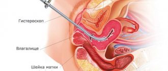
: using a special apparatus (endoscope), the doctor examines the internal cavities and organs (gastroscopy, bronchoscopy, colonoscopy, rectoscopy, hysterosalpingoscopy, etc.; - Magnetic resonance imaging
: This is a study that uses nuclear magnetic resonance. With MRI there is no X-ray radiation or radiation exposure to the patient; - functional diagnostics
: ultrasound, ECG, EEG, EchoCG.
Features of dental x-ray
Dental radiography has several types. The choice of a specific type of dental x-ray during pregnancy, as well as without it, is influenced by the purpose of the study (whether it is possible to do x-rays in principle is decided by the doctor).
If you need to see the structure of the jaw, examine the entire upper and lower dentition, assess the condition of the gums and bone tissue, it is best to do orthopantomography. In another way, such an X-ray is called a “panoramic image.”
If the doctor is only interested in one specific tooth (or several adjacent ones) and its features, it is better to take a targeted picture using a computer visiograph.
Both of these X-ray diagnostic methods are characterized by minimal radiation exposure, so if there is an urgent need, one of them can be prescribed to a pregnant woman (preferably in the second trimester).
Security measures
Before X-rays are taken, the patient must wear a protective vest or apron. Inside it is a lead plate that blocks the X-rays and prevents them from affecting areas that are not being diagnosed.
Before any type of x-ray, the patient should remove metal objects from himself, as they can interfere with the passage of x-rays, which will negatively affect the quality of the image.
Contraindications
It is generally accepted that the first trimester of pregnancy is a contraindication for dental x-rays. It is in the initial period that all the organs and systems of the child are formed, and in no case should you interfere with this process or interfere with it. Even if a woman has a certain pathology of the oral cavity that requires detailed diagnosis, x-rays are usually postponed until the second trimester.
When can you do without a photo?
When dental caries is visible to the naked eye or gum pathology is clearly visible, the dentist can begin treatment without an x-ray.
If the patient is diagnosed with cementoma, an abscess has developed, or multiple cysts have formed, the x-ray may turn out dark and uninformative. In this case, there is no point in going through such a procedure.
Visiograph - an alternative to x-ray
A visiograph is a modern analogue of an x-ray machine. Its advantages:
- Allows you to take an image instantly, high-quality data is immediately displayed on your computer monitor.
- A narrow beam of rays is directed exclusively at the diseased tooth, does not affect adjacent tissues, and certainly cannot “touch” the abdomen.
- The duration of exposure (irradiation) is 10 times lower than when using old-style X-ray machines and is only 0.05–0.3 seconds. Radiation exposure is only 2 microsieverts (when using old devices - from 7 to 80 microsieverts).
For comparison, just sitting in front of the TV for three hours and enjoying your favorite series, a mother receives a radiation dose of 5 microsieverts. The safe radiation dose per year is 1,000 microsieverts (that’s 500 shots). Of course, you shouldn’t take the procedure lightly, but you shouldn’t panic and overestimate the danger to the baby either.
Photo of teeth at different times
In early pregnancy (1st trimester)
Why pregnant women should not have dental x-rays done in the first trimester of pregnancy was stated earlier. If a woman had an X-ray examination of her teeth at a time when she did not yet know about her situation, she should not worry too much about this, much less think about what pathologies this will cause in the fetus. In any case, a routine ultrasound will be performed in each trimester of pregnancy, which will show how the child is developing.
In the 2nd trimester
The second trimester has always been considered the safest period of pregnancy. The baby’s organs and internal systems have already formed, and now they are gradually developing and growing. It is during this period that women can be prescribed x-rays (but choose methods with minimal radiation) if there are certain indications.
What is the tactic for referring a pregnant woman for an x-ray?
I want you, as patients, to understand the entire logical chain of a doctor making such a decision.
1. We make a presumptive diagnosis and understand that in this case an x-ray is needed.
2. We make sure that in a particular situation we cannot replace x-rays with another research method (for example, ultrasound).
3. We weigh the risk-benefit ratio for mother and fetus during X-ray examination. We calculate all possible options for the development of the disease and complications.
4. Once again we are convinced that an x-ray is still needed.
5. We send the pregnant woman for an x-ray according to vital indications. During x-rays, we use protection (lead apron) for maximum protection of the fetus - this is called shielding.
6. If a woman refuses an x-ray, we take a written refusal. We explain all possible consequences, complications and variants of the course of the disease if you refuse this study.
7. We draw up a treatment and examination plan in such a way as if our fears, which an x-ray could show, were confirmed. Or we choose a wait-and-see approach (observe), if possible.
7. We use all other research methods to maximally confirm the diagnosis and prescribe correct and adequate treatment!
MRI - magnetic resonance imaging
The second safest method after ultrasound is MRI - magnetic resonance imaging. This research method is based on the phenomenon of nuclear magnetic resonance. Given that the MRI machine does not provide radiation exposure, the procedure is relatively harmless during the second and third trimester. But it should be carried out with caution, since during the study some heating of the body occurs, which can affect some fetal tissues and stimulate labor.
In general, MRI has been used for a long time around the world to diagnose both maternal health problems and congenital malformations in the fetus. In an infant, tomography is often the only means of confirming changes suspected by ultrasound. A study conducted by Philips in a hospital in India showed that in women who, according to ultrasound results, induced termination of pregnancy was recommended due to detected developmental anomalies, after an MRI, the diagnosis was refuted in 21% of cases, and in 16% of cases the detected anomalies were not required termination of pregnancy.
It is important that magnetic resonance imaging did not show false positive or negative diagnoses, which was later confirmed.
Article on the topic
Pregnancy test: when to do and which one to choose
Risks of dental caries
A child is a key task for a pregnant woman’s body.
If he needs something, the body would rather give up resources to its detriment than allow a shortage for the future baby. A huge amount of calcium is required for the formation of bone and muscle structures of the fetus. Normally, if mom continuously chews cottage cheese pancakes, washing them down with whole milk, calcium will not be a problem. In the case of pathology of calcium metabolism or poor nutrition of the mother, the concentration of calcium in saliva may decrease. This leads to a decrease in its remineralizing properties, since normally saliva replenishes calcium ions washed out under the influence of bacterial acids. As a result, if nothing is done, during nine months of pregnancy you can not only get new foci of acute caries, but also have time to lose several teeth, where the carious process from chronic will turn into a rapid acute course.
In the first trimester, we try not to touch small carious lesions and deal only with remineralization. But in the future, caries must be treated.
Questions about the intricacies of screening
One of the most pressing issues in pregnancy management is screening, which can determine genetic abnormalities in the child. Screening also determines the degree of risk of abnormalities that are incompatible with life. For many mothers, this can be an alarming moment. We asked our expert some particularly concerning questions about screening.
Article on the topic
CT, MRI, ultrasound, x-ray: what types of studies are there and why are they needed?
— The screening procedure consists of a blood test and ultrasound examination (ultrasound). Analysis of the results of these two studies determines the risk of developing genetic abnormalities in the baby (for example, Down syndrome). However, screening sometimes gives false results. More than one such case has been described when a mother was recommended to have an abortion, but she refused and gave birth to a normal child. What causes the errors? How to avoid them?
— There is not a single 100% diagnostic method, so in the first trimester they conduct a blood test and an ultrasound to determine the degree of risk, after which women at high risk undergo a consultation. It is attended by an obstetrician, a geneticist and an ultrasound diagnostician, whose task is to determine the indications for chromosomal analysis (karyotyping) of the fetus. If chromosomal abnormalities are detected, the doctor discusses the possibility of terminating the pregnancy with the woman and her family, who make the final decision. Only parents can make the final decision about the fate of a potentially sick child, in accordance with their moral, religious beliefs, family circumstances or financial capabilities.
Unfortunately, errors are possible. They can be objective and subjective. The first include individual characteristics of the development of the embryo and fetus and some “dark spots” of our general knowledge, as well as inappropriate equipment and working conditions of the doctor, mainly the limitation of research time. The second includes insufficient competence of the doctor.
— Is there any way to check the screening results? Go through it again?
- Of course, if the diagnosis is unfavorable, the mother can (and is probably to some extent obliged) to check it and undergo additional research. The main method of verification is expert ultrasound examination by a specialist of a higher professional level, as well as karyotyping. For some abnormalities, MRI may be used.
Article on the topic
Nuances of tomography. What is more effective: CT or MRI?
— From a human point of view: is it worth the risk of carrying a potentially sick child?
It all depends on the disease. Now the attitude towards children with Down syndrome has changed a lot, they are successfully socialized. But there are defects that are incompatible with life or that significantly limit physical and mental capabilities, which parents should be aware of. Everyone decides for themselves.
How is a sinus x-ray performed?
The procedure is not painful and does not require preparation. Receive a referral at an appointment with an otolaryngologist, infectious disease specialist or surgeon.
How to do a sinus x-ray:
- The patient removes metal jewelry, glasses, and dentures.
- To protect against radiation, he wears a lead apron or vest with a collar.
- The laboratory assistant indicates how to position yourself correctly relative to the apparatus. Depending on the required projections, the position is changed at the command of a specialist.
- While taking the photo, you should not move and hold your breath.
- After development and decoding, the film with the description is given to the patient.
The conclusion is not considered a final diagnosis, but provides clarifying information for the treating specialist.
X-ray of the sinuses for children
Pediatricians try to protect preschool children from the harm caused by X-ray radiation to the fragile skeletal system. The procedure is allowed to be performed on patients over 7 years of age.
Before this period, a clear justification for the importance of intervention is necessary - for example, severe facial trauma or severe sinusitis with a risk of inflammation of the meninges.
It is difficult for a young child to sit or stand still during an x-ray. To distract him, they use toys, sedatives, and in emergency cases, anesthesia. At an older age, you can captivate your child with a game in which you should freeze for a short time.
Sign up for the study
Risks for mom
You can lose a tooth
We try not to use antibiotics, especially in the early stages of pregnancy.
This greatly changes the treatment tactics if we discover a chronic or aggravated purulent focus that has arisen due to an affected tooth. In a normal situation, we can try to save the patient's tooth under the additional cover of antibiotics that accumulate in the bone tissue. For example, this is a fairly common fluoroquinolone - ciprofloxacin. Everything is great, but it is strictly prohibited for use by pregnant women and children under three years of age due to the fact that it can cause defects in the formation of joints and cartilage. In a situation where the prospects for a tooth without antibiotic therapy in a pregnant woman are questionable, and the risks of developing an infection are high, we are usually inclined to remove such a tooth. It is much better to remove a potentially problematic tooth than to risk the health of the baby or the expectant mother. You cannot leave a chronic purulent lesion: it is dangerous for the child and for herself if it eventually worsens. If periostitis or, especially, phlegmon develops, the mother’s life will have to be saved. Then you will definitely have to prescribe antibiotics and other drugs.
Orthodontics
We stop the orthodontic treatment process if the patient reports pregnancy. First of all, we do this due to the impossibility of X-ray monitoring of the treatment process. Working with a bite blindly is a very bad idea and can end in even bigger problems. Most often, the patient remains at the stage when she found out about pregnancy. Either we continue to use the same aligner, or we simply fix the current condition in the case of braces.
Periodontal problems
The pregnant woman's body directs all efforts to pregnancy.
At the same time, he may ignore some of his own needs and problems. For example, large doses of sex hormones ensure good blood supply to the uterus and the desired condition of the mucous membranes. But in parallel, these same mechanisms lead to gum hypertrophy in the oral cavity. This is especially pronounced in the last trimester, when it becomes noticeable. According to various authors, gingivitis in pregnant women occurs in 25–100% of women, usually from the second to the eighth months of pregnancy. Moreover, in the region, 80% of women experience bleeding gums and have at least chronic catarrhal gingivitis. For the most part, all these symptoms will go away after childbirth and normalization of hormonal levels. However, without high-quality professional hygiene, there is a risk of developing more severe forms of periodontitis and damage to its structures. This also creates additional risks of accumulation of subgingival dental plaque and the development of multiple cervical caries.
To prevent such problems, we always recommend professional hygiene and give recommendations on what to do at home.
Importance of gestational age
Before sending a pregnant woman to take an X-ray, the doctor must find out how far along she is. All dentists at the Planet of Childhood clinic in Moscow interview their pregnant patients in detail and find out the duration of their pregnancy. You can prescribe any x-ray examination or draw up a treatment plan only in accordance with this information.
In the first trimester, dental photographs are usually not taken, since at this time the fetus is not yet fully formed, except for emergency indications (for example, acute pain).
In the second trimester, X-ray examination is no longer dangerous for the fetus, and the dentist can safely refer the woman for diagnostics. The same applies to the third trimester.
Taking a picture of the teeth is much safer than taking a picture of the pelvis or torso, since in a dental clinic a woman’s vulnerable spots are necessarily covered with special protective elements that do not allow X-rays to pass through.
What's the result?
Pregnant women are very complex, fragile and emotionally vulnerable patients.
We always try to be especially attentive to their treatment. If a difficult situation arises that requires the prescription of some potent drug, we often arrange a consultation with the obstetrician who is caring for the patient’s pregnancy in order to jointly make the least risky decision for her and the baby. Pregnant women have other specific difficulties. For example, we cannot keep them in a chair in one position for a long time. Expectant mothers should not lie on their backs for a long time, as this position compresses the blood vessels supplying the uterus. After some time, the child usually begins to push indignantly and demand to change the position. It is necessary to give the opportunity to turn on one side or even stand up and walk around, if the treatment protocol allows.
In pregnant women, the volume of the bladder is greatly reduced, which is compressed by the growing uterus. This also has to be taken into account when planning long-term manipulations. In short, there are a lot of nuances, and they are all important. The most important thing is to remember that refusing dental treatment and support often carries greater risks than using local anesthesia or even x-rays.
Risks for the baby
The general approach of dentists generally coincides with doctors of other specialties.
In the first trimester, we try not to perform any unnecessary manipulations or medicinal effects without emergency indications. If a pregnant woman has developed pulpitis and is suffering from acute pain, we will treat it urgently. If during the examination we find a carious cavity, we will postpone it until the second trimester, but we will take all preventive measures to reduce the risks of further development: we will professionally clean the teeth, remove tartar and carry out remineralization therapy. Starting from the second trimester, most manipulations can already be done.
X-ray
X-rays are always very scary for expectant mothers.
Ionizing radiation does pose a potential danger to the unborn child. Like most harmful effects, X-ray radiation in the first trimester of pregnancy leads either to its termination, or the pregnancy continues without pathologies. If exposed to X-ray radiation at a later date, there may be a risk of problems in the development of the nervous system and other organs. Therefore, we try to never use x-ray examination without serious indications, when it is impossible to do without it. The most dangerous for the fetus are studies that directly examine the pelvic and abdominal area. The maxillofacial area that the dentist works with is one of the least risky options. If the doctor is forced to decide on the need to take an image, then you need to remember that the child is reliably protected by a lead apron, and the beam of the device for taking targeted images is quite narrow and directed almost horizontally. The dose received by the mother is approximately 0.02-0.03 mSv. This is 1/50 of the permissible annual dose. In this case, the child will receive an even lower dose.
However, until the very end of pregnancy, we try not to use x-rays at all, if the clinical situation allows it. For example, x-rays are highly desirable when diagnosing hidden cavities on the contact surface between two adjacent teeth.
Diagnocam by Kavo. It illuminates the tooth with powerful side lighting and simultaneously takes a photo from above.
In pregnant women, we instead use auxiliary types of examinations such as transillumination. With this method, questionable areas of enamel are illuminated with a powerful point source of light.
Superficial caries using the Diagnocam device.
Based on refraction and absorption defects, the doctor can determine the hidden cavity.
X-rays are absolutely necessary for any endodontic manipulation, when we treat pulpitis or periodontitis, working in the canals of the tooth. The classic option involves a CT scan of the dentofacial area and a targeted control image after filling. For pregnant women, we change tactics. If the patient has already been with us before, then her earlier CT scan or other X-ray images will probably be stored in the database. It will be possible to use them to clarify the topography of the channels.
But even without earlier photographs, a dentist can perform high-quality treatment if the clinic has the appropriate equipment. For example, we use high-quality microscopes from Carl Zeiss for endodontic treatment. They allow you to see all the details of the anatomy of the root canal mouth and accurately control the process. In addition, we make sure to use an apex locator. This is a device that, by changing the resistance of the probe, informs the doctor that it has reached the root apex. Thus, we can perform treatment without the use of x-rays, following all protocols. But we will still do an x-ray later, when the girl has successfully given birth.
There are some situations where the risks of working without x-rays are too great. For example, if a late pregnant woman comes to the clinic with an acute purulent process in the area of the eighth tooth, we will insist on taking an image. The risks for the child’s development are very low, but complex tooth extraction with unknown root topography carries risks of bleeding, trauma to bone structures and a sharp deterioration of the infectious process. Of course, x-rays will be taken in case of jaw fractures or other life-threatening situations.
Anesthesia
Anesthetic activity, duration of action, toxicity and maximum permissible dose of various local anesthetics.
Anesthesia is a very important issue in dentistry. It was Soviet punitive medicine that created a layer of patients with terrible dental phobia. All because of the wonderful concept “Be patient, little one, everyone is in pain.” At the same time, there were no problems with the availability of anesthetics, at least lidocaine and novocaine.
If you look at the table above, you can see that typical anesthetics vary greatly in their systemic toxicity and effectiveness. The time of action is also expressed in two columns - with and without a vasoconstrictor. Now I'll tell you how it works.
To begin with, several anesthetics are rarely used in dentistry today. Novocaine is the least toxic and can actually be administered in large quantities. Previously, sometimes even abdominal surgeries were performed using it. But it has a big disadvantage: it is at the same time the least effective both in terms of anesthesia strength and duration. Enzymes quickly break it apart.
Bupivacaine and ropivacaine are powerful and last up to six hours. But they are also the most toxic. Bupivacaine is eight times more toxic than novocaine in equal doses.
As a result, we usually use the following anesthetics:
- Lidocaine for topical local anesthesia in the form of a gel. This is only necessary for pain relief of the mucous membranes. In compulsory health insurance it is sometimes used for tooth extraction, but this is not the best option.
- Mepivacaine is a low-toxic anesthetic that works very well for infiltration and conduction anesthesia.
- Articaine is a “workhorse” in dentistry. It has an ideal balance of action time, low toxicity and high activity.
In addition to the anesthetic itself, a vasoconstrictor - a vasoconstrictor substance - is added to the capsule with it. Most often it is adrenaline or its analogues. If you do not add adrenaline, the anesthetic will cause vasodilation at the injection site and will quickly be washed out of the area of action. In this case, you will have to either work very quickly or continuously finish off new portions. Everything here is individual and depends on the patient. With a vasoconstrictor, vasospasm occurs and the anesthetic remains at the injection site for a long time.
It is often believed that the use of a vasoconstrictor in late pregnancy can provoke preterm labor. However, research does not support this opinion. Moreover, if we hurt a woman due to the insufficient effectiveness of the anesthetic, then her adrenal glands will release such an amount of adrenaline that they will exceed any dose in a standard capsule with a reserve. However, in order not to worry the patients themselves, we try to use the minimum working doses of anesthetics with a minimum dosage of adrenaline. It all depends on the clinical situation. The most common drug of choice is mepivacaine.
Antibiotic therapy
The use of antibiotics in expectant mothers is possible, but only for vital indications.
Moreover, the later the date, the greater the likelihood that the doctor will allow their use. The baby’s body is already formed at this point, most of the drug will not pass the hematoplacental barrier, but a severe infection of the mother can potentially greatly harm the child. In dentistry, such situations usually include acute purulent diseases, for example, periostitis, resulting from an exacerbation of periodontitis. If we do not apply adequate antibiotic therapy, we will endanger not only the life of the baby, but also the mother.
In all other cases, we will try to make do with local antiseptics and other methods that will avoid the use of antibiotics. In some cases, this carries additional risks for the pregnant woman herself.
Conclusions:
We always weigh the risks and benefits for the woman and the fetus. What's more dangerous? That we do not diagnose a disease (for example, tuberculosis), and do not prescribe timely treatment, or POSSIBLE consequences in the fetus from x-rays?
- If possible, avoid exposure to X-rays during pregnancy;
- If it is possible NOT to do an x-ray, we DO NOT do it or replace it with another research method (ultrasound, MRI);
- If an x-ray is necessary for health reasons, then we do it!
What does a sinus x-ray show?
An x-ray shows the human skull with bone structures, cavities and septa. The method is used at the preparatory stage in surgery.
What can be seen on a sinus x-ray:
- Foreign object in the nasal passages.
- Inflammatory process, thickening of the mucous membrane of the infected area.
- Consequences of injuries to the face and head.
- The presence of exudate (mucous, blood, purulent) in the paranasal and frontal cavities.
- "Airiness" of the sinuses.
- Neoplasms: polyps, tumors, cysts.
- Anomalies in the structure of the facial skeleton.
X-rays help to correct the diagnosis in case of fever or headache of unknown etiology.


