Angiography of the vessels of the eye - what is it?
Angiography is one of the radiographic methods used to study blood vessels. Regardless of the purpose and object of the study (vessels of the brain, limbs, eyeballs), it is carried out using fluorescence. This means that a drug is injected into the patient’s circulatory system that can change color when exposed to an X-ray machine. The contrast agent is distributed throughout all vessels and capillaries, so that they can be examined with maximum accuracy.
The development of fluorescein angiography (FA) was predicted by the physiologist Bekhterev back in 1896. He stated that there are solutions that do not transmit x-rays, fill the vessels and can be photographed. In 1931, Forsman implemented this idea; he performed the first angiography in history. But almost 40 years remained before its introduction into medical practice. Today, this procedure is prescribed for any vascular examination, including ophthalmological examination. Let's find out what are the indications for eye angiography, how it is performed, and whether the examination is accompanied by side effects. These questions are especially often asked by patients to doctors.
Magnetic resonance angiography (MRA)
Like CTA, magnetic resonance angiography is a non-invasive study, that is, it does not penetrate the body. But it allows you to obtain a highly accurate image of the vascular network of the organ under study. There is no need for an operating room or any preliminary preparation of the patient, everything is simple: come and do it. For greater accuracy of the study, and when studying the vascular bed of a tumor without fail, the manipulation is carried out with the introduction of a contrast agent, which highlights the vessels against the general background. The contrast agent for MRA does not contain iodine, but it can also damage the kidneys, although to a lesser extent.
The power of a magnetic resonance imaging scanner and, consequently, the accuracy of the study is determined by the magnetic field created by the device. The higher the magnetic field voltage, measured in Tesla units, the better the image. The simplest “low-field” MRI scanners correspond to 0.5 Tesla, the most accurate are “ultra-high-field” 3 Tesla, but in clinical practice “high-field” 1.5 Tesla are quite sufficient. MRA is more accurate than all other angiographic techniques when examining calcified vessels, but the degree of vessel narrowing may exaggerate.
There are limitations in the use of magnetic resonance angiography in the form of the presence of pacemakers and defibrillators in the patient’s body, which go astray from the program, which threatens the patient with irreparable consequences. It is impossible to study obese patients and those who are terrified of closed spaces - claustrophobia; a magnetic resonance imaging scanner is a tube of limited diameter where the patient is located. The main difference between MRA and CTA is the absence of ionizing radiation - radiation, so it is possible to study children and pregnant women.
Book a consultation 24 hours a day
+7+7+78
Indications for fluorescein angiography of ocular vessels
This research method is prescribed not only for suspected ophthalmological diseases. Indications may include other diseases that affect the condition of blood vessels and can lead to the development of ophthalmopathology. Thus, angiography is recommended for people with diabetes. In this case, there is a high probability of diabetic retinopathy, a disease that is accompanied by damage to the vessels of the retina. It becomes the main cause of complete loss of vision.
The main indications for angiography of ocular vessels are the following pathological processes and conditions:
- Primary signs of retinal dystrophy: decreased visual acuity and narrowing of its fields, scotomas, impaired color vision, “spots” before the eyes.
- Tumors of the choroid: leiomyoma, melanoma.
- Disruption of blood flow to the vessels of the retina.
- Inflammatory processes in the retina.
- Diseases of the optic nerve head: congestive disc, pseudocongestion, neuritis.
Angiography of the eye can reveal other pathological changes:
- Thrombosis of the central retinal vein. With this ophthalmopathology, the veins become more tortuous, they expand, and the retina swells. Hemorrhages occur. The patient's vision deteriorates sharply. Pain appears, flashes and lightning flash in the eyes.
- Occlusion of the central retinal artery is a violation of the patency of the vessels of the inner lining of the eye. The disease manifests itself in a severe decrease in vision, the appearance of a veil before the eyes, pain and stinging.
- Vasculitis is an inflammatory process in blood vessels. The patient complains of poor vision, decreased visual acuity at night, and dry eye syndrome.
- Optic nerve atrophy is the gradual drying and death of fibers. Thanks to the work of the optic nerve, the image received by the retina enters the cerebral cortex. With atrophy, all visual functions deteriorate, which cannot be restored.
- Retinal tumors: melanoma, retinoblastoma. These diseases affect not only the quality of vision, but also the mobility of the eyeballs. It is important to detect tumors in the early stages, when they are treatable.
- Retinal detachment. Rupture, detachment and degeneration of the retina can occur for various reasons. There is a high probability of developing this pathology in patients with severe myopia. Retinal detachment without proper treatment leads to blindness.
All of these pathologies are considered very dangerous. Because of them, a person can become visually impaired. The advantage of vascular angiography is that it allows not only to identify the disease, but also to determine the nature of its development, stage and possible ways of worsening the condition.
Our clinic specialists perform:
- angiography of the coronary, brachiocephalic, renal arteries, aortography, angiography of the vessels of the lower extremities, panangiography,
- permanent and temporary embolization of the uterine arteries,
- cerebral angiography,
- angioplasty and stenting of coronary, brachiocephalic, renal arteries,
- recanalization, angioplasty and stenting of the arteries of the lower extremities,
- embolization of intracranial vessels,
- implantation of stent grafts (prosthesis of the abdominal aorta),
- ventriculography,
- angiopulmonography,
- phlebography,
- cholangiography,
- drainage of bile ducts,
- targeted administration of drugs, chemoembolization,
- transesophageal electrophysiological study,
- transesophageal ischemic test,
- programming artificial pacemakers (pacemakers), cardioverter-defibrillators, devices for cardiac resynchronization therapy,
- endocardial electrophysiological study,
- implantation of a temporary and permanent pacemaker (one- and two-chamber pacemaker),
- implantation of a cardioverter-defibrillator (one- and two-chamber),
- implantation of devices for cardiac resynchronization therapy,
- catheter ablation of arrhythmogenic zones of the heart for tachyarrhythmias (radiofrequency catheter ablation),
- implantation of devices for long-term ECG recording,
- transesophageal restoration of heart rhythm during reentrant tachycardia.
Rehabilitation measures after angiography
It is important to apply a pressure bandage to the puncture site to minimize the risk of bleeding. After the procedure, the patient must be under the supervision of medical personnel for at least 6-8 hours, during which time the doctor monitors his well-being. If the condition is satisfactory, you can continue rehabilitation at home. It is necessary to refrain from physical activity for several days, and also follow all the instructions of the attending physician. In general, specific rehabilitation measures are not required during this period.
Minimally invasive (not requiring incisions) x-ray endovascular technologies are a new alternative in the diagnosis and treatment of many diseases, often superior in their effectiveness and safety to conventional surgical and therapeutic methods.
Do I need to prepare for the procedure?
There is no need to take tests in advance, undergo preliminary studies, or adhere to diets before fluorescein angiography. However, allergy tests may be taken. During the conversation with the doctor, he will ask if you are allergic to any drugs, what medications you use, whether you have had eye surgery, or whether you wear contact lenses. They will have to be removed before the procedure. After this, the ophthalmologist instills mydriatic drops into the patient’s eyes. They act on the ciliary muscle and cause artificial mydriasis - dilation of the pupils. This will allow you to enlarge the image of the inside of the eyes and conduct a more detailed examination. The drug will take effect in about 10-15 minutes. After this, the immediate examination of the vessels begins.
How is the procedure done?
How angiography is done can be described as follows:
- administration of antihistamines (even if a preliminary test showed that there is no allergy to iodine);
- antiseptic treatment of the area of the body where the puncture will be made to introduce contrast;
- anesthesia (usually lidocaine is used);
- incision of the skin to provide access to a vein or artery;
- installation of an introducer (hollow tube) to facilitate further insertion of the catheter;
- administration of a drug that prevents vascular spasm;
- inserting a catheter into a hollow tube and moving it to the beginning of the vessel to be examined;
- administration of contrast;
- shooting.
After the shooting is completed, the catheter is removed, hemostatic measures are carried out (if there is bleeding), and then a tight bandage is applied to the puncture site.
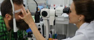
Performing fluorescein angiography
The patient sits in front of the device with the camera and places his chin on the stand. The doctor injects a dye through a vein in the arm. It reaches the blood vessels of the eye in 10-15 seconds. The camera starts taking pictures. Usually 1 photo is taken per 1 second. In total there are about 25-30 of them. The ophthalmologist may ask you not to move or blink during the examination. Next, the doctor takes a break for 20-30 minutes, after which several more pictures are taken. It is necessary to monitor how the substance affected the retina and whether hemorrhages occurred. An examination of the second eye, if required, is carried out after half an hour.
Where is the best place to do angiography of the cerebral arteries?
Such examinations are carried out mainly by large clinical and diagnostic centers that have the appropriate equipment, in particular:
- a fluorography unit operating at high speed;
- angiograph - a specialized system for X-ray studies of the vascular bed;
- camera for X-ray photo and video recording of results.
The most modern approach in this area is CT angiography. Devices for its implementation provide higher quality and more detailed images, and therefore more reliable results.
Find out the price
Find out the price
Error! Please fill in all required fields
Thank you! We will contact you shortly
✕
How are the results interpreted?
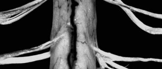
The images should show the vascular network. An experienced doctor will draw many conclusions from it. In almost any vascular disease, they change their shape, narrow or widen, and become tortuous. In the presence of aneurysms—expansion or protrusion of arterial walls—the fluorescent substance extends beyond the vessels, forming luminous spots on the retina. Collaterals may be observed - new vessels that provide blood supply bypassing the main channel. The condition of the blood vessels can also tell us about other pathological processes. Thus, low luminosity of the dye indicates one of the following diseases:
- neoplasm of the retina;
- hemorrhage;
- thrombosis;
- degeneration of the iris.
A very bright glow of the vessels, hyperfluorescence, indicates the onset of atrophic processes and damage to the optic nerve. Normally, it shows the absence of degenerative changes. In other words, based on the images obtained, the ophthalmologist is able to identify a variety of diseases. Their causes can also be determined.
How is pulmonary angiography performed?
Pulmonary angiography begins with puncture of the right subclavian or jugular vein, into which a 6 FR introducer is installed. The right parts of the heart are catheterized through the introducer on the guidewire using diagnostic catheters. A diagnostic catheter is installed in the pulmonary trunk in the area of bifurcation of the right and left main pulmonary arteries. Using an automatic injector, contrast is injected into the pulmonary artery. Next, to visualize the right PA, the catheter is inserted into the right main PA and 40 ml of contrast is injected. To visualize the left PA, a catheter is placed in the left main PA and the same volume of contrast is injected. During the administration of contrast, the patient holds his breath, and the surgeon performs an examination of the pulmonary artery in two projections.
The next step is to insert a Swan-Ganz catheter through the right side of the heart into the pulmonary artery. The distal end of the Swan-Gantz catheter is installed in one of the branches of the pulmonary artery. After measuring the indicators, the average pressure in the pulmonary artery is calculated. Next, air is pumped into a special port of the catheter with a syringe, thereby jamming one of the branches of the pulmonary artery. In this way, pulmonary artery wedge pressure is measured. Also, during angiopulmonography, cardiac output is measured using the thermodilution technique.
Next, the resistance of the vessels of the pulmonary circulation is calculated. A mandatory stage of the study is cavagraphy - the catheter is lowered below the renal veins to detect possible blood clots and search for the mouths of the renal veins.
Undesirable consequences
The doctor warns you in advance about all possible side effects. The dye is eliminated from the body quickly, it is safe, but it is impossible to predict 100% how the body will react to it. Undesirable consequences of angiography include:
- dizziness, nausea, vomiting;
- fainting;
- hives;
- attack of coughing or sneezing;
- blurred vision, tinnitus;
- swelling of the larynx;
- unpleasant metallic taste in the mouth;
- paresthesia (tingling sensation, goosebumps);
- anaphylactic shock.
Side effects occur extremely rarely, occurring in 5% of patients. In most cases, all these symptoms go away on their own within a short period of time. Drops to dilate the pupil may cause some discomfort. They act for about 4-5 hours, during which photophobia and problems with focusing are bothersome. Bring sunglasses with you to the clinic.
What preparation is needed before the procedure?
Before angiography, the patient undergoes a series of laboratory and instrumental studies. This is necessary to know for sure whether the body is ready for the study and whether there are contraindications to the procedure or not. For example, will the kidneys be able to remove the contrast agent, will there be an excessive reaction to it. It is necessary to inform your doctor about chronic pathologies, allergies, and pregnancy. A week before the procedure, you must give up alcohol and also stop taking medications that thin the blood, such as warfarin, but only in consultation with your cardiologist. Angiography is performed on an empty stomach. The area in which the puncture will be made is freed from hair.
Are there any contraindications for angiography?
It is not always advisable to carry out this diagnostic method due to the fact that a dye is introduced that can cause side effects. There are contraindications for angiography:
- glaucoma;
- diseases in which clouding of optical media is observed;
- cardiovascular pathologies;
- neurological problems;
- renal failure;
- allergy to a fluorescent substance;
- inflammatory diseases of the veins;
- asthma;
- inability to cause artificial mydriasis;
- suffered a stroke or heart attack;
- psychical deviations;
- epilepsy;
- regular use of antidepressants;
- age up to 14-16 years.
Angiography is also not recommended for older people over 65 years of age. Test results may not be accurate. In addition, at this age people often suffer from various chronic diseases. Usually, the procedure for examining blood vessels using dye is not prescribed for pregnant women, especially in the first three months of gestation.
An exception may be the suspicion of a neoplasm. Nursing mothers will not be able to feed their baby breast milk for 4 days after the procedure, until the dye is completely removed from the body. Almost all contraindications are relative. Even after a stroke or heart attack, the procedure can be prescribed, but only after the patient has undergone rehabilitation.
Computed tomography angiography
This technique is fast. To carry it out, there is no need to invade the patient’s body. The patient is injected with a contrast agent containing iodine into a vein. Using a computed tomograph, the necessary images are obtained.
CT angiography makes it possible not only to visualize the bed of the vessel, but also to give an idea and reconstruction of the blood supply at any time, to obtain a picture of the lumen of the vessel “in its natural form,” and to accurately assess the degree of its narrowing. Difficulties can arise when calcium is deposited in an atherosclerotic vessel, since it is impenetrable to X-rays.
The number of images in one projection per second is the main principle of classification of computed tomographs. Today, 16-, 32-, 64-, 256- and 320-slice tomographs are used. The more pictures you take, the clearer the image. In addition, increasing the number of images minimizes the negative impact of the patient’s breathing on the image and reduces the amount of contrast agent in the body.
With all the advantages of CT angiography, there are contraindications and limitations:
- Patient intolerance to iodine preparations;
- Serious renal impairment;
- Dangerous for children and pregnant women;
- Within 24 hours after the examination, a nursing mother should not feed her baby breast milk;
- It is difficult, and sometimes impossible, to perform the study if the patient is hypermobile or overweight;
- It is impossible to carry out therapeutic actions during the study.
What happens after a pulmonary angiogram?
After the procedure, you will lie in the recovery room for 1 to 2 hours. Your blood pressure, pulse and breathing will be monitored. The groin or arm puncture site will be checked for bleeding. You will need to keep your leg or arm straight. If necessary, you will be given pain medication. You may be able to return home the same day. Or you may need to stay overnight.
At home, you can return to your normal diet and activities if directed by your doctor. Drink plenty of water. This will help wash the contrast dye off your body. Avoid strenuous physical activity for several days. Avoid taking hot baths or showers for a day or two. Check the piercing site in your groin or arm several times a day. Check for bleeding, pain, swelling, change in color or temperature. A slight bruise is normal.