As you will learn from this article, nail dystrophies are a very heterogeneous group of pathologies that require different approaches to diagnosis and treatment.
.
Often years or even decades pass from the onset of symptoms to the correct diagnosis. The specialists of our clinic have extensive experience in working with onychodystrophies and can quickly guide you in which direction to move just from photographs. Don’t be lazy to send us a photo of your problem on Whatsapp (link in the banner above) and we will advise you for free
. Don't waste time and money on unnecessary tests and searching for specialists!
Nail dystrophy - diagnosis, treatment, prevention, care
Onychodystrophy (nail dystrophy) is a trophic disorder (i.e., nutritional disorder) that occurs in the nail apparatus under the influence of external or internal factors. Deformation of the nail plates is often a manifestation of diseases of the internal organs, which can be important when making a general diagnosis. Also, nail dystrophies can be both independent diseases and manifestations of skin pathology. Based on some changes in the nail, many serious diseases can be suspected, and by ordering an additional examination, these diseases can be identified at an early stage of their development. Changes in the nail plates occur primarily as a result of trophic disorders of various origins. Onychodystrophy accounts for approximately 50% of nail pathology, the remaining 50% is fungal pathology.
How to disguise ribbing?
As a temporary solution to the problem, special medicinal compounds are used to level the ribbed surface of the plate. They are applied under varnish instead of a base coat. Which remedy is right for you and whether it is safe to use, you need to check with your podiatrist. Among decorative varnishes, varnishes with mother-of-pearl or sparkles will better hide a cosmetic defect, while plain glossy coatings, on the contrary, will emphasize unevenness.
Under no circumstances should you try to level the surface of the plate with a sanding file, as the nail will become thinner and more vulnerable. It is also not recommended to do extensions or use gel polish until the nail has recovered.
Causes of development of nail dystrophy
Before moving on to an analysis of the types, symptoms, treatment and prevention of onychodystrophies, it is necessary to take a closer look at the structure of the nails.
The nail apparatus consists of:
- nail plate;
- nail matrix and bed;
- accumulations of blood vessels and nerve fibers;
- skin ridges.
The nail plate is a dense horny structure consisting of keratin protein, lipids, minerals and water. Ultimately, the nail plate is a product of onychogenesis (“nail production”), which occurs in the matrix and nail bed. Specific cells that are located in the matrix and nail bed are constantly divided into mother and daughter cells, which, when separated, actually turn into the nail plate, going through a series of biochemical transformations. These cells are called onychoblasts. It is worth noting that the larger of these cells are located in the matrix area, and the smaller ones are located in the nail bed. Due to large cells, the nail grows in length, and due to small cells, it grows in thickness. Cell division occurs constantly as a result of neurotrophic processes, so the nail matrix and bed contain a large number of nerve fibers and blood vessels that nourish onychoblasts. The skin ridges are in direct contact with the nail plate and are often involved in the pathological processes of the nail apparatus. On the posterior skin fold there is a thin skin - the eponychium (cuticle), which performs an important function in protecting the largest onychoblasts, so injuries to this area often lead to serious deformations of the nail plates.
The domestic classification reflects the factors that determine the development of destructive changes in the nail plates:
- infectious diseases of the nails (fungal, bacterial);
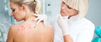
- onychia and paronychia in skin diseases (psoriasis, lichen planus, eczema, neurodermatitis, alopecia, pemphigus, etc.);
- nail lesions due to internal, infectious, neuropsychic, endocrine and other general diseases;
- traumatic and occupational onychia and paronychia;
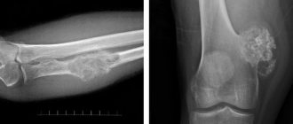
- neoplasms of the nail apparatus;
- congenital, hereditary onychia.
Clinical manifestations of onychodystrophy
Hippocrates nails
The form of the lesion, in which the nails are somewhat thickened and deformed, is often combined with drumstick-shaped fingers. This symptom was first described by Hippocrates in the 1st century BC. e. in patients with empyema (suppuration) of the pleura. Clinically, an expansion of the terminal phalanges of the fingers is noted, they increase in size, become wide, convex, rounded, and the nails acquire a dome-shaped or watch-glass shape, retain a shiny surface and normal color, but their elasticity often changes and they become softer. The posterior and lateral nail folds usually have a bluish, stagnant color.
Previously, it was believed that drumstick-shaped fingers were a characteristic symptom of skin tuberculosis. Today, this peculiar dystrophy is often found in emphysema, bronchial asthma, tumor processes in the lungs, and cardiovascular pathology. It is also observed in patients with leukemia, ulcerative colitis, with neuropsychiatric disorders, as well as with venous congestion in the extremities. It is believed that in 10% of cases this deformity is a variant of the norm and is inherited.
Onycholysis
A type of nail dystrophy often encountered in practice, which is characterized by a disruption of the connection between the nail plate and the nail bed while maintaining the integrity of the latter. We are talking, therefore, not about the dissolution or melting of the nail, but only about its inability to grow to the nail bed. This type of dystrophy was first described by Geller in 1910.
Separation of the nail plate from the bed begins at the free distal edge and gradually progresses towards the proximal edge to the nail lunula area. In most cases, the part of the nail separated from the bed is no more than half the length of the entire nail plate; relatively rarely, the entire nail is separated. The part of the plate that has separated from the nail bed usually retains its normal consistency and smooth surface, but acquires a whitish-grayish color. The exception is cases of onycholysis of fungal and bacterial etiology, when the nail plate can become deformed, its surface becomes uneven, and the color changes.
Depending on the size of the separated part of the nail, partial and total onycholysis are distinguished. Onycholysis develops in systemic diseases, endocrine disorders, many skin diseases, and as a result of injuries.
Koilonychia
Koilonychia (koilonychias; Greek koilos - hollow nail) is characterized by the formation of a more or less deep saucer-shaped depression on the surface of the nail plate. The nail plate with true koilonychia usually remains smooth, of normal thickness, with the gradual formation of a saucer-shaped, spoon-shaped or cup-shaped depression in the central part. If you place a drop of water in this area, it will not flow out.
Koilonychia usually develops on the nails of the hands, most often on the 2nd and 3rd fingers, and very rarely on the toenails. More often than not, several nail plates are affected, but sometimes all nails are involved in the pathological process. Koilonychia can be congenital or acquired. This symptom is observed in iron deficiency anemia, peripheral circulatory disorders such as Raynaud's syndrome, severe infections, and prolonged exposure to alkalis and acids. We have observed koilonychia in rheumatoid arthritis and ulcerative colitis.
Onychogryphosis
Sharp thickening, hypertrophy of the curved nail. Onychogryphosis usually affects single nail plates, mainly on the big toes.
Changes in the nail with onychogryphosis include sharp hypertrophy, changes in texture, color, and direction of growth. The nail becomes convex, first grows upward, then begins to lengthen beyond the top of the finger, bends, and bends like a bird’s beak, hence the name. Sometimes a deformed nail takes the shape of a horn or twists in a spiral so that the length in advanced cases can reach 6–8 cm or more. The surface of the nail is usually uneven, rough, often lumpy, but can sometimes be smooth. The nail plate becomes dirty yellow, brown, often almost black; becomes very dense, like animal horns. Often, with severe onychogryphosis, soft tissue pressure sores are observed.
In the pathogenesis of onychogryphosis, a large role is given to various mechanical, physical, chemical injuries, as well as biological factors. In such cases, we are talking about constant chronic irritation, to which the nail bed and nail matrix react with excessive formation of quickly keratinizing onychoblasts.
This type of onychodystrophy is much more common in elderly and senile people. Flat feet have a certain significance in the formation of finger deformation and onychogryphosis. Poor circulation in the extremities due to frostbite of the fingers can also lead to onychogryphosis. It is known that sharp hypertrophy and deformation of the nails, especially the toes, like onychogryphosis, often occurs with onychomycosis.
Scleronychia
Characterized by the special hardness of the nail plate. In this case, we are talking about acquired onychodystrophy - a unique type of nail hypertrophy. The clinical picture consists of thickening of the nail plates, complete loss of elasticity and separation of the nail plate from the bed like onycholysis. In this case, the nails become yellowish or brown, and the lunula disappears. The transverse axis of the nail becomes curved, while the longitudinal axis becomes only slightly arched. Nail growth is sharply slowed down, the nail skin disappears. The process usually begins on all fingers at the same time and lasts from several months to many years with a tendency to self-heal. It is often observed in people who have been treated for a long time for onychomycosis of the feet using various keratolytics [4].
Transverse groove of the nail (Beau's groove, Beau-Reil's groove)
A transverse, or rather arcuate, groove that crosses the surface of the nail plate from one side ridge to the other is one of the most common types of nail dystrophy. A transverse groove, sometimes with a slightly raised ridge along its posterior edge, appears on the surface of the nail plate after inflammation or trauma to the posterior nail fold or after damage to the nail skin during manicure. The appearance of furrows is associated with eczema and psoriasis, especially if the rashes are localized on the dorsum of the hands. Bo's furrows may appear within 1–2 weeks. after suffering neuropsychic, infectious or systemic diseases in which the function and nutrition of the nail matrix are disrupted. At this moment, the division process of onychoblasts stops, as a result of which the nail plate becomes thinner and a depression in the form of a groove appears on the surface of the nail. The appearance of Bo's furrow has been described in children who have had measles, scarlet fever and other childhood infections. With a minor injury, Bo's groove is mostly superficial, but with severe damage to the nail matrix it can be deep, dividing the entire thickness of the nail into two halves. In such cases, the distal part of the nail plate gradually loses its connection with the nail bed, while the proximal part of the nail continues its normal growth. Thus, by the depth of Bo’s groove one can judge the severity of damage to the nail matrix.
If the damage to the matrix is repeated at short time intervals, then several transverse grooves appear, located sequentially, one after the other, as a result of which the surface of the nail plate becomes wavy. Knowing the rate of nail growth (about 1–2 mm per month, depending on age), you can determine the time of the illness by the distance from the nail fold to the Bo line.
Onychomadesis
(onychomadesis; onycho + Greek madesis - baldness). A relatively rare type of onychodystrophy, which is characterized by separation from the entire nail plate from the bed, not from the free edge, as with onycholysis, but from the proximal part. In contrast to slowly progressing onycholysis, onychomadesis usually develops in a short time. Onychomadesis of the nails of the hands and feet occurs on one, several, and occasionally on all fingers. Mostly the nails on the big toes are torn off.
The process of separating the nail plate from the matrix depends on the nature of the disease and can occur acutely, with inflammatory phenomena, accompanied by pain and a visible inflammatory reaction, or without subjective sensations. Onychomadesis can occur after severe trauma to the nail phalanx of the finger. Cases of relatively rapid rejection of the nail with rapidly occurring paronychia with onychia caused by fungi of the genus Candida or pathogenic bacteria (streptostaphylococcal flora) have been described. Onychomadesis can be observed with scarlet fever (during the period of active peeling of the skin of the hands), rotavirus infection, severe alopecia areata, and psoriasis. In some cases, the mechanism of onychomadesis remains unclear, although the cause is usually associated with circulatory disorders and pathology of the nail matrix. When matrix function is restored, a new, healthy nail plate grows.
Longitudinal grooves of the nail
The formation of longitudinal grooves occurs in cases of peripheral circulation disorders, traumatic damage to the matrix or nail bed, nerve endings in the phalanges of the fingers, as well as lichen planus, psoriasis, gout, chronic rheumatoid polyarthritis and other chronic diseases.
The grooves on the nail plates can be single, located mainly in the central part of the plate, or multiple, occupying the entire surface of the nail. Cases of the formation of two grooves along the lateral edges of the nail in arterial hypertension, coronary insufficiency, and diseases of the spinal cord have been described; at the same time, the central part of the nail plate becomes wider and somewhat flattened with two narrow zones on the sides. Longitudinal lines and grooves may not be continuous, but consist of several component parts, resembling beaded beads - a symptom of beads. We observed such symptoms in patients with liver cirrhosis and HIV infection.
Mi Lines
Transverse white stripes, less deep than Bo's lines, but of the same origin. They are observed in patients with various forms of polymorphic dermal angiitis, periarteritis nodosa, and vibration disease. Usually the stripes cover only part of the nail plate and do not last long.
Median canaliform nail dystrophy
This type of onychodystrophy has a polyetiological nature and a polymorphic clinical picture. More often, a deep channel-like groove 4–5 mm wide is observed in the central part of the nail plate, originating at the root of the nail, gradually moving towards the free edge and dividing it into two equal parts. This dystrophy is characterized by a herringbone symptom - strips extending from the central crack at an angle, which resemble the branches of a Christmas tree. The grooves are localized on the lateral parts of the nail plate. The nail plates of the first fingers of the hands are most often affected, less often - all the rest. Cases of the development of this pathology in members of the same family are described.
Onychorrhexis; onycho + Greek rhexis - withdrawal)
Splitting of the nail plate in the longitudinal direction. A crack can easily form at the bottom of a nail groove, especially a deep one, even with minor and rarely repeated injuries. Initially, the groove splits at the free edge of the nail, then the crack increases in length towards the nail matrix. Onychorrhexis is often combined with thimble-shaped dystrophy, onycholysis in eczema, psoriasis, and is especially pronounced in lichen planus. The development of onychorrhexis can also be caused by constant contact with alkali solutions, formaldehyde, weak acids and other active chemicals that dry out the nail plate [2].
Onychoschisis (onychoschizis; onycho + Greek schisis - splitting)
Nail dystrophy in the form of their splitting in the transverse direction, parallel to the free edge of the nail. In this case, the nail grows normally until the free edge, after which it begins to split (2-3 layers or more), breaks off, or continues to grow in the form of 2-3 very thin plates lying one on top of the other. There are no inflammatory phenomena of soft tissues.
The nails of the 2nd, 3rd and 4th fingers are most often affected. If the nails are cut short, they take on a normal appearance, but the free edge that grows back becomes exfoliated.
In the pathogenesis of onychoschisis, the main role belongs to exposure to chemicals and detergents. This type of onychodystrophy occurs mainly in women who often do manicures using various varnishes and acetone to remove them.
Trachyonychia
A peculiar onychodystrophy, in which the nail plate becomes dull, rough, and may peel off in small thin scales. Trachyonychia is rarely observed in patients with eczema, especially in the presence of a large number of pinpoint impressions on the surface of the nails.
Nail pigmentation disorder
Discoloration of nails varies from white (leukonychia) and light yellow, orange, brown to red, blue, green and black. Leukonychia is one of the most common types of dystrophic disorders and disorders of nail pigmentation, mainly on the hands, rarely on the feet. This term refers to the presence of white areas of different sizes and shapes in the thickness of the nail plate.
The following clinical forms of leukonychia are distinguished:
- dotted, spotted in the form of small, sometimes dotted white spots of various sizes and shapes;
- stripe-like in the form of stripes: one wide transverse stripe or several narrow ones located transversely with respect to the long axis of the nail;
- total, characterized by whitening of the entire nail plate;
- partial, subtotal, in which only part of the nail plate becomes white.
On the same nail plate there can be both spotted (dotted) and stripe-like forms of leukonychia at the same time. With dotted and stripe-shaped leukonychia, spots and stripes gradually move toward the free edge as the nail grows and disappear, almost unchanged in size. With total and subtotal leukonychia, the clinical picture remains constant and persistent. Total leukonychia develops mostly in early childhood, with the nails losing their normal color, starting from the lunula. All clinical forms of leukonychia, except macular, can occur after severe illness, neuritis, poisoning, including arsenic, as well as after measles, scarlet fever, dysentery, etc.
Mührke lines
- these are two white stripes on the nail, parallel to the lunula. They are clearly visible against a pink background and do not move as the nail grows. Muhrke lines are a sign of hypoalbuminemia (low blood protein levels); after normalization of serum albumin levels they disappear. They are especially common in nephrotic syndrome. The reason for the appearance is unknown.
Terry's sign and two-color nail
. Terry's sign: The proximal two-thirds of the nail is white, the distal third is pink. The symptom is quite rare, mainly in heart failure and liver cirrhosis, accompanied by hypoalbuminemia. Two-tone nail: The pink or brown distal half is sharply separated from the milky white proximal half of the nail. The hole is not visible. A two-color nail is found in 10% of patients with uremia. The severity of the symptom does not depend on the severity of renal failure. Some experts consider both symptoms to be manifestations of the same pathology [7].
Hyperpigmentation
. Changes in nail color may be due to the accumulation of melanin, hemosiderin and other pigments. The entire nail plate or part of it can be pigmented (in the form of longitudinal and transverse spots and stripes). The color of nails can change due to various reasons of an exo- and endogenous nature. Hyperpigmentation of the nail plate occurs with primary adrenal insufficiency, hemochromatosis, treatment with gold preparations, and arsenic poisoning. The most important causes of brown nails in whites are primary adrenal insufficiency and Nelson's syndrome. In whites, a single dark stripe usually turns out to be a pigmented nevus, and if the stripe involves the posterior nail fold, melanoma can be suspected. With subungual melanoma, black-brown coloration of the posterior and lateral nail folds, matrix, entire nail bed and nail plate is possible. The lunula is not visible. The nail is gradually destroyed.
When infected with the Trichophyton fungus, the nails become dirty gray in color; with some trichophytosis, they become yellow or ocher-yellow. Many molds, when they penetrate the nail plate, can color it black, yellow, dark gray, or brown. When the nail plate is infected with Pseudomonas aeruginosa, the nail may turn green - a symptom of green nails.
Yellow nail syndrome
. The syndrome includes a triad of signs: dystrophy and yellow coloration of the nails; pathology of the lymphatic system (aplasia, lymphangiectasia, lymphedema, lymphangitis) and any disease of the internal organs (usually the respiratory system, malignant neoplasms) [7].
Pigmentation of nails as a result of exposure to drugs is quite common. Tetracycline antibiotics can cause a brownish discoloration and onycholysis of the nail plates on the hands. Phenolphthalein preparations, when used for a long time, can cause the appearance of bluish or blue stripes on the nail bed and dark blue pigmentation in the area of the nail craters. Silver preparations cause argyria, a bluish-gray coloration of the nail bed. Resorcinol, when applied topically, can cause nails to turn yellow, orange, or orange-red. Sometimes persistent nail coloring occurs after using low-quality varnishes (Fig. 8).
Longitudinal subungual hemorrhages
. Multiple hemorrhages in the form of brown or red thin stripes most often occur after injury and are localized in the distal part of the nail bed. Another cause is infective endocarditis, in which case the center of the nail bed is usually affected.
Why do nails become ridged horizontally or crosswise on the hands or feet?
Horizontal ribbing of the nail plate is a reason to consult a dermatologist or podologist, as it is often a sign of a fungal infection or other diseases. And only properly selected treatment will help here.
Horizontal stripes on the nails that are not associated with a fungal infection are called Beau's lines. They look like waves or ridges on the nails and are formed as a result of a sudden cessation of keratin synthesis.
Bo lines appear due to:
- nail plate injuries;
- acute kidney disease;
- diabetes mellitus;
- undergoing chemotherapy;
- disturbances of blood supply and metabolic processes;
- frostbite;
- iron deficiency, anemia;
- infections.
To determine the cause, consider additional symptoms and other changes in the nails.
Therapy of onychodystrophies
Treatment of onychodystrophies causes a lot of difficulties for a number of reasons. Firstly, it is not always possible to find the cause of the disease, and secondly, nail plates grow slowly and treatment, as a rule, is very long, which significantly reduces adherence to treatment and compliance with recommendations by the patient. Many patients quit treatment in the early stages without seeing the effect. It is very important to realize and accept the inevitability of long-term treatment, since fingernails grow completely in an average of 6 months, and toenails - in 9. Treatment of nail plates, like skin, is carried out according to the standard scheme:
- general mode;
- diet;
- general therapy;
- local treatment;
- physiotherapy;
- Spa treatment.
In general mode
It is recommended to limit contact with detergents, alkalis, acids, varnishes and nail polish removers. For frequent contact with water and detergents, it is imperative to use rubber gloves with a cotton backing. During the consultation, the doctor gives explanatory recommendations on the correct treatment of the nail plates and cuticles. The diet includes fresh vegetables and fruits, protein products of plant and animal origin, boiled meat, fish, and nuts. It is useful to take products containing gelatin - jelly, jellied fish, etc.
General therapy
plays a vital role in the treatment of nail pathology, since all biological processes, nutrition and reproduction of onychoblasts take place under the nail plate in the matrix and nail bed. The main goal of therapy is to influence precisely these zones, the pathological processes that occur in them. External preparations penetrate very poorly through the nail plate and, as a rule, do not reach the growth zone. In the general treatment of onychodystrophies, drugs of different pharmacological groups are used. Depending on the causes, these may be vascular/antihistamine drugs. For severe types of dystrophies, quinoline drugs, systemic corticosteroids, aromatic retinoids and even cytostatics can be prescribed.
But the basic therapy for all onychodystrophies is the so-called “nail plate growth accelerators” - combined vitamin and mineral complexes that force onychoblasts to divide with greater intensity. The B complex of vitamins improves blood supply to the nail bed, thereby improving the trophism of onychoblasts. Biotin is a biological source of sulfur in the body, is involved in collagen synthesis and, together with L-cystine, strengthens the nail plate. A mineral complex containing iron, selenium, magnesium and other elements improves the cosmetic appearance of the nail plates. Mega-3 polyunsaturated fatty acids moisturize the skin from the inside and provide optimal levels of hydration and protection. Lycopene and lutein, having antioxidant properties, prevent premature photoaging.
In external therapy of onychodystrophies, nourishing ointments and oils are used. Ointments are usually applied to the skin of the posterior skin fold; application under occlusion is possible. Oils are rubbed into the nail plate. It should be remembered that a nail changed as a result of a dystrophic process is a good environment for the development of bacterial or fungal flora. Therefore, antibacterial and antifungal solutions are often used in a complex of external therapy. For inflammatory processes and severe types of onychodystrophy (nail psoriasis, onychorrhexis), corticosteroid ointments are prescribed externally. Physiotherapy is also widely used in the treatment of onychodystrophy: ultraviolet irradiation, PUVA therapy, electrophoresis, acupuncture.
It is important to note that trophic disorders of the nail apparatus require a more in-depth study using new methods - dermatoscopy, confocal microscopy, angiography and others. This will contribute to a more accurate diagnosis and will allow the cause of a trophic disorder to be identified at an earlier stage. Treatment of onychodystrophy varies greatly depending on the type and the cause that caused it. In some cases, you can limit yourself to prescribing only vitamin-mineral complexes, while in others you have to use drugs that have a lot of side effects. It is very important to identify at the early stages those types of dystrophies that are combined with pathology of internal organs and carry out treatment in tandem with related specialists.
Our clinic has accumulated vast experience in treating this group of pathologies. Contact us, we will be happy to help you. You can make an appointment at the clinic with a doctor in person or online by phone
Correction methods at home
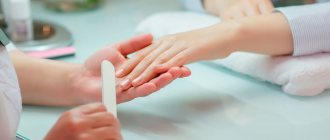
If the relief (ribbing) of the nail is not subject to serious medical intervention, a number of restorative procedures are recommended. They are often advised to be carried out in combination, using vitamin therapy and external procedures according to traditional medicine recipes - baths, compresses, rubbing .
Treatment of ribbed nails
Sage bath
Nail bath
- sage – 2 tbsp. l.
- honey - 3 tbsp. l.
- warm water – 0.5 cups
- olive oil – 0.5 cups
- lemon – 0.5 pcs.
The cooled part of the sage broth should be honey melted in a water bath , add warm olive oil . The mixture should be mixed until smooth, gradually adding the juice of ½ lemon .
Keep your hands in the prepared substance for half an hour . For this compress, a person’s body temperature is recommended - this is how the greatest effect is achieved.
All components should not be overheated: their therapeutic effect is lost.
Lemon compress for nails
- Lemon compress
- Lemon – 1 pc.
- Sea salt – 0.5 – 1 tsp.
Cut the lemon in half , sprinkle sea salt , let it soak for 15 minutes . Dip in lemon and leave for 15 minutes .
Preventive measures
- Review your diet . Eliminate a diet that could cause a deficiency of one or another nutritional component: calories, vitamins, microelements.
- Protect your hands from contact with things that may cause the problem: cold, tight gloves, chemicals .
- Replace softening baths with oil compositions before the manicure procedure .
- To care for your hands, switch to more gentle types of manicure - European, Japanese, hardware.
- Eliminate metal manicure tools , give preference to orange sticks and glass files.
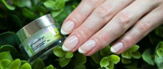
- to apply special protective medicinal base varnishes under a layer of colored varnish .
Even more recipes and tips for getting rid of grooves on nails are presented in this video:
The condition of the nail is an important indicator by which you can monitor the condition of the body as a whole. Don't ignore these signals. A problem that is resolved in a timely manner is the key to your health.
More interesting articles: