Non-ossifying fibroma (NOF) is a common benign bone lesion in children and adolescents, consisting of proliferating elongated fibroblasts, combined with a variable number of multinucleated osteoclast-like giant cells [7]. There are several synonyms: metaphyseal fibrous defect (MFD), cortical fibrous defect (CFD), histiocytic fibroma and fibrous xanthoma. When we are talking about small lesions of the cortical layer, the term MFD or KFD is more often used. When the lesion spreads medullarily, and even more so when there is a threat of pathological fracture, the term NOF is used.
In most cases, NOF has no clinical symptoms and, as a rule, is an incidental finding during examination. With actively enlarging lesions, complaints of local pain sometimes appear [10]. Some researchers consider NOF not as a true tumor, but as a local defect of osteogenesis during the growth of skeletal bones [1]. Multiple lesions are rare, more often within the framework of Jaffe-Campanacci syndrome, which, in addition to multiple NSF, is characterized by the presence of light brown spots on the skin (Café au lait spots), may be accompanied by mental retardation, hypogonadism or cryptorchidism, malformations of the cardiovascular system, eyes, and also be combined with neurofibromatosis [3, 4].
The detection rate of MFD/NOF reaches 30%. Since clinical symptoms are most often absent, the true prevalence of this pathology remains unknown. MFD/NOF is registered more often in children and adolescents, rarely in those under 5 years of age and in adults over 20 years of age.
Despite the high prevalence of this pathology, the awareness of surgeons, radiology specialists and pathologists regarding the latter remains extremely low.
Etiology
Metaphyseal bone defects are usually detected in childhood during the period of active bone tissue growth and are most common in male patients. The lesions are most often localized in the cortical layer of the metaphyses of long ribbed bones. The most common sites are the femur (distal metaphysis) and the tibia (with equal frequency in both the proximal and distal metaphysis). Up to 80% of all observed lesions were identified in this location. Localization in the fibula is also common. When this pathology is combined with skin pigmentation like coffee spots, Jaffe Campanacci syndrome is stated (Combination of multiple lesions in the bones, skin pigmentation like coffee spots, mental retardation, hypogonadism, cryptorchidism, eye abnormalities, pathology of the cardiovascular system). Currently, only isolated cases have been described.
According to observations, a large number of MFCDs undergo spontaneous regression. They often do not manifest themselves as any obvious orthopedic picture in children and, for the most part, are a finding during examination using radiographs. In some cases, the defect reaches a significant size and manifests itself as pain and requires surgical intervention.
With NF, pain occurs more often than with MFCD. With non-osteogenic fibroma, pathological fractures are possible in 40% of cases. Also, in addition to pain, a swelling of a dense consistency appears in the area of the lesion.
New bone formation - symptoms and treatment
The diagnostic algorithm includes interviewing the patient, detailing complaints and clarifying the medical history. Particular attention should be paid to fever, weight loss, loss of appetite and other general symptoms. The time that has passed since the discovery of a bone tumor, the presence and intensity of its growth are also specified.[8]
During examination, the size of the formation, its structure, localization and skin changes are assessed. The areas of regional lymph nodes are palpated to assess possible changes - enlargement of the lymph nodes, their adhesion to the surrounding tissue and to each other.
A laboratory search begins with a general and biochemical blood test: determination of alkaline phosphatase, phosphate and calcium.[1] These analyzes provide evaluative information.
There are laboratory markers of bone resorption (dissolution) that confirm the predominance of bone breakdown (possibly at the tumor site).[9]
But their diagnostic value, unfortunately, is limited, since bone resorption can also occur in other conditions not associated with a bone tumor.
Instrumental diagnostics
The most accessible, fastest and most revealing instrumental diagnostic method is radiography. An x-ray taken in two projections gives an idea not only of the size and location of the bone tumor, but also of its type.[3]
A more detailed and sensitive visualization of new bone formations is computed tomography.[16] It is especially relevant for joint tumors and small bone tumors that are poorly visible on ordinary X-rays.[8]
Scintigraphy is used as a screening (search evaluation method) - bone scanning after intravenous administration of a radioactive isotope. The isotope accumulates in the bone tumor, causing it to “glow” on the scanner screen. This method provides information about the presence of skeletal tumors and their number. It is also effective for the early detection of bone metastases in cancer patients.[6] However, it is impossible to distinguish a benign tumor from a malignant one, much less determine the type of tumor on a scintiogram.[16]
It is noteworthy that radiation methods for diagnosing neoplasms do not provide the right to reliably diagnose a specific type of bone tumor. Even with all the X-ray signs that speak in favor of a certain tumor, only examination of the tumor under a microscope is decisive.
For microscopy, cells are obtained using a biopsy - taking part of the tumor. For this, a puncture biopsy is used, when the bone is obtained with a wide needle, or an incisional biopsy - surgical removal of the tumor, i.e. cutting off part of it.[16]
Often a biopsy is performed immediately during surgery to obtain a preliminary result. This makes it possible to determine the volume of the operation.[2]
Treatment
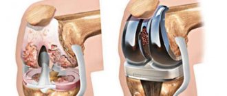
If there is a defect measuring less than 1/3 of the diameter of the bone tissue (no pathological fracture), conservative treatment and observation is possible with the exception of physical activity until the lesion is closed. If there is pain and the lesion is more than 1/3 in diameter, surgical intervention in the form of marginal resection and bone grafting (auto- or allografts) is necessary. In the presence of a pathological fracture, extrafocal osteosynthesis is performed.
MFDC always proceeds benignly; after adequate surgical intervention, the disease does not recur.
You can obtain more detailed information about the MFKD and the procedure in your particular case by contacting our call center by phone.
General information
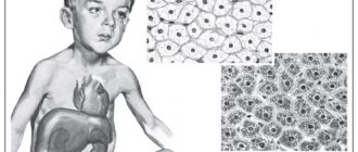
Single or multiple fibromas can affect the lungs, mammary glands, liver, skin, oral mucosa, etc. The proliferation of connective tissue occurs under the influence of various factors: unfavorable environmental conditions, severe injuries, bacterial or viral infections. When making a diagnosis and developing a treatment strategy for a patient, dermatologists, oncologists and surgeons take into account laboratory data. Information about the morphology of the tumor allows doctors to assess the risk of its malignant degeneration.
Features of education in children
Non-osteogenic fibroma reduces bone strength, and this is very dangerous due to the extreme activity of children.
The process of development of non-osteogenic fibroma in children begins with a slow thinning of the outer layer of bone tissue. The growth of the tumor provokes painless resorption of bone tissue. At the final stage of thinning, the density of bone tissue is radically transformed, which negatively affects the overall structure of the bone. The plates cease to perform their functions and are no longer aligned along the lines of compression and tension. This condition causes a decrease in resistance to physical activity.
Bone fibroma is considered a defect in the outer layer of bone, which is located in a long, tubular, hard formation in the human body. The defect looks like multi-chamber formations, similar to small holes. Treatment of non-osteogenic fibroma is prescribed in rare cases. Since the porous tissue gradually heals spontaneously. At an early stage, the clinical picture will not make it possible to diagnose the disease due to the absence of symptoms and spontaneous resorption of fibroids.
Prognosis and prevention
When patients seek medical help in a timely manner, doctors can formulate a favorable prognosis in 75–80% of cases. The likelihood of a complete recovery of a child or adult is reduced when fibromatosis is advanced or secondary pathologies are attached to the disease.
Preventive measures are simple - patients are advised to avoid repetitive trauma to the skin, soft tissue and bones. Persons working in hazardous industries must undergo regular preventive examinations. Children and adults should eat a healthy diet and include adequate amounts of vitamins and minerals.
Types of fibroids
There are two types of tumors: soft and hard.
Soft fibromas in a child are characterized by very slow growth, rather flabby consistency, mobility and clear localization. Such growths occur mainly in the genital area and anus, sometimes forming multiple clusters. Solid neoplasms are smooth or slightly rough pink and flesh-colored nodules with dense contents. Such fibromas are located on a wide base and, as a rule, do not exceed 1 cm in size.
Predictions of why fibroids are dangerous
Dermatofibroma does not have serious complications.
Complications of plantar fibroids usually occur as a result of surgical interventions. These include flattening of the arch of the foot, postoperative nerve entrapment, and postoperative growth of larger and recurrent fibromas. Article sources:
- Desmoid fibroma. Staged, organ-preserving surgical treatment. Khvastunov R.A., Mozgovoy P.V., Lukovskova A.A., Yusifova A.A. Volgograd scientific and medical journal No. 2, 2021. p. 54-57
- https://www.ncbi.nlm.nih.gov/pmc/articles/PMC2927520/ Central odontogenic fibroma: a case report with long-term follow-up. Marco T Brazão-Silva, Alexandre V Fernandes, Antônio F Durighetto-Júnior, Sérgio V Cardoso, Adriano M Loyolacorresponding
- https://www.ncbi.nlm.nih.gov/pmc/articles/PMC3351722/ Literature survey on epidemiology and pathology of cardiac fibroma. SuguruTorimitsu, Tetsuo Nemoto, Megumi Wakayama, Yoichiro Okubo, Tomoyuki Yokose, Kanako Kitahara, Tsukasa Ozawa, Haruo Nakayama, Minoru Shinozaki, Daisuke Sasai, Takao Ishiwatari, Kensuke Takuma, Kazutoshi Shibuya
The information in this article is provided for reference purposes and does not replace advice from a qualified professional. Don't self-medicate! At the first signs of illness, you should consult a doctor.
Diagnostic measures
Diagnosis of fibroids is performed by doctors of various specializations - dermatologists, oncologists, surgeons, gynecologists, etc. Methods for confirming the primary diagnosis are determined by the location of the benign neoplasm. At the first stage of the examination, patients receive a referral for radiography. Computed tomography allows you to determine the exact size of the tumor and identify signs of its invasion into adjacent organs.
Fibrous formations in bone and cartilage tissue are detected during scintigraphy. If necessary, the doctor can take a biopsy sample for laboratory tests. Microscopy of the obtained biomaterials will allow doctors to assess the morphological structure of tumor cells and the likelihood of their malignant degeneration.
Benign neoplasms of the female reproductive system are often detected during ultrasound examinations. The focus of the pathological process has less echogenicity compared to adjacent tissues.
Diagnostics
The examination begins with a visual examination: the doctor listens to the complaints of children and parents, carefully palpates the formation, and collects a detailed medical history. To determine the nature of the lesion, a number of instrumental studies are carried out:
- ultrasound examination of soft tissues;
- radiography;
- computed tomography or magnetic resonance imaging.
A biopsy and cytological examination of biological material help to identify the nature of the pathology and clarify the diagnosis.
Reasons for the development of pathology
The causes of fibroids are varied. Doctors identify several factors that contribute to the formation of connective tissue tumors in patients’ bodies:
- systematic use of alcohol and tobacco;
- repeated injuries to the skin, muscles, bones;
- unfavorable environmental conditions in the region of residence;
- the predominance of fatty, spicy and sweet foods in the diet of children and adults;
- viral and bacterial infections;
- immunosuppression due to the use of immunosuppressants.
Doctors often diagnose fibromatosis in people working in chemical production. Benign skin tumors can develop under the influence of excessive sun exposure. Damage to internal organs is often a consequence of endocrine disorders.
Symptoms
Non-osteogenic fibroma does not have characteristic manifestations that indicate the presence of a specific problem. In most cases, the lesion does not appear until the affected bone is fractured. Sometimes it is detected during an x-ray examination prescribed for other reasons.
A characteristic sign of fibroma is a fracture of the limbs in an unnatural shape. More often this disease affects the lower extremities, so this symptom is characteristic of leg fractures. Other possible manifestations include:
- porosity of bone tissue, which is detected when examining an x-ray image;
- a general analysis of urine and blood indicates a reduced level of calcium in the body;
- disturbance of phosphorus-calcium metabolism, as well as other organic disorders, leading to the inability to prevent bone destruction and restore them after damage.
Otherwise, bone diseases occur without additional symptoms.
Fibroma: origin and essence of the disease
The causes of dermatofibroma and other forms of this type of tumor are not known. Some researchers believe that fibroids can form as a localized tissue reaction after minor trauma. Sometimes fibroids can have a genetic component, especially in people of Northern European descent. Some medications, including beta blockers, have been reported to cause changes in fibrous tissue. In addition, some fibroids may be influenced by hormonal imbalances or pathologies of endocrine organs, including problems with the thyroid and pancreas. Hyperhidrosis (increased sweating), inflammatory processes on the skin, especially chronic ones, as well as the influence of prolonged UV radiation, poor nutrition and bad habits can have a certain impact.