- Deltaclinic
- Services
- Ophthalmology
- Xanthelasma (flat xanthoma)
Prices
Xanthelasma (flat xanthoma) is a slowly growing benign formation in the form of yellow, flat, slightly elevated, clearly demarcated plaques with a smooth or somewhat wrinkled surface.
There is essentially no difference between xanthoma and xanthelasma. Their formation is associated with a violation of fat metabolism. Most often, xanthelasma of the eyelids is formed (they are also called cholesterol plaques on the eyelids), they are symmetrically located around the eyelid. In the photo:
xanthelasma
Xanthelasma is accompanied by a general disorder of lipid metabolism, increased cholesterol and blood sugar levels. Xanthelasma is often combined with xanthomas on other parts of the body, especially on the elbows and knees, and sometimes found on the mucous membranes.
Flat single and multiple plaques of yellow color, located on the eyelids, the size of a pea to a bean, soft consistency, tend to merge and form lumpy elements. Multiple xanthomas are called xanthomatosis.
Xanthelasma is detected mainly in middle-aged and elderly women suffering from lipid metabolism disorders (hyperlipidemia) or liver diseases with normal lipid levels. The sizes of such xanthomas range from a few millimeters to 5 cm or more. Appearing suddenly, they remain unchanged for a long time.
Xanthelasma is a localized form in which identifying disorders of fat metabolism, in most cases, seems to be a rather difficult task, although these patients, as a rule, suffer from obesity, diabetes or hypertension.
Danish researchers have discovered that cholesterol deposits in the eyelids (xanthelasma) can be an excellent indicator of the risk of developing heart disease.
Scientists from the University Hospital of Copenhagen and the University of Copenhagen found that half of people with deposits have completely normal blood cholesterol levels. Thus, xanthelasma may be an important independent indicator of developing arterial disease.
Researchers suggest that xanthelasma can be used to determine the risk of developing cardiovascular diseases where other risk factors for such diseases are not sufficiently manifested. Xanthelasma predicted a 51% increased risk of developing a heart attack, and a 40% increased risk of developing coronary heart disease. Those with xanthelasma had a 17% increased risk of death from heart disease after adjusting for other well-known risk factors, including blood cholesterol levels.
The data obtained by Danish scientists can be used for therapeutic purposes. And if doctors find xanthelasma in patients during regular examinations, they will be advised to pay special attention to the functioning of the heart and blood vessels.
What is Xanthomatosis?
A xanthoma is a skin lesion caused by the accumulation of fat in macrophage immune cells in the skin and, less commonly, in the layer of fat under the skin.
Some types of xanthoma indicate lipid disorders (eg, hyperlipidemia or high blood fats), where they may be associated with an increased risk of coronary heart disease and sometimes pancreatitis.
Xanthomas are classified into the following types depending on where they are found on the body and how they develop.
Forecast
The prognosis of the disorder directly depends on the factors that provoked it. This is due to the fact that in some cases xanthomas can disappear on their own, while in others they can lead to consequences such as atherosclerosis of the coronary or cerebral vessels, heart disease and an increase in liver size. However, the most common danger of the disease lies in its frequent relapses.
In addition, patients should not forget about the possible formation of complications of the underlying disease. Often, in addition to cosmetic inconveniences, xanthomas do not cause any other discomfort, which is why the prognosis of the disease is favorable.
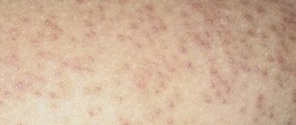
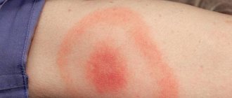
Eruptive xanthoma
- Lesions usually appear as seedlings of small red-yellow papules
- They most often occur on the buttocks, shoulders, arms and legs, but can occur throughout the body
- Rarely the face and inside of the mouth are affected
- Lesions may be tender and usually itchy
- Lesions may resolve spontaneously within a few weeks
- Associated with hypertriglyceridemia (increased levels of triglycerides in the blood) often in patients with diabetes mellitus (diabetes mellitus)
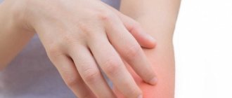
Clinical picture
Xanthomas of the eyelids
The symptoms of xanthomatosis are quite characteristic:
- The rash looks like papules or plaques of soft consistency. The color of the nodules varies from yellowish to orange, in rare cases to red-brown. Dimensions – from 1 mm to 2 cm. Prone to fusion.
- There is no pain syndrome.
- The most common location (face, limbs, buttocks, elbow and knee joints).
- May be accompanied by thinning of the skin and itching.
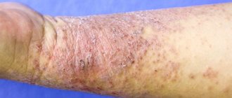
Common xanthoma
- Xanthoma-like lesions are caused by a rare form of histiocytosis.
- Lipid metabolism is normal.
- The skin lesions usually consist of hundreds of small yellowish-brown or reddish-brown bumps that are usually evenly distributed on both sides of the face and torso. They can be especially hard on the armpits and groin.
- Small bumps may join together to form sheets of thickened skin.
- In 30% of affected people, the mucous membranes of the mouth, respiratory tract or eyes (mucous membranes) are affected. Warty plaques in the mouth are called verruciform xanthoma.
- 40% of affected people develop diabetes insipidus, a condition that results in an inability to control water loss (leading to constant thirst and excessive urine production). This occurs due to the overgrowth of histiocytes on the lining of the brain (meninges).
- May affect internal organs (eg liver, lungs, kidneys, etc.)
- Self-limiting and eventually improves on its own, but may persist for many years.
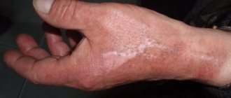
Squamous cell carcinoma
It is less common than basal cell carcinoma, the second most common type of skin cancer, and has a slightly less favorable prognosis. However, it should be noted that the course of the disease much less malignant than that of melanoma.
Metastases occur relatively rarely - on average in 16% of cases [1]. In patients with squamous cell skin cancer less than 2 cm in size, the 5-year survival rate is about 90%; for larger sizes and tumor invasion into the underlying tissue, it is less than 50% [1].
It can occur on any part of the body, including the genitals and mucous membranes, but most often in places exposed to sunlight.
Symptoms and signs
What squamous cell skin cancer looks like depends largely on the clinical form of the disease.
The keratinizing form is a raised or flat surface covered with horny scales that can grow and fall off. If damaged, it may bleed.
Keratinizing form of squamous cell skin cancer
It must be remembered that it is the keratinizing form of squamous cell carcinoma that may be hiding under the mask of the cutaneous horn . In this regard, such formations should always be removed only with histological examination:
The cutaneous horn should be removed with histology - a keratinizing form of squamous cell carcinoma may be hidden under its mask
Non-keratinizing endophytic form (growing towards surrounding tissues). Most often it looks like a long-term non-healing wound or ulcer, which can deepen and expand over time.
Non-keratinizing endophytic form of squamous cell skin cancer
The exophytic non-keratinizing form of squamous cell skin cancer appears as a nodule that rises above the level of the skin. The surface of the node may be eroded or wet.
Exophytic nonkeratinizing form of squamous cell skin cancer
Photos in the initial stage
The initial stage of squamous cell carcinoma refers to a condition when the malignant process is limited to the epidermis - the outermost layer of the skin. It is referred to in the diagnosis as in situ or intraepidermal squamous cell carcinoma. This disease is not life-threatening if completely removed.
There are 2 forms of this phase of the disease:
Bowen's disease
Most often it is represented by single flat plaques, with clear boundaries, an asymmetrical shape, and uneven edges. The size reaches 7–8 mm. The formation may gradually increase, and peeling or crusting is often observed on the surface.
The color is red or brown, located on any part of the body. [3]
On my own behalf, I will add that in my practice, histologically confirmed Bowen’s disease occurred only once. It looked like a small (3 x 4 x 3 mm) flesh-colored lump with a smooth surface on the skin of the shaft of the penis in a 43-year-old man.
Bowen's disease
Erythroplasia Keira
The second form of early-stage skin cancer, which develops most often on the skin of the foreskin of the penis or the glans. Much less commonly, the disease affects the female external genitalia.
The most common appearance of Queyre's erythroplasia is a bright red spot with clear boundaries and a moist, shiny surface [3].
Erythroplasia Keira
Treatment of squamous cell skin cancer (NCCN, 2018)
As in the case of basal cell carcinoma, squamous cell carcinoma is divided into groups of high and low risks of recurrence and metastasis.
Area H: Facial mask (including eyelids, eyebrows, skin around eyes, nose, lips [skin and red border of lips], chin, lower jaw, skin/grooves in front and behind the auricle, temples, ears), genitals, palms and feet .
Area M: cheeks, forehead, scalp, neck and legs
Region L: trunk and limbs (excluding shins, palms, feet, nails and ankles)
Notes
- The rim of hyperemia should be taken into account when measuring size.
- Excisional biopsy is preferred over incisional biopsy.
- The modified Breslow thickness measurement should exclude parakeratosis and crusting and should be taken from the base of the ulcer, if present.
- Localization, regardless of size, may be a sign of high risk.
- Area H implies high risk regardless of size.
The basic principles and methods of treatment for squamous cell carcinoma are the same as for basal cell carcinoma.
The main goal is to maintain functionality and cosmetic qualities. method is considered to be removal of the tumor, including 4–6 mm of healthy tissue with a low risk of recurrence and metastasis. For high-risk tumors, Mohs micrographic surgery or wider excision is recommended than for low-risk tumors.
Radiation therapy is useful in cases where other methods cannot be used. Platinum drugs (cisplatin, carboplatin) as well as EGFR inhibitors (cetuximab) can be used in chemotherapy for squamous cell carcinoma.
What causes xanthoma?
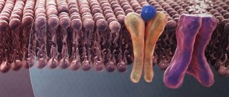
There are several main disorders in which xanthoma is caused by a disorder of lipid (fat) metabolism. Because lipids are insoluble in water, they combine with proteins to form compounds called lipoproteins. Lipoproteins transport lipids and cholesterol in the blood to various parts of the body. Based on their size and weight, common lipoproteins are classified as chylomicrons, very low-density lipoproteins, low-density lipoproteins, and high-density lipoproteins (Fredrickson classification). They all play a role in maintaining the metabolic functioning of the body.
Changes in lipoproteins may result from a genetic defect (eg, primary hyperlipoproteinemia) or from an underlying systemic disorder such as diabetes mellitus, hypothyroidism, or nephrotic syndrome. These underlying diseases can cause elevated levels of certain lipids and lipoproteins, which then manifest as cutaneous xanthoma.
Monogenic familial hypercholesterolemia: type IIa
Mutation in the LDL (Low Density Lipoprotein) receptor
- High LDL level
- Total cholesterol in heterozygotes is 9-14 mmol/l
- Total cholesterol in homozygotes is 15-30 mmol/l
Polygenic hereditary hypercholesterolemia type IIIA
Diagnosis of the disease
The characteristic appearance of the rash, as a rule, does not cause difficulties in diagnosis. To confirm the diagnosis, the following types of studies are performed:
- Examination of the patient and collection of personal and family history;
- Biochemical blood tests;
- Study of lipid spectrum;
- Biopsy.
A lipidogram evaluates the level of the main indicators of fat metabolism in the body: cholesterol, “bad” and “good” cholesterol and triglycerides.
Biochemical blood parameters will tell you the direction in which to look for the cause of lipid disorders.
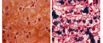
A plaque biopsy is performed if necessary to differentiate xanthomatosis from other diseases:
Elastic pseudoxanthoma Darier is a congenital pathology that affects the skin, eyes and internal organs. Most often it is localized symmetrically on the lateral surfaces of the neck, in the inguinal folds, armpits, on the inner surfaces of the elbows and knees, and sometimes in the navel area. It never affects the area of the face, soles and palms.
Histiocytosis X is a disease characterized by excessive production of histiocytes. Actively multiplying in combination with eosinophils, they can give a similar rash in the spine and chest.
Reticulohistiocytoma , which is based on benign dysplasia of reticular tissue. Visually similar to tuberous xanthoma.
Wide range of results
Polygenic familial combined hyperlipidemia: type IIb
- Mixed genetic and life history causes
- Elevated total cholesterol levels
- Elevated triglyceride levels
- Low HDL (High Density Lipoprotein) Cholesterol
- Elevated LDL cholesterol levels
- LDL may be normal levels but denser and more likely to cause atheroma (sebaceous cyst)
Moderate hypertriglyceridemia
- Often also associated with high blood pressure, obesity, diabetes, metabolic syndrome, high insulin levels, high uric acid levels
- This may be due to alcohol or medications such as systemic steroids, isotretinoin, acitretin
- Triglycerides 2_10 mmol/l
- Often associated with low HDL cholesterol levels
Severe hypertriglyceridemia: Types 1 and V
- Mixed genetic and life history causes
- Diabetes
- Familial LPL (lipoprotein lipase) deficiency
- Triglycerides > 10 mmol/l
- Elevated total cholesterol levels
- Raised chylomicrons
- Elevated LDL cholesterol levels
Wide beta hyperlipoproteinemia: type III
- Rare mutation of the apo E gene
- Triglycerides 5-20 mmol/l
- Total cholesterol 7-12 mmol/l
The reason for the appearance of xanthoma with normal levels of fat in the blood is currently unclear.
Traditional medicine methods
Traditional methods of treatment are used as an aid. The essence of the method is to take herbal infusions and decoctions, as well as external influence on the elements of the rash.
Take internally:
- for a choleretic effect: yarrow herb, birch buds, dandelion roots, corn silk, hellebore roots, rose hips;
- to facilitate the work of the liver: chicory stems and leaves, immortelle flowers, milk thistle herb, artichoke leaves, celandine herb;
- to stimulate pancreatic function: valerian roots, St. John's wort, dill seeds, elderberry flowers, elecampane rhizomes, blueberry leaves.
For external treatment use:
- juice of fresh immortelle (aloe);
- baked onion;
- Golden mustache;
- a mixture of vegetable oil and salt;
- badger fat.
Before using traditional medicine methods, you should first consult a doctor.
What examination is required to treat xanthoma?
A skin biopsy may be required to confirm the clinical diagnosis of xanthoma.
Appropriate blood and urine tests and x-rays are performed to determine the cause of abnormal lipoprotein levels, if present. The risk of cardiovascular disease, including heart attacks, peripheral vascular disease and stroke, increases with elevated levels of certain lipoproteins. It is important to identify contributing factors so that appropriate therapy can be instituted.
How does xanthoma heal?
The primary goal of treating xanthoma that is associated with an underlying lipid disorder is to identify and treat that lipid disorder. In many cases, treatment of the underlying disorder will reduce or resolve the xanthoma. In addition, treating hyperlipidemia will reduce the risk of heart disease, and treating hypertriglyceridemia will prevent pancreatitis. Lipid disorders are treated with changes in diet and lifestyle, with or without medications.
Dietary measures should include:
- Prepare most dishes from vegetables, salads, grains and fish
- Minimize saturated fat (found in meat, butter, other dairy products, coconut oil, palm oil)
- Minimize your intake of simple refined sugars found in carbonated drinks, sweets, cookies and cakes
- If you are obese or overweight, aim to lose weight slowly by reducing your calorie intake and increasing exercise.
Very effective medications may also be prescribed. These may include:
- Statins are pharmaceutical drugs designed to combat high levels of cholesterol in human blood (HMG-CoA reductase inhibitors). Such as simvastatin and atorvastatin reduce the production of cholesterol by the liver, resulting in lower LDL cholesterol, increased HDL cholesterol and a mild reduction in triglyceride levels. Treatment should be monitored by regular blood tests to check lipid levels and ensure that liver and muscle enzymes are normal, as statins sometimes cause problems, especially when given at higher doses.
- Fibrates, such as bezafibrate, may be added to reduce triglyceride production by the liver, lower triglyceride levels, and increase HDL cholesterol levels. They may cause gastrointestinal side effects.
- Ezetimibe may be added in high-risk patients or those who do not tolerate higher doses of statins. It reduces the absorption of cholesterol from the intestines, lowering total and LDL cholesterol.
- Nicotinic acid lowers cholesterol, LDL cholesterol and triglycerides, and increases HDL cholesterol. At therapeutic doses of at least one gram per day, it causes redness of the skin. Its analogue, acipimox, is better tolerated.
- Cholestyramine and colestipol are rarely used because they are not as effective as the drugs listed above and are poorly tolerated.
Surgery or locally destructive techniques may be used to remove xanthomas that do not resolve spontaneously or with treatment of the underlying cause. Advanced xanthoma that affects vital organ functions can be treated with chemotherapy drugs or radiation therapy.
Prevention and recommendations
Prevention of xanthoma is carried out only with the awareness that the patient suffers from hyperlipemia or other primary diseases. Recommended:
- maintain normal weight;
- practice regular physical activity, especially aerobics;
- follow a proper diet, limiting the consumption of foods that are not recommended for consumption with high cholesterol, increasing the amount of foods that are useful for reducing high cholesterol;
- use medication prescribed by your doctor.