In this article we will tell you:
- Causes of iridocyclitis
- Main types of iridocyclitis
- Symptoms and complications of iridocyclitis
- Diagnosis of iridocyclitis. Treatment
- Prevention of iridocyclitis
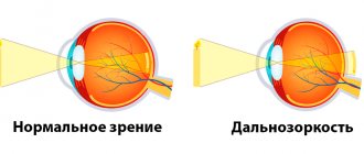
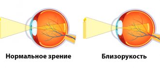
It happens that eye diseases concern one particular department, tissue or structure of an organ. So, with cataracts we talk about clouding of the lens, with keratitis - with inflammation of the cornea, with macular degeneration - with damage to the retina. Something else happens when several departments are involved in the pathological process at once. Such diseases are called combined, and it is to this group that the disease discussed in this article, iridocyclitis, belongs.
Iridocyclitis
is an inflammation of both the ciliary (ciliary) body and the iris of the eyeball.
The name of the disease comes from the Greek words iris - rainbow, and kykios circle, eye. Iridocyclitis is also called anterior uveitis. The disease is distinguished from uveitis proper by the localization of the inflammatory process: with ordinary uveitis, the choroid of the eye (uveal tract) is affected. The combination of lesions characteristic of ocular iridocyclitis occurs due to the anatomical similarity: the blood supply and innervation of the departments are closely related, and when one of them is affected, the inflammation quickly spreads to the other. Sometimes there are inflammatory processes in only one of the mentioned sections - the iris or the ciliary body. In these cases, we will talk about individual diseases: cyclitis and iritis.
You can get iridocyclitis at any age. Somewhat more often it affects young people 20-40 years old.
Causes of iridocyclitis
There are many reasons why iridocyclitis can develop. Among them are both exo- and endogenous. With the exogenous nature of the disease, the reason for its development lies in external factors. Endogenous iridocyclitis is a consequence of internal problems in the body and is more common.
The blood-ophthalmic barrier is normally responsible for the health of the eyes: it traps harmful bacteria, microbes, poisons - all those foreign objects that can cause diseases of the eye structures. When the permeability of this natural filter increases, harmful substances enter directly into the iris and then into the ciliary body, turning both sections into foci of inflammation.
The following are the main groups of causes of iridocyclitis:
- Infections: bacterial, viral, fungal.
- Helminthic infestations.
- Allergy.
Herpes, influenza, ARVI measles, tuberculosis, some sexually transmitted diseases (syphilis, gonorrhea, chlamydia), staphylococcal and streptococcal infections, malaria, brucellosis: all these ailments can affect the permeability of the blood-ophthalmic barrier and cause inflammation.
Seasonal hay fever or year-round allergies (to dust, food, household chemicals, medications, etc.) can also cause the disease.
- Chronic foci of inflammation in the oral or nasal cavities, otitis.
- Rheumatic diseases.
In persons suffering from rheumatism and syndromes associated with it, the incidence rate can reach up to 40%.
- Metabolic diseases (diabetes, sarcoidosis, gout, etc.).
- Injuries (contusions, wounds) and postoperative complications.
Risk factors for the occurrence of this disease may include:
- traumatic, stressful situations;
- hypothermia of the body;
- too intense physical activity;
- bad habits, unbalanced diet;
- immune failures.
It happens that even specialists fail to find out the cause of the disease: then they talk about iridocyclitis of unknown etiology.
Risk factors
The risk of developing iritis increases if: 6
- There is a specific genetic change. This change is referred to as HLA-B27 and causes the immune system to not function fully.
- There is a sexually transmitted infection. Syphilis and HIV are associated with a significant risk of iridotskilitis.
- I have an autoimmune disease. Iridocyclitis often develops in people with ankylosing spondylitis and rheumatoid arthritis. About 40% of cases of iridocyclitis occur against the background of a systemic disease.
- There are pockets of chronic infection of the ear, throat, nose or mouth.
- There are bad habits. Tobacco smoke has a negative impact on eye health.
Main types of iridocyclitis
According to the nature of the appearance and course of iridocyclitis, they are divided into:
- acute (begin abruptly. Symptoms of acute iridocyclitis of the eye are pronounced, easily diagnosed, duration – 3-6 weeks);
- chronic (they are more sluggish in nature compared to acute forms, symptoms are more difficult to relieve, duration is up to several months);
- recurrent - periods of attenuation of the disease are replaced by exacerbations (more often in winter).
According to the nature of inflammation, iridocyclitis is:
- serous. Iridocyclitis of this form is characterized by the accumulation of serous secretion in the chambers of the eye.
- hemorrhagic. Such iridocyclitis is characterized by severe damage to the vessels supplying blood to the ciliary body and iris.
- exudative. The main difference between this form is the accumulation of pus in the form of a crescent on the lower part of the iris.
- fibrinous-plastic. Iridocyclitis of this type occurs with the formation of fibrin threads. With fibrinous iridocyclitis, synechiae form in the anterior chamber of the eye, and fibrinous exudate accumulates.
What it is?
Iridocyclitis is an intraocular inflammation that affects the iris and the anterior part of the ciliary body.1 It is a fairly rare disease - about 0.3 cases per 1000 population. In the structure of eye pathology they occupy 5-12%.2
If iridocyclitis is not chronic, it can be cured in about 1-3 months. If we talk about all forms of intraocular inflammation, this pathology is one of the most common forms. Quite often, acute iridocyclitis develops into chronic recurrent forms.
Complications may also occur, including glaucoma and cataracts. To avoid such a scenario, it is important to diagnose iridocyclitis early and treat it.
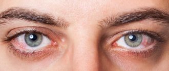
Symptoms and complications of iridocyclitis
The main symptoms of iridocyclitis are nonspecific: they are inherent in many ophthalmological (and not only) diseases. Patients with inflammation of the ciliary body and iris complain of:
- headache;
- blurred vision;
- photophobia;
- lacrimation;
- blurred vision;
- hyperemia.
Signs characteristic of acute iridocyclitis are:
- sharp pain in the eyeball, radiating to the head;
- increased pain when touching the eye in the projection area of the ciliary body;
- thickening of the iris, narrowing and immobility of the pupil;
- increased pain at night;
- swelling of the eyelids;
- the vessels visible along the outer edge of the iris are first bright red, later violet.
In chronic iridocyclitis, smoothing of the iris pattern, pronounced exudation, and the formation of protein tubercles, adhesions, and synechiae are observed. The pain is less pronounced than in the acute form.
Untimely or ineffective treatment of the disease often leads to complications. The inflammatory process can spread to other parts - then the patient develops chorioretinitis, scleritis, keratitis, vasculitis, endo- or panophthalmitis. In addition, complications of iridocyclitis include:
- the appearance of cataracts, glaucoma;
- retinal disinsertion;
- adhesions, pupil fusion;
- corneal dystrophy.
In difficult cases, we can talk about irreparable loss of vision and even removal of the eyeball.
Iridocyclitis: treatment methods
Drops that dilate the pupil are prescribed as first aid. The specialist prescribes medications that act on the causative agent of infection, increase immunity, and eliminate inflammation. Usually these are drugs that contain antibiotics. Corticosteroids, cytostatics, immunomodulators, enzyme agents, and absorbable exudates can be used.
With timely initiation of therapy, the prognosis is favorable. If treatment is prescribed late or the disease is severe, then various consequences are possible: sympathetic inflammation of the second eye, glaucoma, atrophy of the eyeball, blindness.
Diagnosis of iridocyclitis. Treatment
Patients with any form of the disease should undergo treatment under the supervision of a specialist. Self-diagnosis and self-medication in this case are unacceptable: after all, the consequences can be too severe.
To clarify the diagnosis, the ophthalmologist will prescribe general tests: blood tests for various parameters, X-ray and ultrasound examinations, and, if necessary, tests for allergens and rheumatism. Local examination will consist of examination with a slit lamp, palpation, measurement of intraocular pressure, and biomicroscopy of the eye.
The doctor will develop a treatment plan. Most likely, it will be carried out in a hospital. The set of drugs will depend on the cause of the disease. In addition to medications, a specialist may prescribe a course of physiotherapeutic procedures, for example, electrophoresis.
Coloboma of the choroid proper
| It is detected during ophthalmoscopy in the form of a white sector of varying size and shape with distinct and uneven edges, over which the retinal vessels pass without changing (see.
|
Toxoplasmosis uveitis
| May be congenital or acquired. Eye disease in congenital toxoplasmosis is often bilateral (70-80% of cases). The process is localized mainly in the choroid itself and occurs in the form of chorioretinitis. The anterior part of the eye, as a rule, is not involved in the process. The first indirect symptom of uveitis is deterioration of central or twilight vision and a distorted image of the objects in question. A characteristic ophthalmoscopic picture of choroiditis in congenital toxoplasmosis is a central lesion, combined with changes in the retina. The resulting inflammatory focus has a yellow-white or whitish-brown color (see.
|
In cases of an acute course of the process, there may be hemorrhages, exudate, and opacities of the vitreous body. Concomitant diseases such as progressive myopia, ptosis, cataracts, and choroidal coloboma are often observed.
Acquired toxoplasmosis occurs at any age through infection from domestic animals (cats, dogs) and birds. The clinical picture is characterized by severe redness of the eyeball, clouding of the posterior epithelium of the cornea due to many precipitates, hyperemia of the iris, posterior synechiae and opacification in the vitreous body. The process is usually one-sided, acute with an increase in body temperature. Vision drops sharply.
Heterochromia of the iris
Heterochromia of the iris is a different color of the iris of the right and left eyes or unequal coloring of different parts of the iris of one eye (see).
Inflammation of the choroid (iridocyclitis, cyclitis, uveitis) is one of the common causes of low vision and blindness (up to 25% of cases). The relatively high incidence of damage to the vascular tract of the eye is due to the pronounced branching of blood vessels and, in connection with this, slow blood flow in the choroid. The peculiarities of the blood supply to the eyeball determine that the anterior and posterior sections of the choroid are usually affected separately. Accordingly, iridocyclitis, or anterior uveitis, occurs (isolated iritis and cyclitis are rare) and choroiditis, or posterior uveitis.