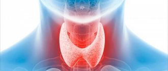
The most useful information from the article about thyroid nodules on the TUT.BY
from endocrinologist
Tatyana Pugavko :
What is a nodule on the thyroid gland?
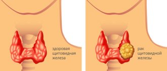
A nodule (in medical terms, a nodular formation of the thyroid gland) is an area of altered tissue of the thyroid gland, which is detected by palpation (by palpation), ultrasound or other methods of studying the thyroid gland. Nodules vary in size (even the size of a chicken egg), and in consistency (either dense or soft). They can be painful or painless (the latter is more common). Nodes with signs of malignancy are also isolated. All this is determined by an endocrinologist when examining the patient, palpating the thyroid gland, looking at the thyroid ultrasound ,
studying thyroid hormone tests and the results of other studies if necessary.
What could be causing the formation of a nodule? Is it dangerous?
The most common benign nodes found in people are colloid nodes and cysts. The main reason is iodine deficiency
and hereditary predisposition (overgrowth of individual tissue areas, thyroid cells).
A provoking factor may also be selenium deficiency ,
which is necessary for the normal functioning of the thyroid gland. Cigarette smoke, insecticides, and herbicides also have an adverse effect on the thyroid gland (some of their components can cause the formation of nodules).
The reasons for the development of a tumor (cancer) of the thyroid gland are more complex. Among them, for example, there may be mutations of various genes ,
which lead to disruption of the formation of thyroid cells. In any case, detection of a nodule on the thyroid gland is a reason to consult an endocrinologist.
Causes
The reasons why diffuse nodular goiter develops are not exactly clear. The most likely predisposing factors are considered to be age-related changes in the body associated with changes in the endocrine system and an unbalanced diet. The risk of developing diffuse nodular goiter increases with frequent and chronic stress, reduced immunity, and unfavorable environmental conditions.
The disease often develops against the background of:
- psychological injuries;
- infectious processes in the body;
- inflammatory diseases;
- autoimmune disorders;
- genetic predisposition;
- insufficient consumption of iodine-containing products;
- diseases of the central nervous system;
- bad habits;
- brain injuries;
- hormonal disorders.
Medical practice also shows that signs of a nodular process in the tissues of the thyroid gland are often found in elderly patients. This suggests that the development of diffuse nodular goiter may be associated with the natural mechanism of aging.
Can a nodule on the thyroid gland be detected during self-examination?
Sometimes a person himself can actually detect (palp or see in the mirror) a nodular formation in himself, but more often in cases where it is quite large in size
. Sometimes nodules can be identified by a doctor during examination, but in most cases they are found during an ultrasound of the thyroid gland. Much depends on the size of these nodules, their location in the thyroid gland, and the patient’s physique. Typically, the doctor can palpate nodules larger than 1 cm located on the surface of the thyroid gland. The nodes are easier to palpate in people of thinner build and in children, then sometimes even smaller formations can be detected.
What is a goiter
In medicine, this term refers to a pathological change in the thyroid gland, expressed in the formation of nodules. A node, in turn, is a neoplasm of any size with a capsule, determined by palpation or during a visual examination. The diffuse type of the disease involves uniform tissue proliferation. And mixed cases in which both of these pathological processes are combined are called diffuse nodular goiter.
With this disease there is no connection with tumor, or neoplastic, or inflammatory processes. Enlargement of the thyroid gland with diffuse nodular goiter is not an oncological pathology. This is a consequence of other, independent pathological conditions or changes.
Diffuse nodular goiter is more often diagnosed in women than in men. According to medical statistics, among patients with this disease there are 3 times more females. The vast majority of them are of middle age.
How are such formations treated?
Everything is individual.
Treatment of thyroid nodules depends on the type of nodule itself (benign or malignant), its size (small or very large, compressing the trachea, esophagus, when there are complaints of difficulty swallowing, breathing, etc.). Treatment may include both observation for benign nodes with monitoring of ultrasound, hormones, puncture of the thyroid gland and other studies, as well as the prescription of various drugs, for example, thyroid hormones - everything is determined by a specialist.
For thyroid cancer, treatment is carried out by oncologists (this can be surgical or chemotherapy treatment, radiation therapy with a radioactive isotope of iodine).
Prevention
Since the exact reasons why diffuse nodular goiter of the thyroid gland develops are not yet clear, preventive measures are precautionary in nature.
Endocrinologists strongly recommend enriching your diet with foods rich in iodine:
- sea fish;
- crustaceans;
- seaweed;
- whole milk;
- beef.
Adequate nutrition is especially important when the immune system is weakened, in childhood and old age. If one of your close relatives has been diagnosed with diffuse nodular goiter, it is recommended to regularly visit an endocrinologist and monitor hormone levels through testing - at least once a year. Good sleep, a properly organized rest regimen, and an active lifestyle will also help reduce the risk of developing the disease.
How often should I check for these nodules?
If you have any complaints (fatigue, drowsiness, weight loss, rapid or slow heartbeat, impaired hair growth, discomfort in the neck) and have close relatives with thyroid cancer, you have had episodes of radiation exposure in your life head or neck, if your age at the time of the Chernobyl nuclear power plant accident was from 0 to 18 years, then you should consult a doctor and do an ultrasound of the thyroid gland
. And even if, during the research, “something was discovered and they told you about it in a scary voice,” please do not panic, but wait for a consultation with an endocrinologist who will prescribe further tests.
When self-examining the thyroid gland, the minimum list of tests, in addition to ultrasound, includes hormonal testing
(TSH, free T4, AT-TPO) and general blood test.
The most popular questions from patients with answers from Romanov Georgy Nikitich
Galina: I have had nodules on my thyroid gland for 7 years now. Some say to have surgery, others say to wait. I do not know what to do.
Romanov G.N: Indeed, this is a controversial issue, even among doctors. The previous generation of doctors saw a period when ALL nodes in the thyroid gland had to be removed. They were very afraid of degeneration into cancer. Times change, science does not stand still. Today, the practice of node puncture has been actively introduced. If the node is benign, then the tactic is watchful (just observation). If there is the slightest suspicion of an oncological process, prepare for surgery. In addition to oncology, today there are two more indications for the removal of nodes - a cosmetic defect and compression of surrounding tissues and organs (trachea and esophagus). In addition, your consent is also required...
Galina: Is it true that puncture provokes the growth of nodes in the thyroid gland and should not be done often?
Romanov G.N: Such data does not exist in the world. It has never been proven anywhere that puncture of nodes stimulates their growth, much less degeneration. The doctor will offer you repeated punctures only in two cases: when a new node has appeared or the old node has changed during a repeated ultrasound examination (it has grown quickly, changed its contours or its characteristics).
Elena: Hello, doctor! I have been under observation for thyroid nodules for 20 years. In 2003, I had a consultation with you. There were always 2 nodes in the right lobe, which did not tend to increase, they grew a little only with me. They punctured it 2 times - everything was fine. Hormones are normal - the golden mean, the last ultrasound showed that one node (13*11*13mm) became fuzzy, heterogeneous, with uneven edges. I donated blood for hormones - again the golden mean, the thyroid gland itself is not enlarged. I made an appointment for a puncture, wait a week. I'm very worried. I have a question: can a 20-year-old quiet nodule suddenly degenerate into a malignant one? And why do such rebirths occur??
Romanov G.N: This happens extremely rarely. If it does, it will be a follicular tumor, which very rarely behaves aggressively. I think everything is going to be alright!
Tatyana23: Age 40 years old, ultrasound of the thyroid gland size 9.7 mm on the right, almost 5 mm on the left, tests for hormones tsn-1.2 ft4-16.2 anti-tpo 15.8 puncture of the gland (they said it was good), the doctor did not give any advice: see for yourself. But he said in a month and a half to do the puncture again, is this necessary?? Please advise me when to perform a repeat puncture and whether surgery is necessary. Do I need to take any medications or vitamins???
Romanov G.N: You don’t need to take anything! Ultrasound monitoring of the thyroid gland after 6 months and that’s it!
Oksana (Odessa): Hello! I have been diagnosed with a nodule in the thyroid gland, 3 cm. There are ultrasound tests, a test for thyroid hormones, and a biopsy test.
1. Ultrasound results: the thyroid gland is located in a typical manner, the lobes are asymmetrical, with clear contours. Dimensions according to Brunn (age norm 12 cc): right lobe - 22x26x50 mm (volume - 13.7 cc); left lobe - 15x17x44 mm (volume - 5.4 cc), total volume 19.1 cc. The isthmus is 4mm thick. The structure of the glandular tissue is homogeneous, the tissue is isoechoic. Occupying the lower and middle segments of the right lobe, an oval-shaped node of moderately increased echogenicity with clear contours measuring 31x21 mm is determined, containing an oval-shaped anechoic focus measuring 6.5x4 mm. Enlarged lymph nodes are not detected.
2. Study of the content of thyroid hormones and antibodies to thyroid peroxidase in human serum using the RIA method:
Determination of free thyroxine (FT4) - 19.0 nmol/l
Determination of total triiodothyronine (TTZ) - 2.1 nmol/l
Determination of thyroid-stimulating hormone (TSH) - 3.2 mIU/l
Determination of antibodies to thyroid peroxidase - not detected.
3.FNA of the thyroid gland under ultrasound control
Diagnosis: stage 2 nodular goiter
Material: Thyroid node punctate.
Cytogram of a follicular tumor (hypercellular colloid goiter? adenoma?)
Cytodiagnosis is recommended.
Please tell me your opinion, maybe there are some signs that you don’t understand?
Romanov G.N: Remove the right lobe with the node and forget... Unfortunately, there are no other options here.
Yulia: Hello. After laser destruction of the node, on the 4th day my throat began to hurt inside at the level of the node itself. This is fine? I am very worried.
Romanov G.N: This is a burn and it should hurt. Until the wound inside heals...
Oksana: Good evening, ultrasound results: in the lower third on the right there is an isoechoic node with clear contours, heterogeneous structure, size 5x6 mm, with signs of blood flow. This is with normal hormones and normal tissue. Are there indications for puncture?
Romanov G.N: It is necessary to consider other risk factors for cancer to solve (Chernobyl, irradiation of the head and neck, relatives, etc.).
Nadezhda: Hello! Please decipher the results of the test, what does it mean - no signs of malignancy were found? I have a node of almost 2 cm, in the right lobe.
Romanov G.N: This means that everything is fine.
Marina: Hello, doctor. In 2013, I was diagnosed with a thyroid nodule. The gland itself is enlarged. The right lobe was 23x22x50, the left lobe was 18x16x42, the total volume was 19.5. And the knot was 16.1 by 16.8 by 22 mm. Being a coward (age 42 at the moment), I did not do a biopsy. And this year I had a repeat ultrasound. The size of the right lobe is length 56, width 29, thickness 31. The left lobe is length 50, width 18, thickness 16. Total volume 31.6. The node in the right lobe is 36 by 23.5 by 29, with intra and perinodular blood flow. And in the left lobe a small one appeared, 4.7 mm. TSH is now 0.5. I went to the surgeon, and he scolded me, said that I should have operated on three years ago. That I had low TSH (although I thought it was within normal limits). Now I’ll only go for a biopsy, and the surgeon said that regardless Based on its results, I have no other options other than surgery. Please tell me your opinion. Is it really as bad as the surgeon said? What normal recommendations can you give?
Romanov G.N: The growth is really significant. Your history also matters (Chernobyl, heredity?). A biopsy will dot all the i's... It should definitely be performed in your case.
Natalya: Good afternoon! My ultrasound of the thyroid gland: Total volume 7 cm cubic. The contours are smooth, the structure is homogeneous. Knot: 6mm*7mm*5mm, volume 0.13 cm cubic. the shape is round, the contours are smooth, the echogenicity is hypoechoic, the structure is heterogeneous. There are no regional lymph nodes. Please tell me whether it is worth doing a biopsy now or if we can observe.
Romanov G.N: After 6 months, ultrasound control and then a decision on TAB.
Olga Basharenkova: Good afternoon, Georgy Nikitich! I am 36 years old. I examined the thyroid gland a long time ago, the result was a total volume of 18.3, hormone tests were all normal, but in the right lobe there was a hypoechoic 3*2*2 node with mixed blood flow. They advised me to do an ultrasound every year and take hormone tests. In 2021, I did an ultrasound - in the segment of the right lobe - a hypoechoic heterogeneous node 4.5 * 2.4 * 3.5 mm with mixed blood flow, in the IV segment - a cyst 1.4 * 2.7 mm. In the s/segment of the left lobe there is an isoechoic node with a hypoechoic “halo” 6.7 * 12.2 * 8.4 mm with perinodular blood flow. TPO tests - 7.98; TSH-1.370,FT3-4.37, FT4-14.1. I'm worried about the new node, and I'm going to the doctor next week. Can you recommend something on how to reduce it? Thanks a lot.
Romanov G.N: You were born in 1980, which means you are at risk for Chernobyl. All nodes larger than 1 cm will have to be punctured.
Anna: Good day! The background is this: I did a general blood test in the fall - TSH was increased to 6, while the normal level was 4, all other hormones were normal, dizziness started a month ago, my cycle was off (or rather, it hasn’t been there for a couple of months). The endocrinologist diagnosed hypothyroidism and prescribed L-thyroxine 25 mg, an x-ray of the sella turcica and an ultrasound of the thyroid gland. I had an x-ray, the results will be out next week, the ultrasound found a nodule 8x11mm, hypoechoic, smooth fuzzy contours, perinodular blood flow, everything else is normal, he advises doing a biopsy, I doubt it, I’m thinking of visiting an endocrinologist again. I am 28 years old, my mother also has nodes (normal), the men in the family do not have such problems, there are no hereditary diseases. What do you advise? Thanks in advance)
Romanov G.N: Restore TSH with thyroxine and observe.
Svetlana: Good afternoon! Answer please. An ultrasound scan found two nodes in the left lobe. One is 15mm in size with low density, the other is 10mm calcified. They did a biopsy of which node I don’t know. Conclusion - colloid goiter. Is it necessary to do a biopsy of another node? Can we say that the second node is also good? Thank you.
Romanov G.N: They are different in structure, so it is necessary to puncture two nodes.
Olga K: Hello! About 5 years ago, primary hypothyroidism was discovered due to A&T. Eutirox dosage 50 was prescribed. Latest tests: Antibodies to TPO = 5.68, Thyroid-stimulating hormone = 5.46, thyroxine 14.3. Ultrasound: 12/17/14 thyroid lobe right lobe 7.5, left 6.0. echostructure is heterogeneous. mixed hyperechogenicity. In the right lobe there is hyperechogenicity, structure 4.4*3.8 mm. The shape is round, the contours are clear, halo. The structure is homogeneous.
Ultrasound 7.09.12: thyroid gland - right lobe 6.0, left lobe 4.2. The echostructure is heterogeneous. There is no dilatation of blood vessels. Hyperechoic heaviness - no. Echogenicity - isoechoic, uneven. There are no anomalies. The nodular formation in the right lobe is 12.7*8.1 mm. The shape is round. The contours are unclear. Echogenicity - hypoechoic. The structure is homogeneous. What are the forecasts? (they did not suggest performing a puncture; repeat ultrasound in 3 months)
Romanov G.N: Forget about ultrasound - start normalizing TSH.
Elena: Hello, Georgy! I am 57 years old. For the purpose of prevention, an ultrasound scan of the thyroid gland was performed. The right lobe is 4.6 cm cubic. Left - 6.6 cm cube, gland volume 11.2 cm cube. In the middle third of the left lobe there is an oval hypoechoic formation with clear contours, a solid-liquid structure, measuring 2.8-2.1 cm. With CDK, pronounced peripheral and intranodal blood flow TIRADS 3 is determined, mobility during swallowing is preserved. In the lower third of the left lobe, there is an oval hypoechoic formation with clear contours of a solid-liquid structure, measuring 0.9-0.5 cm; with color circulation, moderate peripheral blood flow is TIRADS 3. The mobility of the lobes during swallowing is preserved. Lymph nodes without signs of pathological changes. Hormones are all within normal limits. Is it worth doing node puncture? Or can I be observed?
Romanov G.N: Very similar to a simple cystic nodular goiter. You can still watch.
Are there any ways to protect yourself from these formations?
The most important thing is a healthy lifestyle :
proper and nutritious nutrition, sufficient physical activity. You need to eliminate trigger factors - bad habits. Do not consult with “all-knowing” neighbors and friends, do not engage in self-diagnosis and self-medication - but consult a doctor in a timely manner if you have complaints. It is also important not to take iodine preparations and other vitamins and microelements without a doctor’s prescription.
You can make an appointment with Tatyana Pugavko or another endocrinologist by calling the center: +375 29 311-88-44 ;
+375 33 311-01-44 ; +375 17 299-99-92 .
Or through the
online registration
on the website.
Diagnosis and treatment of diffuse nodular goiter of the thyroid gland in Moscow
The danger of diffuse nodular goiter is that in the early stages it practically does not manifest itself externally. Symptoms become apparent only after changes in thyroid hormone levels and significant enlargement of the thyroid gland. The later diffuse nodular goiter is diagnosed, the more radical methods of therapy may be needed.
"Alpha Health Center" recommends checking the condition of the thyroid gland, and if necessary, undergoing treatment in a modern medical center.
For more information or to make an appointment, please contact the front desk. Share:
Symptoms of the disease
Often, provided that the thyroid gland is of normal size and its function is optimal, patients do not report any complaints. Clinical manifestations make themselves felt only if excessive enlargement of the organ leads to compression of the surrounding anatomical structures, as well as in case of dysfunction of the gland itself.
Mechanical compression of nearby organs causes various complaints depending on which organ is affected. Thus, compression of the larynx and trachea leads to breathing problems, foreign body sensation, a constant dry cough and a hoarse voice. Compression of the esophagus makes swallowing difficult. Compression of blood vessels is fraught with the appearance of general cerebral symptoms, as well as difficulty in the outflow of venous blood from the upper parts of the body. Pain may also be observed at the location of the thyroid gland due to the development of an inflammatory process in it or a rapid increase in the size of the pathological focus.
Violation of the functional activity of the organ leads to the appearance of hyper- or hypothyroidism. Hyperfunction is manifested by characteristic symptoms of thyrotoxicosis: prolonged low-grade fever, trembling at the fingertips, increased heart rate, protruding eyeballs, increased irritability, insomnia, pronounced appetite, accompanied by weight loss.
Reduced thyroid function or hypothyroidism is manifested by clinical symptoms opposite to thyrotoxicosis: decreased body temperature, bradycardia, drowsiness, and lack of appetite. Patients are concerned about dry skin, pain in the cardiac region, a decrease in blood pressure, a depressive state develops, disorders of the gastrointestinal tract, genital area, patients often become susceptible to diseases of the upper respiratory tract and acute respiratory viral infections.
Treatment
If the disease has not had a negative impact on the condition and performance of the thyroid gland, the size of the nodes is small, and the hormonal levels are normal, treatment is not prescribed. The patient is registered with a doctor and periodically sent for monitoring, which allows the pathology to be tracked over time.
The need for surgical intervention
Indications for surgical treatment of colloid goiter of the thyroid gland are:
- pathology with the formation of functional autonomy (proliferation of functioning gland cells) and obvious signs of thyrotoxicosis;
- a disease with signs of compression that threatens the life and health of the patient;
- colloid goiter with a cosmetic effect, giving a noticeable curvature of the neck;
- rapid growth in the number and volume of nodes;
The need for surgical intervention is assessed individually in each specific case. At the same time, the method of performing surgical procedures is selected: standard, video-assisted intervention or endoscopic method without making incisions on the patient’s neck. The optimal volume of surgery is considered to be:
- with bilateral damage to the thyroid lobes, complete removal;
- if one lobe is affected - partial removal.
To remove a small node, it is punctured with puncture of the contents and subsequent sclerosis of the walls using ethyl alcohol. A larger operation is performed under general or local anesthesia and lasts no more than an hour. The result of surgical treatment is the normalization of the functions of the gland and the elimination of the effect of compression of the larynx and esophagus. The rehabilitation period is short.
