The causative agent of leishmaniasis is parasitic protozoa of the genus Leishmania, which has more than 20 species. It has been established that more than 90 species of mosquitoes can carry Leishmania parasites. There are 3 main forms of the disease:
- Visceral leishmaniasis (VL), also known as kala-azar, is fatal in 95% of cases if left untreated. It is characterized by irregular bouts of fever, weight loss, enlarged spleen and liver, and anemia. Most cases occur in Brazil, East Africa and India. It is estimated that between 50,000 and 90,000 new cases of VL occur annually worldwide, but only 25–45% of these are notified to WHO. This form of leishmaniasis remains one of the parasitic infections with the highest epidemic potential and mortality. In 2018, more than 95% of new cases notified to WHO were reported in 10 countries: Brazil, China, Ethiopia, India, Iraq, Kenya, Nepal, Somalia, South Sudan and Sudan.
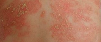
- Cutaneous leishmaniasis (CL) is the most common form of leishmaniasis and is accompanied by skin lesions, mainly ulcers, on exposed areas of the body. Skin lesions can leave permanent scars and lead to disability or stigmatization. About 95% of cases of CL are observed in the countries of the Americas, the Mediterranean basin, the Middle East and Central Asia. In 2021, more than 85% of new cases of CL were reported in 10 countries: Afghanistan, Algeria, Bolivia, Brazil, Colombia, Iran (Islamic Republic of), Iraq, Pakistan, and the Syrian Arab Republic and Tunisia. There are an estimated 600,000 to 1 million new cases of the disease each year worldwide.
- Mucocutaneous leishmaniasis leads to partial or complete destruction of the mucous membranes of the nose, mouth and larynx. More than 90% of cases of mucocutaneous leishmaniasis occur in Bolivia (Plurinational State of), Brazil, Ethiopia and Peru.
Transmission mechanism
Leishmania parasites are transmitted by the bites of infected female mosquitoes that feed on blood to lay eggs. The epidemiological characteristics of leishmaniasis may vary depending on the species of parasites and mosquitoes, the ecology of the areas where transmission occurs, the population's current or past exposure to the pathogen, and behavioral factors. It has been established that the natural reservoirs of Leishmania parasites are about 70 animal species, including humans.
Features of the epidemiology of the disease in different WHO regions
African region
Visceral, cutaneous and mucocutaneous forms of leishmaniasis are highly endemic in Algeria and East African countries. In East Africa, outbreaks of visceral leishmaniasis are common.
Americas region
The epidemiology of cutaneous leishmaniasis in the Americas is highly complex and highly heterogeneous with regard to transmission cycles, reservoirs, mosquito vector species, clinical manifestations and treatment response; in addition, different Leishmania species may circulate in the same geographic area. In 2021, more than 97% of VL cases in this region were reported in Brazil.
Eastern Mediterranean Region
This region accounts for 70% of all cases of cutaneous leishmaniasis in the world. Visceral leishmaniasis is highly endemic in Iraq, Somalia and Sudan.
European region
This region is endemic for cutaneous and visceral leishmaniasis. In 2018, more than 200 cases were reported in the region, mainly imported from Africa and the Americas region.
Southeast Asia region
The most common form of the disease in this region is visceral leishmaniasis, but the region is also endemic for cutaneous leishmaniasis. It is the only region with a regional initiative to eliminate visceral leishmaniasis as a public health problem by 2021. The region reported fewer than 5,000 cases in 2021, an all-time low. The region is confidently moving towards achieving its goal, and countries in the region intend to receive confirmation from the WHO of eliminating the disease by 2023.
Infectious diseases.
Infectious diseases have occupied a major place in human pathology for many centuries and have been a scourge for the population. In pre-revolutionary Russia, millions of people fell ill with various infectious diseases every year; there was no year free from epidemics. Infectious diseases were the main cause of child mortality and general mortality of the population; they caused enormous damage to the population and the country's economy because they often left severe and irreversible consequences. Tens of thousands were blind after smallpox, deaf, deaf-mute after scarlet fever, and had persistent paresis and paralysis after meningitis and polio. The Soviet state faced a difficult situation with regard to infectious diseases from the very first days of its formation. It was further aggravated by the previous world war, the civil war that followed the revolution, foreign intervention, famine and devastation, and the huge migration of the population in the country. All this taken together led to the emergence of epidemics of unprecedented scale. Already in those days, doctors attached great importance to the fight against infectious diseases. Already among the first acts of the Soviet state were decrees and resolutions to combat these diseases. At the beginning of the 20th century, the fight against infectious diseases unfolded on a broad front. First of all, appropriate personnel were required. This problem was solved thanks to the organization of appropriate departments at medical institutes. No less importance was attached to the study of infectious diseases, which included specialists from various fields. Gradually, corresponding laboratories and institutes were created, in which (together with the departments of medical institutes) issues of infectious pathology were developed. Children's infectious diseases were isolated in a separate course because the most common infections, more often or almost exclusively, occur in childhood and even mainly in early or preschool age (they were even considered obligatory for children). For example, the incidence of measles was almost universal among children and was observed more often under the age of 5 years. Among those sick with whooping cough (the second most common childhood infection), half were children under 3 years of age. The incidence of chickenpox and scarlet fever is highest between the ages of 1 and 9–10 years. Infectious diseases took first place in the structure of child mortality, which was very high. Finally, all infectious diseases, including those that are common among both children and adults, such as dysentery, in children have a number of pathogenetic and clinical features due to the anatomical and physiological differences of the child’s body. In the 1920s, a consistently expanding study of infectious diseases in children began. It is necessary to note the leading importance of the work carried out by teams of scientists led by A. I. Dobrokhotova, M. G. Danilevich, A. A. Koltypin, V. I. Molchanov, and subsequently their students and followers. An exceptionally large contribution to the study of the pathogenesis of infectious diseases was made by pathomorphologists (V.D. Tsinzerling, A.A. Skvortsov, etc.). Various studies were carried out by domestic immunologists, microbiologists, virologists and other specialists; The most active work was carried out by epidemiologists. A huge amount of research has been carried out, unique literature has been created, and a coherent system for combating childhood infectious diseases has been developed, as a result of which unprecedented successes have been achieved in history. In our country, the incidence of morbidity has sharply decreased - even to the point of eliminating certain nosological forms; for all infectious diseases, the mortality rate has decreased, the mortality rate has decreased hundreds and tens of times; Childhood infections have almost lost their significance in population mortality. The sharply increased life expectancy in our country is generally recognized as the result of a decrease in child mortality and mainly due to childhood infections. A coherent system of combating infectious diseases has a state character. The work of healthcare workers, in particular their fight against infectious diseases, is an important part of the general creative activity of modern society. This fight is not over; the tasks of further neutralizing childhood infections remain to be solved. Currently, intensive development in the field of infectious diseases continues and leads to the discovery of new aspects of known diseases, the identification of new pathogens and even new nosological forms. Infectious diseases, under the influence of numerous factors, have undergone evolution, especially noticeable in recent decades. Therefore, the presentation of the course is characterized by increasing complexity and versatility when considering each nosological unit. A description of the classic manifestations of infections, characteristics of new forms of infectious diseases that have appeared in the process of evolution, the reason for their formation and, finally, modern features of pathogenesis and clinic are presented. Infectious diseases are diseases caused by microorganisms. They are distinguished by their contagiousness, the specificity of the pathogen, clinical patterns in the form of cyclicity and the formation of immunity during the disease process. Contagiousness consists in the transmission of the pathogen from the patient to surrounding people. The specificity of the pathogen is that this microorganism, when infecting other people, causes the same disease in them and this nosological form cannot be caused by other microorganisms. For example, typhoid fever, dysentery, diphtheria, and measles are caused, respectively, by typhoid bacilli, dysentery, and measles virus. Some other microorganisms may cause similar individual symptoms, syndromes, but not the entire complex of the disease. An infectious disease is often referred to as “infection.” However, these are not synonyms; the concept of “infection” is much broader and, along with infectious diseases, includes the so-called infectious inflammatory processes. The latter are also caused by microorganisms and are contagious, but do not have pronounced clinical patterns in the form of cyclicity divided into certain periods. The main difference is that the pathogen does not cause any specific clinical form, but a variety of diseases with their own clinical picture. Similar, up to complete clinical identity, diseases can be caused by other types of pathogens, for example, streptococcal, staphylococcal processes. Both streptococci and staphylococci have the ability to infect any tissue, any organ of the human body and cause various diseases: rhinitis, tonsillitis, otitis media, lymphadenitis, pneumonia, pleurisy, meningitis, sepsis, etc. Many of these diseases can be caused by other microorganisms. Thus, pneumonia can be caused not only by streptococci and staphylococci, but also by Pfeiffer’s bacillus, Klebsiella, etc. These infectious processes have not taken shape in the form of a specific infectious disease with its inherent cyclicity, their nosological characteristics are not sufficiently delineated. They usually occur in the form of inflammatory processes, their infectious nature is immutable. There are also differences regarding immunity. Infectious diseases provide predominantly strong immunity not only for years, but often for life (measles, chicken pox, mumps, whooping cough, typhoid fever, scarlet fever, etc.). Infectious processes either do not provide immunity at all or it is short-lived; in some cases there is even a tendency to recurrence of diseases, as is observed in relation to streptococcal and staphylococcal diseases. The division between infectious diseases and infectious processes is often very arbitrary; as we study, the lines between them become noticeably blurred. Individual infectious processes are united on the basis of common clinical, morphological and other signs, and most importantly - on the basis of the unity of the pathogen. Thus, streptococcal and staphylococcal infections were isolated and included in textbooks on infectious diseases. The main differences between infectious diseases are contagiousness, the specificity of the pathogen, the peculiarity of clinical patterns, and the formation of immunity. During infectious diseases, there is an incubation, or latent, period, a prodromal, or period of precursors, periods of disease development and convalescence. Infectious pathology must be understood in a broad sense, including all diseases caused by microorganisms - both infectious diseases and infectious processes.
Post-kala azar cutaneous leishmaniasis (PCCL)
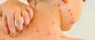
Post-kala azar cutaneous leishmaniasis (PCCL) is usually a consequence of visceral leishmaniasis and manifests as a macular, papular or nodular rash, most often on the face, shoulders, trunk and other parts of the body. This clinical form of the disease is characteristic mainly of East Africa and the Indian subcontinent, where it is reported in 5-10% of patients with visceral (kala-azar) leishmaniasis. Typically, skin rashes appear between 6 months and one or more years after signs of visceral leishmaniasis have disappeared, but it can occur earlier. People with PCCL are considered a potential source of infection.
Leishmaniasis
In accordance with the clinical features, etiology and epidemiology, leishmaniasis is divided into the following types. Visceral leishmaniasis (kala-azar) 1. Zoonotic: Mediterranean-Central Asian (children's kala-azar), East African (dum-dum fever), mucocutaneous leishmaniasis (New World leishmaniasis, nasopharyngeal leishmaniasis). 2. Anthroponotic (Indian kala-azar).
Cutaneous leishmaniasis 1. Zoonotic (rural type of Borovsky's disease, Pendinsky ulcer). 2. Anthroponotic (urban type of Borovsky's disease, Ashgabat ulcer, Baghdad boil). 3. Cutaneous and mucocutaneous leishmaniasis of the New World (espundia, Breda disease). 4. Ethiopian cutaneous leishmaniasis.
Visceral Mediterranean-Asian leishmaniasis. Incubation period. Varies from 20 days to 3-5 months, in rare cases up to 1 year or more. In young children and rarely in adults, long before the general manifestations of the disease, a primary affect occurs in the form of a papule.
Initial period of the disease. Characterized by the gradual development of weakness, loss of appetite, adynamia, pallor of the skin, and a slight enlargement of the spleen. Body temperature rises slightly.
High period. It usually begins with a rise in body temperature to 39-40 °C. The fever takes on a wave-like or irregular pattern and lasts from several days to several months with alternating episodes of high fever and remissions. In some cases, body temperature during the first 2-3 months can be low-grade or even normal.
When examining patients, polylymphadenopathy (peripheral, peribronchial, mesenteric and other lymph nodes), enlargement and hardening of the liver and even to a greater extent of the spleen, painless on palpation, are determined. In cases of development of bronhadenitis, a cough is possible, and pneumonia of a secondary bacterial nature is not uncommon.
As the disease progresses, the condition of patients progressively worsens. Weight loss (even cachexia) and hypersplenism develop. Bone marrow lesions lead to progressive anemia, granulocytopenia and agranulocytosis, sometimes with necrosis of the oral mucosa. Manifestations of hemorrhagic syndrome often occur: hemorrhages in the skin and mucous membranes, bleeding from the nose, and gastrointestinal tract. Fibrous changes in the liver lead to portal hypertension with edema and ascites, which is facilitated by progressive hypoalbuminemia.
Due to hypersplenism and the high position of the diaphragm, the heart shifts somewhat to the right, its sounds become muffled, tachycardia and arterial hypotension develop. These changes, along with anemia and intoxication, lead to the appearance and worsening of signs of heart failure. Possible diarrhea, menstrual irregularities, impotence.
Terminal period. Cachexia, a drop in muscle tone, thinning of the skin, the development of protein-free edema, and severe anemia are observed.
The disease can manifest itself in acute, subacute and chronic forms. • Acute form. Occasionally found in young children. It develops rapidly and without treatment quickly ends in death. • Subacute form. Seen more often. Severe clinical manifestations are characteristic, lasting 5-6 months. • Chronic form. It develops most often, often occurring subclinically and latently.
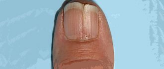
With visceral anthroponotic leishmaniasis (Indian kala-azar), in 10% of patients, several months (up to 1 year) after therapeutic remission, so-called leishmanoids appear on the skin. They are small nodules, papillomas, erythematous spots or areas of skin with reduced pigmentation, which contain Leishmania for a long time (years and decades).
Cutaneous zoonotic leishmaniasis (Pendinsky ulcer, Borovsky disease). Found in tropical and subtropical countries. The incubation period varies from 1 week to 1.5 months, on average 10-20 days. At the site of the entrance gate, primary leishmanioma appears, initially representing a smooth pink papule with a diameter of 2-3 mm. The size of the tubercle quickly increases, and it sometimes resembles a boil, but is painless or slightly painful on palpation. After 1-2 weeks, necrosis begins in the center of the leishmanioma, resembling the head of an abscess, and then a painful ulcer up to 1-1.5 cm in diameter is formed, with undermined edges, a thick rim of infiltrate and abundant serous-purulent or sanguineous exudate; Small secondary tubercles often form around it, the so-called “tubercles of seeding”, which also ulcerate and, when fused, form ulcerative fields. This is how sequential leishmanioma is formed. Leishmaniomas are most often localized on exposed parts of the body, their number varies from a few to dozens. The formation of ulcers in many cases accompanies the development of painless lymphangitis and lymphadenitis. After 2-6 months, epithelization of the ulcers and their scarring begin. The total duration of the disease does not exceed 6-7 months.
Diffuse infiltrating leishmaniasis . It is characterized by pronounced infiltration and thickening of the skin with a large area of distribution. Gradually the infiltrate resolves without a trace. Minor ulcerations are observed only in exceptional cases; they heal with the formation of barely noticeable scars. This variant of cutaneous leishmaniasis is very rare in older people.
Tuberculoid cutaneous leishmaniasis . Sometimes observed in children and young people. It is characterized by the formation of small tubercles around scars or on them. The latter can increase and merge with each other. As the disease progresses, they occasionally ulcerate; subsequently the ulcers heal with scarring.
Cutaneous antroponotic leishmaniasis . It is characterized by a long incubation period of several months or even years and two main features: slow development and less severe skin lesions.
Complications and prognosis Advanced leishmaniasis can be complicated by pneumonia, purulent-necrotic processes, nephritis, agranulocytosis, and hemorrhagic diathesis. The prognosis of severe and complicated forms of visceral leishmaniasis with untimely treatment is often unfavorable. In mild forms, spontaneous recovery is possible. In cases of cutaneous leishmaniasis, the prognosis for life is favorable, but cosmetic defects are possible.
Main risk factors
Socio-economic conditions
Poverty is a risk factor for leishmaniasis. Poor housing conditions and unsanitary conditions (eg, lack of waste disposal systems, open sewers) can increase the number of breeding sites for mosquitoes, as well as their proximity to humans. Mosquitoes are attracted to crowded living conditions that are favorable for feeding on human blood. Behavioral factors such as sleeping outdoors or on the ground may also be associated with an increased risk of infection.
Poor nutrition
Protein-energy malnutrition and dietary deficiencies of iron, vitamin A and zinc increase the risk of developing clinical disease in the event of infection.
Population movement
Epidemics of the two most common forms of leishmaniasis are often associated with migration and the movement of non-immune people into areas where the infection is circulating. Professional activities and large-scale deforestation remain important morbidity factors.
Environmental changes
Urbanization and increased intensity of economic activity in forest areas may be factors for the incidence of leishmaniasis.
Changing of the climate
The epidemiology of leishmaniasis depends on a number of climatic factors:
- changes in temperature, precipitation and humidity can have a significant impact on the distribution area, survival and population sizes of vectors and reservoirs of infection;
- small fluctuations in temperature can have a profound effect on the developmental cycle of Leishmania promastigotes in mosquitoes, which may create conditions for transmission of the protozoa in areas not previously endemic for the disease;
- drought, famine and floods can trigger mass displacement and migration of populations into areas where leishmaniasis circulates, and poor nutrition can have a negative impact on immunity.
Diagnosis and treatment
Diagnosis of visceral leishmaniasis is made on the basis of the clinical picture in combination with parasitological or serological studies (for example, rapid testing). For the diagnosis of cutaneous and mucocutaneous leishmaniasis, serological tests are not of great interest; in these cases, the diagnosis is made on the basis of the clinical picture and the results of parasitological examination.
The choice of treatment for leishmaniasis depends on a number of factors, such as the clinical form, the presence of associated pathologies, the type of parasite and the geographical area. Leishmaniasis is treatable and can be cured completely, however, the effectiveness of drugs depends on the state of the patient’s immune system, and relapses are possible if the immune system is weakened. All patients with visceral leishmaniasis are advised to immediately receive a full course of treatment. Detailed information on the treatment of different forms of leishmaniasis depending on the geographical area is given in the WHO technical report series No. 949 on the control of leishmaniasis.
Publications in the media
Leishmaniasis is the general name for protozoal vector-borne infections caused by intracellular parasitic flagellated protozoa of the genus Leishmania.
Etiology • Various species of Leishmania • Vectors are female mosquitoes of the genus Phlebotomus and Lutzomyia. Pathomorphology • Severe lymphocytic infiltration in the affected organs and tissues in combination with necrotic and degenerative processes, accumulations of Leishmania, fibrosis • Ulcerations on the skin and nasopharyngeal mucosa.
Classification, epidemiology and clinical picture • Visceral leishmaniasis (Leishman-Donovan disease , internal leishmaniasis, cachectic fever, tropical splenomegaly) •• The causative agent is Leishmania donovani •• The main reservoirs in Eurasia and Latin America are rodents, foxes, jackals, porcupines and dogs. In Eastern India and Bangladesh, where the natural reservoir is humans, epidemics of leishmaniasis are recorded every 20 years •• Symptoms develop 3-12 months after infection. In the Asian region, in 75% of cases, symptoms appear in the first month after infection •• Clinical picture: abnormal fever, febrile attacks continue, gradually fading, for 2–8 weeks, then appear at irregular intervals; malabsorption and diarrhea; enlarged liver and spleen; lymphadenopathy; swelling; in persons with weak skin pigmentation, grayish spots are observed on the face and head; anemia and thrombocytopenia with subsequent hemorrhages, agranulocytosis, leukopenia; It is acute and severe with possible death.
• East African visceral leishmaniasis is a type of visceral leishmaniasis. The causative agent is Leishmania donovani subspecies archibaldi. Distributed throughout East Africa (from the Sahara in the North to the Equator). The disease is registered more often in men aged 10–25 years; It is characterized by skin lesions in the form of nodes, often ulcerating, with subsequent damage to internal organs.
• Indian visceral leishmaniasis (kala-azar, Assam fever, dum-dum fever ) is a type of visceral leishmaniasis. The causative agent is Leishmania donovani subspecies donovani. Distributed in Eastern India and Bangladesh; It is distinguished by dark skin coloring due to damage to the adrenal cortex.
• Visceral Mediterranean-Central Asian leishmaniasis (kala-azar Mediterranean childhood) is a type of visceral leishmaniasis. The causative agent is Leishmania donovani subspecies infantum. Distributed in Southern Europe, North Africa, Central Asia, the Middle East and North-West China. Observed mainly in children; characterized by an acute onset with high body temperature and enlarged lymph nodes.
• Cutaneous leishmaniasis of the New World (leishmaniasis mucocutaneous, leishmaniasis of the mucous membranes, American leishmaniasis) •• Pathogens - Leishmania braziliensis, Leishmania mexicana •• The disease is characteristic of the humid forests of Central and South America; reservoir - large forest rodents. Usually recorded among workers engaged in forestry and road work, among the population of forest villages •• Symptoms appear 1–4 weeks after the bite of the vector •• Clinical picture: characterized by painless, deforming lesions of the mouth and nose, metastasizing to neighboring areas with the appearance of mushroom-shaped and erosive ulcers on the tongue, buccal mucosa and nose; relapses are possible several years after the spontaneous disappearance of the primary lesions; destruction of the nasal septum, hard palate and destructive lesions of the pharynx are observed; The disease is accompanied by fever, weight loss and secondary bacterial infections.
• Brazilian mucocutaneous leishmaniasis (Espundia) is a New World type of cutaneous leishmaniasis. The causative agent is Leishmania braziliensis subspecies braziliensis. Distributed in the forests of South America east of the Andes, it occurs with extensive damage to the skin, subsequently the mucous membranes, usually the upper respiratory tract, sometimes with deep destruction of soft tissues and cartilage.
• Leishmaniasis Uta (uta) is a New World variant of cutaneous leishmaniasis. The causative agent is Leishmania peruviana. Distributed in the highlands of South America. Proceeds with the formation of single ulcers, scarring within a year.
• Cutaneous diffuse leishmaniasis is a New World variant of cutaneous leishmaniasis. Pathogens: Leishmania mexicana subspecies amazoniensis, Leishmania mexicana subspecies pifanoi, Leishmania mexicana subspecies venezuelensis, Leishmania mexicana subspecies garnhami. Clinical manifestations do not differ from Asian and African types of cutaneous leishmaniasis. However, cases of spontaneous recovery are observed less frequently. The carriers are mosquitoes of the genus Lutzomyia. Exceptions are lesions caused by Leishmania mexicana subspecies mexicana (rubber canker), common in Mexico, Guatemala, Belize; detected in rubber tappers (chiclero) and lumberjacks; The carrier is the mosquito Lutzomyia olmeca. Characterized by the formation of painless, non-metastasizing chronic (several years) ulcers, usually localized on the neck and ears. As a rule, gross deformations of the auricles (ear chiclero) are observed.
• Cutaneous leishmaniasis of the Old World (Borovsky's disease) •• Causative agent - Leishmania tropica •• Endemic infection with the highest incidence in the autumn months •• Natural reservoir - small rodents (mice, rats, hyraxes), carriers - mosquitoes Phlebotomus papatasi (the main carrier), Phlebotomus duboscqi, Phlebotomus salehi, Phlebotomus longpipes and Phlebotomus pedifer •• Distributed in areas bordering deserts with low groundwater •• Clinical picture: the incubation period lasts from 2 weeks to 5 months; characterized by lesions on open areas of the body, prone to ulceration and centrifugal spread; the bottom of the ulcer is covered with granulation tissue, the edges are inflamed; After 3–12 months, spontaneous healing is observed with the formation of a rough, pigmented scar.
• Cutaneous anthroponotic leishmaniasis (urban cutaneous leishmaniasis, late ulcerating cutaneous leishmaniasis) is a type of cutaneous leishmaniasis of the Old World. The causative agent is Leishmania tropica subspecies minor. Distributed in cities of the Mediterranean, the Near and Middle East, and the western part of the Hindustan Peninsula; characterized by a long incubation period and prolonged necrosis of infiltrates.
• Zoonotic cutaneous leishmaniasis (cutaneous acute necrotizing leishmaniasis, rural cutaneous leishmaniasis) is a type of cutaneous leishmaniasis of the Old World. The causative agent is Leishmania tropica subspecies major. Distributed in oases of deserts and semi-deserts of the Middle East, Central Asia, India and Africa. It is characterized by a short incubation period and rapid necrosis of infiltrates.
• Recurrent (lupus) leishmaniasis is an Old World type of cutaneous leishmaniasis. The causative agent is Leishmania tropica subspecies tropica. It is characterized by partial healing of lesions, intensive formation of granulomas, frequent development of concomitant lesions that contribute to the formation of granulomatous tissue without signs of cure, sometimes for many years.
• Cutaneous lupoid leishmaniasis (cutaneous tuberculoid leishmaniasis, metaleishmaniasis, paraleishmaniasis) is a relapse of cutaneous leishmaniasis, characterized by the formation of dense confluent brownish-red tubercles around healed leishmaniamas. Laboratory tests • Detection of pathogens in Romanovsky-Giemsa-stained smears and in cultures grown after culture of aspiration material from lymph nodes, bone marrow, spleen, or biopsy material from lymph nodes. In macrophages, bean-shaped amastigotes with a dark round nucleus and short rod-shaped kinetoplasts (Leishman–Donovan bodies) are found • The direct agglutination reaction reveals IgM, characteristic of the acute phase • Skin tests with leishmanin (Montegoro test): with the cutaneous form of leishmaniasis, positive 6-8 weeks after recovery, but not with diffuse leishmaniasis. For visceral leishmaniasis, the results are negative. The test is used only for epidemiological studies • For species identification, monoclonal antibodies or the DNA hybridization method are used • ELISA is a highly sensitive and specific method in the diagnosis of visceral lesions.
Differential diagnosis • Malaria • Brucellosis • Tuberculosis • Typhoid • Liver abscess • Syphilis • Leprosy • Yaws. TREATMENT Management tactics • Bed rest, oral hygiene, enhanced nutrition • Blood transfusions for anemia and antibacterial chemotherapy for bacterial complications (in addition to specific therapy) • Periodic monitoring of the functions of the cardiovascular system, liver, kidneys • To prevent relapses, examinations are carried out after 3 and 12 months.
Surgical treatment - splenectomy. Drug therapy • Drugs of choice •• Meglumine antimoniate (glucantim) - 20–60 mg/kg deep IM 1 time per day for 20–30 days •• If the disease relapses or treatment is insufficiently effective, a second course of injections should be given within 40–60 days. Additional administration of allopurinol 20–30 mg/kg/day in 3 doses orally is effective • Alternative drugs for relapses of the disease and resistance of the pathogen to the drugs of choice •• Amphotericin B - 0.5–1.0 mg/kg IV every other day •• In the absence of chemotherapy effect, additional human recombinant -IFN.
Complications • In the later stages of the disease, edema, cachexia, hyperpigmentation are possible • Bacterial superinfections and bleeding from the gastrointestinal tract can cause death in untreated patients with visceral leishmaniasis • 3–10% of treated patients develop skin changes in the form of depigmented spots and wart-like nodules on the face and extensor surface of the extremities • In patients with mucocutaneous leishmaniasis, metastatic destructive lesions of the nasopharyngeal mucosa are possible.
Course and prognosis • With early diagnosis and timely treatment, more than 90% of patients recover • Even with chemotherapy, mortality reaches 15–25% • Without treatment, 95% of adults and 85% of children die within 3–20 months. Prevention • Extermination of insect vectors, rodents and stray dogs in areas adjacent to populated areas • Persons visiting endemic foci are recommended to use personal protective equipment (repellents, mosquito nets, etc.) • Timely treatment of patients with leishmaniasis.
ICD-10 • B55 Leishmaniasis
Prevention and control
Prevention and control of leishmaniasis requires a multimodal approach because transmission occurs within a complex biological system involving the human or animal reservoir (hosts), the parasite and its vector (the mosquito). The main measures to prevent leishmaniasis include:
- Early diagnosis and prompt initiation of effective treatment help reduce the prevalence of the disease and prevent disability and death in patients. This provides an opportunity to reduce transmission and monitor the spread and burden of disease. There are now highly effective and safe drugs for the treatment of leishmaniasis, especially its visceral form, although their use can be difficult. WHO's efforts to harmonize prices and the WHO-brokered free drug program have greatly increased access to medicines.
- Vector control helps reduce disease or interrupt transmission by reducing mosquito populations. Insecticide spraying, insecticide-treated netting, environmental engineering measures and personal protective equipment are used to control vectors.
- Effective surveillance is important because it allows for rapid monitoring and intervention during epidemics and in situations where there are high case fatality rates among patients under treatment.
- Control of animal populations as reservoirs of infection requires a complex set of measures and therefore must be carried out taking into account local conditions.
- Social mobilization and strengthening partnerships: Community mobilization and health education and effective behavior change interventions must always be locally tailored. Working in partnership and collaboration with various stakeholders and programs to control other vector-borne diseases is critical.
WHO activities
WHO's work on leishmaniasis control is carried out in the following areas:
- technical and financial assistance to national leishmaniasis control programs to update guidance documents and develop disease control plans that include sustainable and effective surveillance systems and epidemic preparedness and response systems;
- monitoring epidemiological indicators and assessing the effectiveness of disease control activities, which contributes to increasing awareness and advocacy on the global burden of leishmaniasis, as well as ensuring equitable access to health care;
- developing evidence-based strategies and standards for the prevention and control of leishmaniasis, and monitoring their implementation;
- strengthening cooperation and coordination among partners and stakeholders;
- promoting research and use of effective treatments for leishmaniasis, including safe, effective and affordable drugs, diagnostics and vaccines;
- providing support to national disease control programs to ensure patient access to quality-assured medicines.
Tropical diseases.
The site includes basic information about the etiology, pathogenesis, clinical picture, diagnosis, treatment and prevention of protozoal, bacterial, viral and fungal diseases common in hot countries. In addition, data is presented on the hygiene of hot countries and anti-epidemic measures to combat infectious diseases in countries with a tropical climate. Teaching and personal experience in medical institutions in some countries of Africa and Asia were the basis for the creation of this textbook on the main sections of the course of tropical medicine. Tropical diseases are based on sections of the official program for foreign students studying at medical institutes and universities of the CIS countries; the main sections of the course of tropical medicine are considered here. When writing the textbook, the authors did not pretend to be an exhaustive presentation of all nosological forms of diseases found in countries with tropical and subtropical climate, but they meant. The site covers selected issues of tropical medicine, which included protozoal and helminthic infestations, viral infections, and tropical mycoses. The proposed material reflects particularly dangerous infections (smallpox, cholera, plague) and diseases caused by poisonous animals. The greatest share is occupied by clinical issues, diagnosis and treatment of the actual tropical forms of parasitic and vector-borne diseases and mycosis. When creating this section of the site, we used the data accumulated to date in domestic and foreign literature, as well as personal experience in epidemiology, clinical presentation, diagnosis, and treatment of diseases characteristic of subtropical and tropical climate zones. One of the important issues, the presentation of which requires unification, is the nomenclature of chemotherapeutic drugs.
Medical news in the world:
| Is coffee harmful or beneficial? Regular coffee drinkers do not experience an increase in blood pressure after caffeine administration. In irregular coffee drinkers, blood pressure increases after taking both caffeine and coffee or CBC; therefore, it can be assumed that other ingredients in coffee, rather than caffeine, are responsible for this increase in BP. Read more… | Pregnancy – no AIDS! Scientists have concluded that highly active antiretroviral therapy (HART) significantly reduces the risk of transmitting HIV infection to children (transmission of infection to children from mothers who received ART was observed in only 1% of cases), and also helps today to lead a normal, comfortable lifestyle . Read more… | Raw milk can cause serious pregnancy complications A chemical compound found in unpasteurized food was found at unusually high levels in the red blood cells of pregnant women diagnosed with preeclampsia, a serious pregnancy complication that can lead to heart problems. Read more… | Fish oil can be harmful Fish oil supplements that help some heart patients may worsen the condition of others, reports EurekAlert, citing a study from St. Michael and the University of Toronto, Canada, published in yesterday's issue of the Journal of the Canadian Medical Association. Read more… |
| Treatment for impotence has been found!!! Recently, scientists managed to isolate a substance that enhances erection, but acts differently than Viagra (and similar drugs) from spider venom. Read more… | New methods of dental treatment. Toothache let's say: “NO!” ...the discovery of this gene will help recreate damaged tooth enamel, facilitate dental treatment, and even grow new teeth to replace lost ones. Read more… | Curing cancer in 60 minutes is a reality. CyberKnife works with submillimeter resolution, allowing tumors to be clearly localized and eliminated so that the loss of surrounding healthy tissue is minimal - up to 0.1–0.3 mm. Read more… | Karakurt or Black Widow is already in our area. The Black Widow's venom is stronger than that of one of the most dangerous snakes - the rattlesnake (15 times stronger). Read more… |