Morphological examination of a biopsy is a histology of tissue sections that were obtained during surgery or endoscopy. This analysis allows us to determine the structure of the tissue and sometimes the cause of the disease, the presence of the pathogen. With the correct interpretation of this analysis, the doctor will subsequently select effective treatment.
This type of research is often critical. For example, patients who suffer from chronic gastritis can be thoroughly tested only by determining the morphological structure of the biopsy specimen. The diagnostic method allows you to diagnose precancerous conditions and the presence of other serious diseases in the early stages.
Why is a biopsy needed?
Biopsy refers to one of the diagnostic research methods, which consists of taking histological material from a suspicious area. These can be various seals, tumor formations, as well as long-term non-healing wounds.
Currently, this technique is the main one of the most reliable and informative. It can be used not only for diagnostic, but also for therapeutic purposes.
Thanks to the study of biomaterial using microscopy, it is possible to determine not only the cytological identity of the tissue, but also to establish the nature of its growth with the stage and general idea of the disease.
Its early implementation makes it possible to establish a diagnosis in the early, possibly preclinical stages, which will greatly facilitate treatment and improve the prognosis for life.
Obtaining the most accurate information about the pathological process using a biopsy is necessary to further determine the extent of surgical intervention. At the preparation stage, the doctor can prevent possible complications and also select the safest and most effective way to solve the problem.
Algorithm for conducting morphological examination of biopsy specimens
Content:
- Algorithm for conducting morphological examination of biopsy specimens
- Indications and contraindications for the study
- Methods for taking biopsy samples
- Preparation for taking a biopsy sample
Histological examination is carried out using a high-resolution light microscope. The laboratory technician performs the necessary preparation of the sample, including dehydration and embedding of the sample in paraffin blocks. Next, using a microtome (an instrument for preparing sections of biological tissue), the laboratory assistant prepares a specimen for research (several micrometers thick).
The separated tissues are placed on a special glass and a special staining procedure is carried out, which allows for maximum visualization of individual cells. After the histologist receives the results of the study, he writes his conclusion, on the basis of which the attending doctor makes or confirms the diagnosis and prescribes appropriate treatment.
In clinical practice, urgent morphological examination is often carried out - when tissue is excised for histology during the operation. Urgent diagnosis is done when it is necessary to quickly determine the course of a surgical intervention. The urgent diagnostic method involves flash freezing of tissues (dehydration is not performed). This is followed by standard cell microscopy. It is worth noting that the duration of an instant study is at least half an hour.
Another option for conducting MIB is cytology. Cytology analysis does not imply excision of tissue, but sampling of cells for examination, when a piece of the mucous membrane is not available to make a preliminary diagnosis. This research method is most often used in gynecology - a smear of the cervical mucosa. It is prescribed to detect tumors in the early stages. It is worth noting that in practice, biopsy is much more effective and efficient than cytology.
Indications for use
Indications for a biopsy are determined individually, based on the patient’s complaints, data obtained during laboratory or instrumental diagnostics, as well as symptoms and signs of the disease identified by the doctor during the examination and conversation.
The most common indications include:
- Suspicion of a malignant process. Currently, measures aimed at excluding cancer are among the main ones when diagnosing a patient. A biopsy can be performed from several areas over time. In addition, it can be prescribed dynamically during treatment.
- The presence of neoplasms of unknown etiology. The appearance of space-occupying formations in a patient requires obtaining information about the nature of their origin and the risk of degeneration into a malignant process.
- The presence of an inflammatory process, including a purulent one. Using this method, the type of infectious agent is determined, as well as its sensitivity to drugs.
Recommended video:
What is a cervical biopsy?
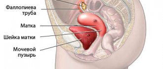
A cervical biopsy is a diagnostic surgical procedure during which the doctor removes a sample of pathologically altered tissue for further examination under a microscope. In gynecology, the technique is carried out with the aim of making a final diagnosis, when there is the slightest suspicion of oncological degeneration of the tissues of the uterine cavity. A biopsy of the cervix is performed after receiving poor results from colposcopy and PAP test, which are necessarily carried out before the procedure.
Indications for the procedure are:
- abnormal changes in the cervical mucosa;
- suspicious formations on tissues;
- endometriosis;
- leukoplakia;
- cervical polyposis;
- dysplasia;
- pre-cancer conditions.
Types and methods of biopsy
In modern medicine, several types of biopsy have been developed, the use of which depends on the nature of the pathological process, its localization, as well as the goal pursued by the doctor.
Table 1. Types and indications for biopsy
| Type of biopsy | Indications |
| Smears—imprints, scrapings, shave biopsy. | Most often, this type of biopsy is used in outpatient practice for the diagnosis and treatment of gynecological, urological and dermatovenerological patients. It is prescribed for the occurrence of itching, burning, discharge, as well as redness of the mucous membrane or skin. The procedure is non-invasive and allows us to identify the nature of the infectious process with possible sensitivity to drugs. In addition, a smear can be taken for the purpose of dynamic examination and prevention of the oncological process on the cervix. |
| Needle biopsy. | The collection of histological material is carried out by puncturing soft superficial tissues with a needle and suctioning out the contents. It is most often used to diagnose mass formations in the mammary glands, thyroid gland, testicle or prostate. The procedure is performed without the control of ultrasonic equipment. Despite its widespread use, there is a high probability of obtaining a false result due to the collection of biomaterial not from a pathologically altered area. |
| Fine needle biopsy. | Tissue collection using a syringe or puncture needle is performed in the presence of a suspicious fluid formation or solid tumor. Its implementation is indicated when there is a high risk of tumor cell spread. During needle advancement, minimal tissue damage and contamination of healthy areas occurs. |
| Core needle biopsy. | The biomaterial is collected using a needle of a larger diameter or a special trocar, which makes it possible to obtain a column of tissue. No incision is required to perform. The procedure is performed on patients to take a sample of skin, bone, as well as breast, prostate or liver. As a result, several layers of tissue are removed, which allows us to give the most complete picture of the pathology. |
| Aspiration biopsy. | The procedure is performed using a vacuum tube and created negative pressure. Several tissue fragments are collected at once. Most often it is performed in gynecological or pulmonological practice to identify pathological changes in the uterine cavity, bronchi and lungs. The resulting secretion or exudate with cells does not come into contact with the external environment. The patient requires less recovery time since in most cases it is not invasive. |
| Scan-guided biopsy. | The stereotactic procedure can be fine-needle, thick-needle, or puncture. It is most often used for deeply located formations or small lesions that cannot be palpated. Tissues are taken from various organs, and the result obtained is highly informative. |
| Biopsy during surgery. | Tissue sampling during surgery is divided into excisional or incisional (with complete or partial removal of an organ or tumor). It can be diagnostic and therapeutic in nature. A similar biopsy is performed on various organs after ensuring free access to the suspicious area. One of the disadvantages is obtaining results after surgery, since in the future it may be necessary to expand the scope of the intervention. Only in large medical centers are tissues examined in a short time before the end of the operation. |
| Biopsy during endoscopy. | During bronchoscopy, hysteroscopy, sigmoidoscopy, cystoscopy or endoscopic operations on the abdominal organs, material is collected. Therefore, there can be many indications, among them the most common is the suspicion of a tumor process, the presence of polyps or inflammatory foci. The procedure is highly informative and causes minimal tissue damage with rapid recovery. Removal of a formation in which the benign nature of the cells has been identified has a therapeutic purpose. |
Methods for taking biopsy samples
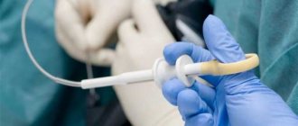
The most minimally traumatic research method today is a puncture aspiration biopsy. To carry it out, a percutaneous puncture is performed with a hollow needle, through which tissue is collected from the pathological focus. Most often, this type of tissue sampling occurs under ultrasound guidance. Among the disadvantages of this method, it is worth noting: there is no absolute guarantee of an accurate hit to the pathological focus; there may not be enough selected material for the study.
Thick-needle trephine biopsy - special threaded needles are used. They seem to be screwed into the tissues being examined and suddenly pulled out.
An incisional biopsy is a type of tissue excision that is performed during surgery. It is worth noting that the procedure does not occur for therapeutic purposes, but exclusively for diagnostic purposes. The surgeon sees the pathogenic focus and excises the biopsy for analysis from several areas.
Excisional biopsy is performed in a surgical setting and involves the complete removal of a tumor or damaged organ.
How long does it take to perform a biopsy?
In most cases, performing a biopsy is a short-term procedure.
The duration of the procedure depends on various factors, including the qualifications and practical experience of the doctor, as well as the location of the tissue source.
The longest are biopsies, which are performed under the control of ultrasound scanning or radiography from hard-to-reach areas where there is a high probability of damage to other organs or vital vessels.
In this case, a violation of integrity becomes the cause of the development of severe complications, so the procedure is performed as carefully and slowly as possible. These include taking biomaterial from the brain, as well as internal organs, using the puncture, fine-needle or aspiration method.
Minimal time is required to perform a biopsy from superficial tissues. This can be taking scrapings from the skin, smears or fingerprints. Obtaining intraoperative material also does not take much time, since it is performed after creating access to the tissues.
How is a cervical biopsy done? Progress of the operation
A cervical biopsy is performed in the treatment room. The patient sits comfortably in a gynecological chair, after which the doctor resorts to an anesthesia procedure. A speculum is inserted into the vaginal cavity and the cervix is exposed. Next, using a colposcope, the doctor determines the exact boundaries of the pathological focus.
Under the control of colposcopy, the specialist carries out all the necessary manipulations, after which the biopath is removed and given to a laboratory assistant for histological examination. The procedure takes 15, maximum 20 minutes. After the biopsy, rehabilitation is required, during which you must strictly follow all the doctor’s recommendations. Diagnostic results are usually available within a few days. Based on the data obtained, the specialist will determine the further treatment plan for the patient and select the most effective treatment.
Test results
Histological or cytological studies determine if the patient has altered cells that act as a potential threat to health or cause the development of cancer.
According to the World Health Organization, all changes obtained are systematized into several groups. Among them are:
- Background. Deviations from the norm in the obtained material act as a cause of various diseases, but do not lead to a malignant process.
- Precancerous. Malignant tumor activity has not been diagnosed, but lack of treatment in more than half of the cases causes the changes to progress to cancer.
- Cancer. Confirmed malignant tumors are divided into preclinical and those with clinically significant changes.
Increasing the accuracy of the results is possible when performing the procedure under the control of ultrasound scanning or radiography.
Purpose of cervical biopsy
The main goal of the procedure is the collection and detailed histological examination of the biopath for the presence of atypical cells. Histology is the study, using diagnostic optics, of a previously prepared and stained section of a biopath extracted from the cervix. The study of the material helps to determine the causes of pathological changes in organ cells, identify exophytic condylomas, and oncological processes. The results of the biopsy will help make a final diagnosis and select the most effective treatment regimen.
Is a biopsy safe? What are the consequences and complications?
The safety of a biopsy depends on the location of the histological material taken. When performing a procedure during surgery, the likelihood of complications may depend on the technique of the procedure, as well as its volume.
With a puncture biopsy performed without the control of ultrasound or X-ray equipment, the integrity of blood vessels or neighboring organs may be compromised. This condition is rare and, due to low-traumatic equipment, no serious consequences are noted, since the bleeding quickly stops.
Cost of cervical biopsy
If you or your loved ones need high-quality, professional medical care, contact the Healthy Family multidisciplinary clinic, where experienced specialists with many years of experience work. Our doctors will quickly establish the correct diagnosis and select the most effective treatment regimen that will help restore women’s health and prevent the development of serious complications.
To find out the cost of a cervical biopsy and make an appointment with a medical specialist, call the number or fill out the feedback form. As soon as the administrators see your request, they will immediately contact you and answer all your questions.
Post-procedure care
Post-procedure care rules depend on the type of biopsy performed. General recommendations include:
- Elimination of physical activity. On the day of the biopsy, the patient is advised to rest and limit heavy lifting.
- Avoiding water getting into the biopsy site. In most cases, the patient is given an aseptic dressing for a day, which allows the tissues to recover, eliminating infection. When applying suture material, a longer aseptic dressing is required.
- Use of medications. These can be non-steroidal anti-inflammatory drugs, which will relieve pain, as well as antibiotics, which will prevent the growth of bacterial cells that have entered the wound.
- Treating the wound with an antiseptic solution to speed up the healing process and prevent infection. The most common are chlorhexidine or brilliant green solution.
Recommended video:
Despite the fact that biopsy is currently one of the highly informative research methods that makes it possible to make an accurate diagnosis, it should be carried out after a thorough examination of the patient and assumption of the diagnosis. This is due to the fact that in some cases it requires repeated execution or dynamic control.
Preparing for cervical biopsy surgery
To prevent serious complications, careful preparation for the cervical biopsy procedure is required. The optimal time frame for diagnosis is days 7–11 of the cycle, starting from the first day of the onset of menstruation.
Before giving a referral for a cervical biopsy, the doctor performs a gynecological examination, which usually includes the following procedures:
- biomanual examination;
- vaginal smear for microflora;
- PAP analysis;
- colposcopy;
- ultrasound examination of the genitourinary system.
If the results of the smear for microflora are positive, additional PAP testing for infections is prescribed. After this, the patient is prescribed appropriate drug therapy, after which a biopsy can be performed.
A woman must also undergo general clinical tests before a cervical biopsy:
- general blood test, urine;
- biochemistry;
- coagulogram;
- blood test for hidden infections, HIV, hepatitis.
Electrocardiography, fluorography, and consultation with specialized specialists are required if there are concomitant diseases.
5–7 days before the procedure, stop taking medications that affect blood viscosity. For 2-3 days, exclude sexual activity, douching, and the use of vaginal suppositories, tampons and other means.
It is also important to follow the rules of hygienic preparation:
- The evening before the biopsy, shave your intimate area.
- In the morning, on the day of the procedure, take a refreshing shower and put on clean underwear.
- After the biopsy, you will need sanitary pads, since after the biopsy is taken, the wound will bleed for some time.