Rules for sputum collection
It is unacceptable to send sputum for examination in containers not intended for these purposes, as they may contain substances that can distort the test results.
Sputum should not be divided into two parts for examination in different laboratories, since important components may end up in only one of the containers.
- Sputum is collected in the morning, on an empty stomach. Before collecting sputum, brush your teeth and rinse your mouth with water to remove food particles and bacterial plaque. Take two deep breaths, holding your breath for a few seconds, and after each deep breath exhale slowly. Then inhale a third time and forcefully exhale (push) the air out. Inhale again and clear your throat well.
- Hold the container as close to your mouth as possible and carefully spit the mucus into it after coughing. Be careful not to get any sputum on the edge of the container. Do not touch the inside of the lid or the sides of the sterile container with your fingers. Close the container tightly with the lid.
- Wash your hands with soap.
- Deliver the morning sputum collected for research to the laboratory no later than 1–1.5 hours after collection. In this case, conditions must be created to prevent its cooling during transportation. Otherwise, changes in sputum qualities and the composition of microbial colonies will quickly occur, which may distort the results of the study.
Attention Do not collect sputum released during expectoration, only when coughing. Saliva entering the sample can significantly affect the test result.
The secretion has bactericidal properties, promotes the removal of inhaled particles, metabolic products and cellular detritus due to the mechanism of mucociliary clearance (purification) provided by the activity of the ciliated epithelium. The volume of tracheobronchial secretion normally ranges from 10 to 100 ml per day; A healthy person usually swallows this entire amount.
Sputum analysis is a laboratory test that allows one to establish the nature of a disease of the respiratory system (bronchi, lungs), determine its causes: identify pathogenic microorganisms (including Mycobacterium tuberculosis), malignant tumor cells, impurities (blood, pus, etc.) characteristic of certain diseases, as well as determine the sensitivity of bacterial flora to antibiotics. Sputum analysis is prescribed for patients suffering from prolonged cough, bronchitis, pneumonia, tuberculosis, lung abscess and other pathologies.
Rules for collecting sputum for analysis:
- Sputum is collected in the morning (since it accumulates at night and is easily removed) before meals in a sterile container, which is provided in the laboratory or can be purchased at any pharmacy. Most often, sputum is collected in two containers, one for general sputum analysis and the other for bacteriological analysis.
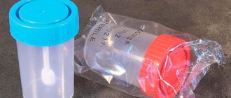
- To safely collect quality sputum samples, a container made of durable, liquid-tight material is required. The hole in the sputum collection container should be wide (diameter 35 mm), which will allow sputum to be coughed up without contaminating the outer surface of the container.
- Sputum is released better if you take expectorants and drink plenty of warm liquids the day before collection. If the mucus does not come out, you need to take a deep breath and exhale several times.
- In order to improve expectoration of sputum, exercise therapy, chest massage, positional drainage, and inhalations can be used.
- Before collecting sputum, you need to brush your teeth and rinse your mouth with boiled water. You need to brush your teeth with a soft brush without damaging your gums. Try to cough up mucus without saliva.
- To conduct the study, you will need at least 3-5 ml of sputum in each container.
- It is necessary to deliver sputum to the laboratory no later than 2 hours after collection. Until the sputum is sent for examination, it must be stored in the refrigerator.
- The result of a general sputum analysis will be ready within 3 days, and a bacteriological analysis will take longer – up to 7 days.
Rules for collecting saliva for studies “Cortisol in saliva”
- One day and during the entire period of saliva collection, avoid physical activity, do not consume caffeine (tea, coffee, caffeine-containing energy drinks, foods, chewing gum), alcohol;
- Do not smoke one hour before saliva collection;
- 30 minutes before collecting saliva, do not eat food, do not brush your teeth, including not using dental floss and mouth rinse, and do not chew chewing gum;
- 10 minutes before collecting saliva, rinse your mouth with water;
- Taking sedatives, cortisone acetate, estrogens, oral contraceptives, glucocorticoid drugs (including ointments) can cause an increase in cortisol levels. Discontinuation of medications is carried out strictly on the recommendation of the attending physician.
For more information about the rules for collecting saliva for studies “Cortisol in saliva”, see the link
Sputum. General clinic
- Sputum is collected in the morning, on an empty stomach. Before collecting sputum, brush your teeth and rinse your mouth with water to remove food particles and bacterial plaque. Take two deep breaths, holding your breath for a few seconds, and after each deep breath exhale slowly. Then inhale a third time and forcefully exhale (push) the air out. Inhale again and clear your throat well.
- Hold the container as close to your mouth as possible and carefully spit the mucus into it after coughing. Be careful not to get any sputum on the edge of the container. Do not touch the inside of the lid or the sides of the sterile container with your fingers. Close the container tightly with the lid.
- Wash your hands with soap.
- Deliver the morning sputum collected for research to the laboratory no later than 1–1.5 hours after collection. In this case, conditions must be created to prevent its cooling during transportation. Otherwise, changes in sputum qualities and the composition of microbial colonies will quickly occur, which may distort the results of the study.
Attention! Do not collect sputum released during expectoration. Saliva entering the sample can significantly affect the test result.
Sputum. Tuberculosis
- Sputum is collected in the morning, on an empty stomach. Before collecting sputum, brush your teeth and rinse your mouth with water to remove food particles and bacterial plaque. Take two deep breaths, holding your breath for a few seconds, and after each deep breath exhale slowly. Then inhale a third time and forcefully exhale (push) the air out. Inhale again and clear your throat well.
- Hold the container as close to your mouth as possible and carefully spit the mucus into it after coughing. Be careful not to get any sputum on the edge of the container. Do not touch the inside of the lid or the sides of the sterile container with your fingers. Close the container tightly with the lid.
- Wash your hands with soap.
- Deliver the morning sputum collected for research to the laboratory no later than 1–1.5 hours after collection. In this case, conditions must be created to prevent its cooling during transportation. Otherwise, changes in sputum qualities and the composition of microbial colonies will quickly occur, which may distort the results of the study.
Attention! You should not collect sputum released during expectoration, only when coughing. Saliva entering the sample can significantly affect the test result.
“Methods for examining sputum for tuberculosis”
Date: 07/19/2018
Sputum is a secretion released during inflammation of the trachea, bronchi and lungs. Its appearance is noted not only with damage to the respiratory organs, but also with disorders of the heart and blood vessels. Methods for examining sputum involve macroscopic, chemical and microscopic determination of its characteristics.
What does the analysis reveal?
Examination of sputum makes it possible to detect microorganisms that cause the pathological process, identify the presence of mycobacteria in tuberculosis, identify cancer cells, blood and purulent impurities, and also determine bacterial resistance to antibiotics.
For what conditions is analysis indicated?
Sputum examination for general analysis is carried out in the following conditions:
- cough;
- pneumonia;
- inflammation of the bronchi;
- suppuration of the lung;
- tuberculosis;
- bronchiectasis;
- pulmonary gangrene;
- tumor in the lungs;
- acute bronchitis;
- chronic bronchitis;
- chronic tonsillitis;
- tuberculosis;
- whooping cough;
- silicosis;
- acute form of obstructive bronchitis;
- pneumonia;
- anthrax.
Preparing for the study
Mucus will be better released if you take an expectorant the day before the test or drink a large amount of warm drink. Before collection, it is recommended to clean your teeth and mouth by rinsing with warm boiled water.
Basic collection rules
It is advisable to collect sputum for bacteriological examination in the morning (it accumulates the night before meals) in a sterile container provided by the laboratory. An amount of 5 ml is sufficient for analysis. The secretion is analyzed no later than 2 hours after its collection. Until sent for testing, the container with the contents should be stored closed in the refrigerator.
The amount of sputum in various diseases
The amount of secretion released varies depending on the nature of the pathological process. Usually it varies from several spits to 1 liter per day. A small amount is released during inflammation of the bronchi, congestive processes in the lungs and at the onset of an attack of bronchial asthma. At the end of the attack the volume increases. It can be up to 0.5 l, and can also be released in large quantities if there is pulmonary edema.
A lot of mucus is released during a purulent process in the lungs when communicating with the bronchi, during suppuration, bronchiectasis and gangrene.
Examination of sputum for tuberculosis shows the breakdown of lung tissue. In particular, this process is provoked by the cavity, which communicates with the bronchi.
What is the reason for the decrease or increase in secretion secretion?
An increase in the amount of secretion secreted may be associated with a deterioration in the patient’s condition and may be observed during an exacerbation. An increase may also refer to positive dynamics in the development of the disease.
A decrease in the amount of mucus secreted may indicate regression of inflammation or a violation in the area of drainage of a cavity filled with pus. At the same time, there is a deterioration in the patient’s well-being.
Nature of the discharge
Mucous secretion is released during acute or chronic bronchitis, bronchial asthma, pneumonia, lung cancer, bronchiectasis, pulmonary echinococcosis accompanied by suppuration, actinomycosis.
Sputum mixed with pus is observed with lung abscess, echinococcosis and bronchiectasis.
Mucus mixed with blood or consisting entirely of blood is characteristic of tuberculosis. The appearance of blood may indicate the presence of oncology, bronchiectasis, or suppuration of the lung. This phenomenon is also observed in middle lobe syndrome, pulmonary infarction, trauma, actinomycosis and syphilitic lesions. Blood can also be released during lobar and focal pneumonia, congestive processes, cardiac asthma and pulmonary edema.
Serous sputum is observed with pulmonary edema.
Sputum color
Examination of sputum reveals its different colors. Mucous and serous discharge has no color or has a whitish tint.
The addition of pus gives the secretion a greenish tint, which characterizes such pathological processes as lung abscess, gangrene, bronchiectasis, and actinomycosis of the lung.
Discharge with a rusty or brown color indicates that it does not contain fresh blood, but a product of its breakdown - hematin. Such a secretion can be released during lobar pneumonia, anthrax, or pulmonary infarction.
A greenish color mixed with dirt or a yellow secretion indicates a pathology of the respiratory system in combination with jaundice.
Sputum turns bright yellow in eosinophilic pneumonia.
Ocher-colored mucus occurs with siderosis of the lung.
A blackish or grayish secretion is noted when there is an admixture of coal dust. With pulmonary edema, serous sputum is observed in large quantities. As a rule, it is colored uniformly pinkish, which is explained by the presence of red blood cells. Such discharge is similar to liquid cranberry juice.
The secretion may also become colored due to some medications. For example, the antibiotic Rifampicin can give it a red color.
Smell
The nature of the pathological process in the respiratory organs can also be indicated by the smell of the secretion. Sputum gives off the smell of rot in cases of gangrene of the lung or putrefactive lesions of the bronchi, oncological neoplasms, complicated by necrosis of bronchiectasis.
Availability of layers
Often, examination of secretions reveals the presence of layers. With a stagnant nature, sputum mixed with pus is observed with suppuration of the lung and bronchiectasis.
The secretion, mixed with rot, contains three layers. The top layer is foam-like, the middle layer is serous, and the bottom layer is mixed with pus. This composition characterizes lung gangrene.
Impurities
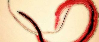
An admixture of food can be observed in the presence of a malignant tumor in the esophagus when it communicates with the bronchi and trachea. When echinococcus enters the bronchi, hooks or scolex of the parasite can be found in the sputum. Very rarely, adult roundworms are found that penetrate the respiratory system of weakened people.
Pulmonary fluke eggs appear when a cyst ruptures, which forms in the lungs in the presence of parasites.
Gangrene and suppuration of the lungs cause the appearance of pieces of pulmonary necrosis. If there is a tumor, fragments may be present in the discharge.
Convolutions containing fibrin are found in patients with fibrinous bronchitis, tuberculosis and pneumonia.
Rice bodies, or Koch lenses, are characteristic of tuberculosis.
Dietrich's plugs, which include decay products of bacteria and fatty acid tissue cells of the lungs, are found in putrefactive bronchitis or gangrene of the lung.
The chronic form of tonsillitis involves the release of plugs from the tonsils, similar to Dietrich's plugs.
Chemical method
Chemical examination of sputum involves determining:
- A protein indicator that can help in the differential diagnosis of chronic bronchitis and tuberculosis. With chronic bronchitis, traces of protein are noted in the secretion, and with tuberculosis, the amount of protein in the sputum will be much higher, and it can be indicated by numbers (up to 100-120 g/l).
- Bile pigments. They are found in sputum when the respiratory system is affected in combination with hepatitis. In this case, the liver communicates with the lungs. Bile pigments are inherent in pneumonia, which is caused by the breakdown of red blood cells inside the lungs and the subsequent change in hemoglobin.
Cytological method for studying secretions
For the differential diagnosis of tuberculosis and many other lung lesions, the cytological method is widely used, which includes two stages: clinical and microscopic examination of sputum.
A clinical study helps determine which method should be used to collect material to obtain the correct test result.
There are two main types of material required by microscopic examination of sputum: spontaneous and reduced. The second type of secretion is obtained by exposure to various irritants (expectorants, inhalations, etc.).
Material obtained from needle biopsy
Cytological examination of sputum involves the study of macroscopic and microscopic analysis of its cells.
The most information for cytological analysis is provided by sputum taken in the morning on an empty stomach. Before testing, it should be stored for no more than 4 hours.
- Sputum contains squamous epithelial cells, which are examined microscopically. But they have no significance for making a diagnosis. Columnar epithelial cells - both individually and in groups - can be observed in diseases such as bronchial asthma, bronchitis and lung cancer. It should be noted that columnar epithelium can also appear due to the penetration of mucus from the nasopharynx.
- Alveolar macrophages are reticuloendothelial cells. Macrophages, which are contained in the protoplasm (phagocytic particles or dust cells), can be found in patients who have inhaled dust for a long time.
- Protoplasmic macrophages (formed during the breakdown of hemoglobin) are called cardiac defect cells. They can occur during congestive processes in the lungs, mitral valve stenosis, and pulmonary infarction.
- A small number of leukocytes are found in any sputum. Their increased content is observed in secretions mixed with pus.
- Eosinophils. The sputum of asthmatics is rich in such cells. Cells can be observed in the eosinophilic form of pneumonia, helminth damage to the body, tuberculosis and pulmonary infarction.
- Red blood cells. Single red blood cells do not reflect the picture of the disease. The appearance of an increased amount indicates the presence of bleeding in the lungs. In fresh blood, unchanged red blood cells are detected. If there is an admixture of blood that has stagnated in the lungs for a long time, then leached red blood cells are detected.
- Cancer cells. They can be found in secret groups. They indicate the presence of a tumor. When single cells are found, diagnostic difficulties often arise. In such cases, a repeat sputum analysis is performed.
- Elastic fibers, the appearance of which is caused by the breakdown of lung tissue, provoked by tuberculosis, abscess, gangrene, tumor. Such cells are not always characterized by gangrene, since due to the action of secreted enzymes, they can be dissolved.
- Kurshman spirals. These are special bodies that look like tubes. They are detected when examined under a microscope. Sometimes visible to the eye. Typically, spirals are associated with diseases such as bronchial asthma, pulmonary tuberculosis and pneumonia.
- Charcot-Leyden crystals are found in sputum with an increased content of eosinophils in diseases such as bronchial asthma, eosinophilic pneumonia. Opening a focus of tuberculosis in the lumen of the bronchi can be characterized by the presence in the secretion of elastic fiber-crystals of cholesterol, MBT and amorphous lime (the so-called Ehrlich tetralogy) - 100%.
Application of bacterioscopy
Collecting sputum for examination by the bacterioscopic method involves analyzing the secretion to detect mycobacteria characteristic of tuberculosis. They look like thin curved sticks of different lengths, thickened on the sides or in the middle, which are located both individually and in groups.
The detection of Mycobacterium tuberculosis is not a dominant sign for diagnosis and requires confirmation by bacteriological methods. Tuberculous mycobacteria are not detected in secretions under normal conditions.
The basis for the analysis is purulent particles, which are taken from forty-six different areas and thoroughly ground to a homogeneous mass using two glasses. Next, they are dried in air and fixed with a burner flame.
Bacteriological examination of sputum using the Ziehl-Neelsen method suggests that it is stained red. In this case, all secretion particles, with the exception of mycobacteria, acquire a blue tint, and mycobacteria acquire a red color.
If the body is suspected of being affected by tuberculosis, after testing three times for the presence of mycobacteria with a negative response, the flotation method (Pottenger analysis) is used.
The usual method of examining a stained smear for MTB gives a positive result only when the amount of MTB is at least 50,000 units in 1 ml of sputum. The presence of tuberculosis cannot be judged by the number of mycobacteria.
Bacterioscopy of patients with nonspecific lung diseases
Laboratory tests of sputum in the presence of nonspecific lung diseases during bacterioscopy can reveal the following bacteria:
- For pneumonia - pneumococci, Frenkel diplococci, Friedlander bacteria, streptococci, staphylococci (100%).
- With gangrene of the lungs, a spindle-shaped rod can be found in combination with Vincent's spirochete (80%).
- Yeast-like fungi (70%), to determine the type of which requires culture of the secretion.
Author: Seytimova Moldir
Regional anti-tuberculosis dispensary
Bachelor laboratory assistant
07/17/2018
Sputum culture for microflora and sensitivity to antibiotics
For bacteriological examination, sputum is collected before the start of antibacterial therapy or after a certain period of time after administration of the drug.
Collection procedure
- Sputum is collected in the morning, on an empty stomach. Before collecting sputum, brush your teeth and rinse your mouth with water to remove food particles and bacterial plaque. Take two deep breaths, holding your breath for a few seconds, and after each deep breath exhale slowly. Then inhale a third time and forcefully exhale (push) the air out. Inhale again and clear your throat well.
- Hold the container as close to your mouth as possible and carefully spit the mucus into it after coughing. Be careful not to get any sputum on the edge of the container. Do not touch the inside of the lid or the sides of the sterile container with your fingers. Close the container tightly with the lid.
- Wash your hands with soap.
- Deliver the morning sputum collected for examination to the laboratory. In this case, conditions must be created to prevent its cooling during transportation. Otherwise, changes in sputum qualities and the composition of microbial colonies will quickly occur, which may distort the results of the study.
Attention! You should not collect sputum released during expectoration, only when coughing. Saliva entering the sample can significantly affect the test result.
Sputum. Anaerobes and antibiotic sensitivity
Attention Sputum is collected on an empty stomach.
First brush your teeth, gums and tongue with a toothbrush and rinse your mouth with boiled water at room temperature.
Collection procedure
- Take two deep breaths, holding your breath for a few seconds, and after each deep breath exhale slowly. Then inhale a third time and forcefully exhale (push) the air out. Inhale again and clear your throat well.
- Hold the container as close to your mouth as possible and carefully spit the mucus into it after coughing. Be careful not to get any sputum on the edge of the container. Do not touch the inside of the lid or the sides of the sterile container with your fingers. Close the container tightly with the lid.
- Wash your hands with soap.
- Deliver the morning sputum collected for examination to the laboratory.
Attention! You should not collect sputum released during expectoration, only when coughing. Saliva entering the sample can significantly affect the test result.
Indications for prescribing analysis
OKAM is prescribed for cough with sputum and suspicion of:
- pneumonia of bacterial etiology;
- bronchial asthma;
- obstructive pulmonary disease in chronic form;
- tuberculosis;
- silicosis;
- whooping cough;
- bronchiectasis;
- lung abscess;
- bronchitis;
- neoplasms in the lungs and other respiratory organs;
- anthrax;
- helminthic, fungal invasion of the lungs;
- Goodpasture's syndrome (autoimmune syndrome with alveolar hemorrhage and glomerulonephritis, the result of which is the development of renal and respiratory failure).
The study can become an additional tool in clarifying the diagnosis if other tests do not remove the doubt.
Sputum. For Candida fungi
Attention! Sputum is collected before starting antibiotic therapy.
Collection procedure
- Take two deep breaths, holding your breath for a few seconds, and after each deep breath exhale slowly. Then inhale a third time and forcefully exhale (push) the air out. Inhale again and clear your throat well.
- Hold the container as close to your mouth as possible and carefully spit the mucus into it after coughing. Be careful not to get any sputum on the edge of the container. Do not touch the inside of the lid or the sides of the sterile container with your fingers. Close the container tightly with the lid.
- Wash your hands with soap.
- Deliver the morning sputum collected for examination to the laboratory.
Sputum. Microscopic mushrooms (complex)
Sputum is collected before starting antibiotic therapy. First brush your teeth, gums and tongue with a toothbrush and rinse your mouth with boiled water at room temperature.
Collection procedure
- Take two deep breaths, holding your breath for a few seconds, and after each deep breath exhale slowly. Then inhale a third time and forcefully exhale (push) the air out. Inhale again and clear your throat well.
- Hold the container as close to your mouth as possible and carefully spit the mucus into it after coughing. Be careful not to get any sputum on the edge of the container. Do not touch the inside of the lid or the sides of the sterile container with your fingers. Close the container tightly with the lid.
- Wash your hands with soap.
- Deliver the morning sputum collected for examination to the laboratory.
Attention! A high-quality material for analysis is considered to be sputum that is mucous or mucopurulent in nature, and also contains dense whitish inclusions.
Results: norm and deviations
Normally (a healthy person) does not produce sputum. More precisely, a small amount of it is released, which is not coughed up, but is swallowed along with food.
Result by quantity
Sputum in a small volume indicates suspicion of:
- pneumonia;
- acute bronchitis;
- congestion in the lungs.
A little sputum is also characteristic of the onset of bronchial asthma attacks.
If a lot of sputum is produced, it is possible:
- suppurative process in the lungs as a result of abscess, gangrene, tuberculosis with the collapse of lung tissue, bronchiectasis;
- pulmonary edema.
Changes in the volume of secretions are used to additionally assess the dynamics of processes.
Color result:
- M. with a greenish tint - the addition of purulent inflammation;
- M. with a reddish tint contains fresh blood, rust-colored - traces of the breakdown of red blood cells;
- M. deep yellow color indicates the accumulation of eosinophils - this happens if a person has bronchial asthma;
- M. with a gray tint, with a tendency to black, is separated from those who smoke and from pneumoconiosis - diseases that occur during prolonged inhalation of dust at work (for example, in a mine);
- M. with various shades can appear as a result of taking certain medications.
Smell results
Sputum with a putrid odor allows us to judge the development of putrefactive infection - a severe complication with necrosis, tissue decay, it is caused by anaerobes, Pseudomonas aeruginosa, B. putrificus, B. sporogenes and other microorganisms. This happens if a person has bronchiectasis, lung abscess, gangrene, putrefactive bronchitis, cancer complicated by necrosis, and some other diseases.
Results based on the nature of the discharge:
- M., secreted as a result of catarrhal inflammation against the background of certain diseases of the respiratory tract (tracheitis, bronchitis);
- serous Sputum. characteristic of pulmonary edema;
- tuberculosis, pneumonia, bronchitis cause the separation of mucopurulent M.;
- gangrene, actinomycosis of the lung (infection with the bacterium Actinomyces bovis), abscess, purulent bronchitis cause the separation of purulent M.;
- M. containing blood is released if the lungs are injured, there are neoplasms in them, or a pulmonary infarction has occurred.
Consistency result
The consistency of M. depends on inclusions and the presence of mucus, and therefore viscous, thick, and liquid M. are isolated.
Contents based on the results of microscopic and macroscopic examination:
- M. with columnar epithelial cells is isolated if a person is sick with chronic or acute bronchitis, bronchial asthma, tracheitis, or malignant neoplasms have appeared;
- M. with a large number of alveolar macrophages is released during chronic processes: pneumonia, bronchitis; containing lipophages - for lung cancer, echinococcosis, tuberculosis; containing siderophages - during congestion in the pulmonary circulation, as a result of pulmonary infarction;
- M. with an increased content of leukocytes is a sign of severe inflammation;
- M. with atypical cells indicate malignant neoplasms;
- the protein content in M. indicates chronic bronchitis (small amount) or tuberculosis (large amount);
- the presence of bile pigments is evidence of jaundice, rupture of a liver abscess into the lung, and in rare cases, pneumonia;
- M. with elastic fibers is the result of the breakdown of lung tissue, which is possible with the development of cancer, tuberculosis, echinococcosis, and abscess;
- M. with coral fibers is the result of chronic diseases;
- M. with calcified (impregnated with calcium salts) elastic fibers is secreted during tuberculosis;
- M. with Kurshman spirals (transparent, whitish, corkscrew tubular bodies) is characteristic of asthma;
- M. with fibrinous convolutions (branched formations of a whitish-reddish color) is characteristic of fibrinous bronchitis;
- M. with “lentils” (Koch lenses) is isolated in cavernous pulmonary tuberculosis;
- M. with purulent plugs (Dietrich plugs) - the result of gangrene of the lung, bronchiectasis;
- dietary fiber can appear in sputum when the trachea or bronchus communicates with the esophagus: this happens with injuries and oncology of the esophagus;
- parts of mature roundworms or migrating larvae are contained in ascariasis;
- in M., pulmonary flukes may appear from the bronchi after the rupture of a cyst that forms in the lung during invasion by a parasite;
Important!
Diagnosis is never made based on sputum testing alone. Interpretation of test results is the job of an experienced physician who considers the data obtained along with other tests and studies.