With an increase in androgens in the blood of women, they begin to exhibit some masculine characteristics (deepening of the voice, abundant hair on some parts of the body). If hyperandrogenism in women is left untreated for a long time, it can lead to infertility.
Hyperandrogenism in women is very rarely asymptomatic, so you should be attentive to changes in your body and the appearance of unwanted hair.
There are several types of this disease:
- ovarian origin - symptoms appear unexpectedly, arise due to a disorder in the ovaries, in which androgens are not converted into estrogens (female sex hormones) under the action of enzymes (often the cause of this dysfunction is tumors in the ovarian zone or polycystic ovaries);
- hyperandrogenism of adrenal origin - can be asymptomatic for a very long time, moving into more severe stages. Most often it develops due to genetic characteristics or a tumor of the adrenal glands, provoked by stressful situations, changes in hormonal levels during pregnancy, etc.;
- mixed genesis - combines disturbances in the functioning of the ovaries and adrenal glands, is the most complex form of hyperandrogenism, since the cause of this condition is disturbances in the functioning of the hypothalamus and neuroendocrine disorders;
- transport - occurs when it is impossible to bind sex hormones due to a lack of globulin;
- central and peripheral - occurs due to disturbances in the functioning of the pituitary gland and hypothalamus, the formation of the hormone FSH, which is involved in the formation of estrogens from androgens, is disrupted, as a result of an increase in androgens and suppression of estrogens.
Causes of hyperandrogenism
Factors leading to the development of hyperandrogenism are:
- genetic predisposition (usually manifests itself in the first years of life);
- dysfunction of organs that produce sex hormones (the pituitary gland and hypothalamus may not work correctly, and this directly affects the functioning of the ovaries and the entire hormonal system);
- With insulin resistance syndrome;
- abnormal development of the adrenal cortex;
- with neoplasms on the ovaries and adrenal glands, an excessive amount of androgens (male sex hormones) is formed;
- PCOS (polycystic ovary syndrome);
- pituitary tumor (a tumor in this part of the brain provokes excessive formation of the hormone prolactin);
- taking steroid hormones;
- in the absence of menstruation in women, the hormonal balance is disrupted and can cause hyperandrogenism;
- pathologies of the thyroid gland;
- pathologies of the biliary tract;
- chronic liver and kidney diseases;
- uncontrolled or incorrect use of oral contraceptives;
- low level of globulin in the blood;
- proliferation of ovarian tissue.
To diagnose hyperandrogenism and determine the causes, a thorough diagnosis of the patient’s body is necessary.
Symptoms
Signs of pathology, depending on the cause that caused it, have varying degrees of severity. In many cases, hyperandrogenism develops extremely slowly and may not be clearly expressed (for example, with polycystic ovary syndrome). The main signs of a violation include:
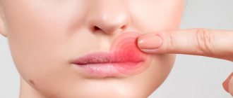
- acne, acne;
- oily scalp, subject to personal hygiene rules;
- irregular menstrual cycle or lack thereof;
- excess hair on the face (mustache, beard, sideburns), arms, back, etc.;
- one of the manifestations of hyperandrogenism in women can be infertility;
- underdeveloped mammary glands;
- deepening of the voice in women;
- late onset of first menstruation.
Diagnosis of hyperandrogenism
The doctor will be able to diagnose hyperandrogenism based on the patient’s medical history and instrumental studies of the body.
The specialist knows that this pathology can have different manifestations at different ages, so he will ask many questions and carefully analyze the answers to them:
- how long ago the patient noticed hirsutism (excess body hair);
- hair growth rate;
- duration of the menstrual cycle, their nature, pain, frequency;
- in which places on the body there is an excess of hair (chest, back, hips, stomach, etc.);
- development of secondary sexual characteristics;
- the quality of the patient’s body, whether there is excess weight, where the greatest accumulation of fat occurs;
- what medications were taken, hormonal therapy.
Based on the data received from the patient, the doctor may prescribe the following options for examining the body:
- taking a blood test to determine the concentration of various hormones in it (testosterone, luteinizing hormone, follicle-stimulating hormone, estradiol, cortisol, progesterone, etc.);
- A genetic blood test may be taken to identify hereditary mutations;
- taking a urine test to determine androgens and their traces;
- ultrasound examination of the pelvic organs and adrenal glands or MRI of these organs.
It is worth paying special attention to all diagnostic procedures prescribed by a doctor, since there are often cases when, with normal levels of hormones in the blood or with a normal state of organs, hyperandrogenism may be present on ultrasound. In such cases, pathology can only be seen based on many indicators.
Prevention
By following simple rules throughout your life, you can prevent the occurrence of adrenal hyperandrogenism or any other type.
Basic preventive measures:
- annual visit to a gynecologist;
- minimizing stressful situations (emotional as well as physical);
- do not abuse smoking, drugs, intoxicants, or alcohol;
- rich diet (fiber, protein, healthy fats without excessive carbohydrates, spicy foods, fried foods, etc.);
- start treatment of diseases in a timely manner to avoid complications and not affect other organs and systems.
Reasons for increased androgens in women
The main reasons for the development of hyperandrogenism include:
- congenital hyperplasia of the adrenal cortex (adrenogenital syndrome), which develops due to deficiency of the enzyme that is necessary for the formation of the hormone cortisol. This deficiency, in turn, is caused by a mutation in the gene encoding the enzyme;
- Itsenko-Cushing's disease, in which the adrenal glands produce excessive amounts of corticosteroids, which is caused by increased production of adrenocorticotropic hormone by the pituitary gland;
- Polycystic ovary syndrome is a polyendocrine pathology, which is characterized by excessive production of androgens by the ovaries. Hyperandrogenism is one of the symptoms of PCOS and is detected in more than half of patients with this diagnosis (the most common external sign is hirsutism - male-pattern hair growth);
- tumors of the ovaries or adrenal glands;
- taking certain medications (for example, anabolic steroids). Progestins can have the same effect, although in modern drugs androgenic activity is maximally reduced.
Normal levels of androgens in human blood
Several androgens are detected in a woman's body. Thus, a woman’s normal testosterone level is 0.2-2.0 ng/ml, cortisol is from 190 to 750 ng/ml, and aldosterone is no more than fifteen nanograms per milliliter. The level of progesterone and estradiol changes depending on the phase of the cycle. So, in the follicular phase, estradiol should be 0.17, and progesterone 1.59 nmol/l. During ovulation, their concentration increases to 1.2 and 4.77 nmol/l, respectively. In the luteal phase, the amount of these hormones is maximum: estradiol 0.57, and progesterone 29.6 mol/ml.
Diagnosis of hyperandrogenism
During diagnostic procedures, the doctor uses various methods.
Anamnesis collection and external examination of the patient
During the initial visit, the doctor finds out what complaints the patient has, when the symptoms appeared, how the menstrual cycle was established and how regular it is, whether there have been pregnancies and childbirths. In addition, it is necessary to inform the specialist about the presence of pathologies in relatives such as polycystic ovary syndrome, diabetes mellitus, and adrenal cortex dysfunction.
Hyperandrogenism is often characterized by a number of signs that are typical for men (male-pattern hair growth on the face and body, narrowing of the hips and widening of the shoulder girdle, bald patches on the forehead and crown, decreased timbre of the voice), as well as the presence of dermatological problems (in particular, acne and oily skin), which are revealed during external examination.
Gynecological examination
Congenital hyperandrogenism causes abnormalities in the development of the genital organs, which include partial fusion of the labia, penile clitoris, and urogenital sinus.
Ultrasound of the pelvic organs and adrenal glands
Ultrasound examination makes it possible to determine the condition of organs, as well as the presence or absence of changes characteristic of pathologies that cause hyperandrogenism. For example, with polycystic ovary syndrome, a bilateral increase in the volume of the ovaries is visualized with the presence of more than 12 follicles in one ovary, located under a thickened membrane. This method also allows us to exclude androgen-producing tumors of these organs. In some cases, a CT or MRI may be prescribed to clarify the diagnosis.
Laboratory research
During the tests, the doctor determines the level of androgens produced by the ovaries and adrenal glands. In addition, determining the concentration and ratio of gonadotropins is important.
Thus, the patient is prescribed the following tests:
- testosterone (total and free) in blood plasma;
- 17-OH progesterone (in blood plasma and urine);
- DHEA-S, plasma cortisol;
- gonadotropins produced by the pituitary gland (LH and FSH).
- AMH, TSH, ACTH, prolactin.
Additionally, the doctor may prescribe an ACTH test and karyotyping. To rule out a pituitary tumor, an MRI of the brain is required. Sometimes you need to additionally consult with an endocrinologist and geneticist.
Androgens play an important role in the life of the body, determining not only male sexual differentiation, but also having a significant impact on various parts of the reproductive and other systems in women. Hyperandrogenism syndrome (HA) is a pathological complex of symptoms caused by the excessive effect of androgens on organs and target tissues in women [1, 3, 6]. GA syndrome is characterized by increased production of androgens in the ovaries, adrenal glands and peripheral tissues (adipose tissue, liver, skin, muscle tissue, etc.), in addition, there may be a disturbance in the sensitivity and number of androgen receptors.
Androgen synthesis in the ovaries occurs mainly in the cells of the ovarian stroma, hilus cells and theca interna cells, in the adrenal glands - mainly in the cells of the zona reticularis cortex, and during pregnancy in the placenta. The main pathways for the biosynthesis of steroid hormones in the ovaries and adrenal cortex are similar, which is explained by the origin of the ovaries and adrenal glands from the same primordium. Androgen synthesis in the ovaries is regulated by LH, insulin-like growth factor and cytochrome P450, the latter is involved in the aromatization of androgens into estrogens and is regulated by FSH.
Impaired biosynthesis, transport and metabolism of androgens causes a variety of clinical manifestations of hyperandrogenism in women, which range from acne and hirsutism to pronounced virilization and reproductive dysfunction.
The following types of androgenic sex steroids are distinguished: testosterone, dihydrotestosterone (DHT), androstenedione, androstenediol, dehydroepiandrosterone (DHEA) and dehydroepiandrosterone sulfate (DHEA-S).
The starting material for the synthesis of steroid hormones, including androgens, is cholesterol, which enters the body mainly with products of animal origin or is synthesized in the liver. Cholesterol produces pregnenolone, which is converted by the enzyme 17α-hydroxylase to 17-hydroxypregnenolone and then to DHEA. DHEA, in turn, can be converted into testosterone, androstenediol, androstenedione and DHEA-S.
Testosterone is characterized by low biological activity and weak binding to androgen receptors (ARc). Testosterone can also be secreted in small quantities by the adrenal glands. The synthesis, secretion and regulation of testosterone levels are carried out under the control of LH and FSH.
In the cells of the inner lining of the follicle, testosterone can be converted into estrogens, promoting the development of mammary glands. Testosterone concentrations increase during pregnancy.
DHT
- a biologically active form of testosterone, formed from it under the influence of the enzyme 5α-reductase. DHT binds to tissue androgen receptors much more strongly than its predecessor testosterone, so that, despite its lower concentration, it has a more pronounced androgenic effect. ARc is a labile protein, accounting for about 0.001% of the total cellular protein content. The receptor is activated when a steroid hormone binds to it.
Androstenedione
is formed in the ovaries, and in small quantities the hormone is secreted by the adrenal cortex. Androstenedione is an intermediate in the formation of both testosterone and estrone in adipose tissue and the liver. Androstenedione has relatively weak androgenic activity (approximately less than 20% of the activity of testosterone).
Elevated levels of androstenedione may indicate congenital virilizing adrenal hyperplasia, polycystic ovary syndrome, stromal ovarian hyperthecosis, 3β-hydroxysteroid dehydrogenase deficiency, etc. High concentrations of androstenedione are common in virilizing tumors of the adrenal gland or ovary.
Androstenediol
- a minor androgen secreted by the ovaries, secreted in small quantities by the adrenal cortex. The androgenic effect of androstenediol is much weaker than that of testosterone. Androstenediol is the main source of androgens aromatized to estrogens in peripheral tissues (more aromatized to estrogens than testosterone).
In the zona fasciculata and reticularis of the adrenal glands, androgens are synthesized from the precursor 17α-hydroxypregnenolone. The most common adrenal androgens are DHEA and DHEA-S, which can be converted into each other. It is important to note that the androgenic activity of the adrenal hormones DHEA and DHEA-S is mainly due to their ability to be transformed into testosterone, although testosterone is produced in small amounts in the adrenal glands themselves.
DHEA
has an effect on ARc, a weak androgen. The metabolism of DHEA is very intense. The concentration of DHEA in the blood is approximately 300 times lower than that of DHEA-S.
DHEA is characterized by a circadian pattern of secretion with maximum levels in the morning. It is important to note that changing the concentration of binding proteins does not affect the DHEA content. DHEA is a source of sex steroids due to peripheral conversion during menopause.
DHEA-S
- a metabolite of DHEA, formed by adding the last sulfate group to the molecule with the participation of sulfotransferase enzymes. DHEA-S is produced almost entirely in the adrenal cortex (95%), and 5% in the ovaries. During the metabolism of DHEA-S, testosterone and DHT are formed in peripheral tissues. As we age, the production of DHEA-S in the body decreases.
DHEA-S has relatively weak androgenic activity (weaker than DHEA), but its activity is enhanced by relatively high serum concentrations, much higher than testosterone levels, and due to its weak affinity for steroid-binding β-globulin.
It should be borne in mind that in patients with congenital adrenal hyperplasia, glucocorticoid drugs inhibit the secretion of DHEA-S, but the level of DHEA-S is not suppressed in patients with adrenal tumors and non-endocrine ACTH-producing tumors. Low levels of DHEA-S can occur with hypofunction of the adrenal glands and thyroid gland.
Androgens circulate in the blood in a free and protein-bound state. The biological activity of androgens remains in the free and albumin-bound state. The leading role in the binding of androgens is played by sex hormone binding globulin (SHBG), synthesized in the liver. Approximately 78-80% of androgens bind to SHBG, 19-20% to albumin, and only a small part of testosterone (1-3%) circulates freely in the blood and exhibits biological activity.
An increase in the level of SHBG in the blood is promoted by estrogens, thyroid hormones, pregnancy, and a decrease in the level of SHBG by glucocorticoids, androgens, growth hormone, synthetic progestins, and insulin.
The concentration of SHBG in plasma increases under the influence of estrogens by 5-10 times and decreases by 2 times under the influence of testosterone. In women, the concentration of SHBG is 2 times higher than in men, since its synthesis is stimulated by estrogens. An increase in the concentration of thyroid hormones in the blood serum leads to an 8-fold increase in the level of SHBG, and a lack of these hormones leads to its decrease.
DHEA, DHEA-S and androstenedione are mostly bound to albumin, while testosterone and DHT are mainly bound to SHBG.
Clinical manifestations
GA syndrome is characterized by a complex of symptoms that can occur in various combinations: deepening of the voice (baryphonia), mammary atrophy, miscarriage, dysmenorrhea, anovulation, amenorrhea, infertility, masculinization, clitoral hypertrophy, severe hirsutism, androgenic alopecia, seborrhea, hyperthecosis, etc. .
GA leads to serious disorders of the female reproductive system. In particular, with GA, miscarriage, anovulation and endocrine infertility, menstrual irregularities, etc. are often observed. Menstrual dysfunction manifests itself in the form of dysmenorrhea, oligo-, amenorrhea, opsomenorrhea, and the development of menometrorrhagia, etc. is not excluded.
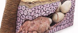
The targets of androgens in the skin are the hair follicles and sebaceous glands.
Androgens, by stimulating the hair follicle, lead to an increase in hair growth and the transformation of vellus hair into terminal hair. Terminal hair is hard, long, usually pigmented, localized around the nipples, on the upper lip, inner thighs, and lower abdomen. The terms hirsutism and hypertrichosis are used to define excess hair growth. Hirsutism is the excessive growth of shaft, dark hair in women according to the male type in androgen-dependent zones. Hypertrichosis is expressed in excess growth and the appearance of coarse pigmented hair in areas of the skin where vellus hair is normally located.
Assessment of the severity of hirsutism and therapeutic monitoring are carried out using the Ferrimana-Gallwey scale (1961) and its modification; the presence of hirsutism is indicated by a score of 8 points or more.
The sebaceous glands are most sensitive to increased androgen activity. HA promotes excess secretion of sebum (sebum) and blockage of the hair follicle, the severity of which depends on genetically determined differences in the sensitivity of the hair follicle to androgens.
Androgens, stimulating receptors located on the surface of the sebaceous gland, lead to increased secretion of sebum and a change in its composition. The skin acquires an unpleasant “oily” shine. Seborrhea on the scalp creates conditions for the activation of opportunistic fungi Pityrosporum ovale
or
Malassezia furfur
. The result of this process is a change in the development cycle of epidermal cells and the accumulation of a large number of greasy whitish-yellow scales, which are nothing more than dandruff.
GA accelerates the formation of new cells in the sebaceous gland (keratonization). The number of desquamated cells inside the follicle increases. Sebum, masses of keratin and desquamated cells can clog the mouth of the follicle. The above contributes to the formation of comedonal plugs (acne). Subsequently, the formation of sebum increases, while the comedonal plug does not allow it to reach the surface, the follicle swells with sebum and bacteria accumulate in it, which causes inflammation of the follicle, in this way inflammatory elements of acne appear.
With GA, the manifestation of alopecia is often noted. Androgenic alopecia accounts for 95% of all types of baldness in women. Hair loss or thinning is usually observed on the crown and parietal region, and rarely becomes diffuse. The mechanism of development of androgen-dependent alopecia is associated with increased production in the follicle of the active form of androgens - DHT, which causes increased protein synthesis in the follicle cell with subsequent atrophy of the latter. Vasospasm and deterioration in follicle nutrition are observed. Due to the shortening of the anagen phase, the hair length becomes shorter and pigment is lost. As a result, the shaft hair turns into vellus hair, the follicle decreases in size and completely atrophies. Moreover, hair loss is often combined with an increase in sebum production.
It should be borne in mind that in the absence of GA, the development of alopecia in women may be due to genetic predisposition, lack of body weight, anemia, thyroid dysfunction, nutritional factors, as well as the use of certain medications.
Cosmetic defects, which are based on androgen-dependent dermopathy, are the cause of deep psycho-emotional disorders in women. Mental changes, especially in young girls, are characterized by symptoms of depression. Patients are depressed and almost always see the reason for their failures in defects in appearance, have difficulty finding contact and prefer to avoid social connections, and often experience problems with establishing family life and career growth at work.
The synthesis of androgens in theca cells is influenced by insulin-like growth factor, epidermal growth factor, etc. Growth factors under the influence of insulin and pituitary growth hormone stimulate the production of androgens, which predetermines the development of insulin resistance/hyperinsulinemia and obesity. In this connection, it is necessary to identify and correct risk factors for metabolic disorders in women with HA: arterial hypertension and cardiovascular diseases, insulin resistance, hyperinsulinemia, impaired glucose tolerance and type 2 diabetes mellitus, dyslipidemia and progressive atherosclerosis.
There are ovarian and adrenal forms of androgenism. Ovarian diseases include polycystic ovary syndrome (PCOS) and androgen-producing ovarian tumors. The adrenal form includes congenital adrenal cortex dysfunction (CAD) and virilizing adrenal tumors. In addition, idiopathic hirsutism and HA are distinguished against the background of hypercortisolism (Cushing's syndrome), acromegaly, hyperprolactinemia and insulin resistance (HAIR-AN syndrome).
GA can be of tumor and non-tumor (or functional) origin. The latter option may be of ovarian, adrenal or mixed origin, and may also be due to increased peripheral tissue sensitivity to androgens.
There are the following forms of GA: true, receptor, transport, iatrogenic. True GA is characterized by an increase in androgen levels, confirmed by laboratory tests. In receptor HA, its clinical manifestations are determined by the number and sensitivity of target tissue receptors to androgens. Transport GA develops as a result of disruption of the binding of testosterone to blood proteins and, as a result, an increase in the concentration of free hormone. Iatrogenic GA is caused by taking medications that have androgenic properties.
The mechanism of HA in PCOS is based on hypersecretion of LH, leading to disruption of folliculogenesis in the ovaries with the formation of cystic atresia of the follicles, hyperplasia of theca cells, and stroma. Approximately 70% of patients with PCOS have elevated LH concentrations, while the rest have normal LH levels and hyperreactivity of ovarian theca cells. Insulin resistance and hyperinsulinemia in PCOS stimulate an increase in LH-dependent androgen synthesis in theca cells and stroma, and also inhibit the production of SHBG by the liver, increasing the amount of bioavailable androgen fractions in the blood.
The cause of the development of idiopathic hirsutism may be an increase in 5α-reductase activity in peripheral tissues or an increase in the number and sensitivity of androgen receptors.
Often the cause of HA in women is CATH, an autosomal recessive disease that occurs due to a deficiency of the enzyme 21-hydroxylase, which results in a disruption in the production of cortisol and, according to the feedback principle, hypersecretion of ACTH occurs, stimulating the synthesis of cortisol precursors and androgens. The non-classical form of CAH (mild severity, characterized by partial impairment of 21-hydroxylase activity) is often combined with insulin resistance and PCOS.
Androgen-producing tumor processes of the ovaries and/or adrenal glands (Leydig cell tumors, gonadoblastoma, Sertoli-Leydig cell tumors, theca and granulosa cell tumors, etc.) are rare cases of GA and, as a rule, appear in early adolescence or reproductive age and rarely in postmenopause.
Diagnostics
Features of the diagnosis of GA include determining the degree, source and pathogenesis of androgen hyperproduction, as well as assessing the degree of impact on reproductive function, the presence of metabolic and cardiovascular risks.
To determine the direction of the diagnostic search, a detailed study of the anamnesis is necessary: features of the menstrual cycle; age of menarche; age of manifestation of acne, hirsutism, seborrhea, alopecia; implementation of reproductive function; identifying hereditary burden, etc.
During the examination, the structural features of the external genitalia are assessed; presence of Acanthosis nigricans
(villous, warty, keratinized growths of slate-black color); severity and distribution of hirsutism according to the Ferrimana-Gallwey scale; degree of severity of alopecia, acne; indicators of height, body weight, body mass index, waist and hip circumference; presence of galactorrhea, etc.
To identify the size and structure of the ovaries, it is necessary to conduct an ultrasound examination (ultrasound), and, if necessary, computed tomography or magnetic resonance imaging. Ultrasound is also performed to determine the ovulatory menstrual cycle (ultrasound folliculometry).
It is advisable to carry out hormonal blood tests on the 3rd day of the menstrual cycle. Blood must be donated on an empty stomach, 2-3 days before blood collection it is necessary to exclude sexual contact, intense physical activity, thermal effects (sauna, steam bath), and 1 hour - smoking. It is recommended to determine the hormonal profile in the morning to eliminate errors associated with small daily fluctuations in hormone concentrations.
Assessment of the hormonal profile may include determination of the following indicators: FSH, LH, prolactin, estradiol, progesterone (analysis is carried out on the 22-23rd day of the menstrual cycle); 17-hydroxyprogesterone (17-OHP), androstenedione, androstenediol glucuronide, DHEA-S, total and free testosterone, DHT, SHBG.
In case of HA syndrome and a normal level of total testosterone, the feasibility of determining the level of free testosterone should be considered. Obesity can be accompanied by high levels of free testosterone due to a decrease in SHBG, often against the background of hyperinsulinemia and insulin resistance.
As a marker of androgen production by the adrenal glands, it is currently more appropriate to determine DHEA-S in the blood rather than 17-ketosteroids in the urine. An increase in DHEA-S may indicate an adrenal origin of HA syndrome, and with a significant increase in the level of DHEA-S, an adrenal tumor should be excluded.
Various exercise tests may be performed to determine the sources of increased androgen production. An LH/FSH ratio of more than 2.5 is characteristic of PCOS. But PCOS can also occur when the LH/FSH ratio is less than 2.5. To diagnose adrenal hypertension due to 21-hydroxylase deficiency (late form of CADC), a 17-OHP test is used. The diagnosis of non-classical or late-onset CDCN is confirmed when the 17-OHP level is above 8 ng/ml. If the 17-OHP level is in the range from 2 to 8 ng/ml, an ACTH stress test should be performed to exclude CATH.
Treatment
Therapy for HA syndrome is aimed at reducing skin manifestations (hirsutism, acne, seborrhea, alopecia), menstrual and reproductive disorders, and correction of metabolic disorders. Treatment tactics should be determined based on identifying the source of increased androgen secretion.
During drug therapy, androgen levels must be determined every 3-4 months for a year.
Options for non-drug therapy include normalization of body weight, surgical treatment (removal of tumors).
Below are the main areas of drug therapy.
1. Suppression of androgen synthesis in the ovaries is carried out using:
- combined oral contraceptives (COCs) - the mechanism of action is based on the antigonadotropic effect, as a result of which the LH-dependent synthesis of androgens in the ovaries is reduced; the estrogenic component contributes to an increase in SHBG [2, 5];
- GnRH agonists - have a strong antigonadotropic effect, block LH-dependent androgen synthesis in the ovaries; their use is advisable in patients with concomitant gynecological diseases (uterine fibroids, adenomyosis, endometriosis, endometrial hyperplasia).
2. Suppression of androgen synthesis in the adrenal glands is carried out using glucocorticoids. As a result, in patients with CAH, the concentration of serum androgens decreases due to ACTH-induced synthesis in the adrenal glands.
3. Correction of metabolic disorders: lipid-lowering and antihypertensive therapy if necessary; the use of metformin, thiazolidinediones (in the presence of impaired glucose tolerance).
4. Blockade of the peripheral action of androgens in target tissues at the level of the hair follicle is one of the most effective methods of treating androgen-dependent dermopathy. During therapy, reliable contraception is necessary.
The following androgen blockers are used.
1. Cyproterone acetate is a 17-OHP derivative that blocks androgen receptors. Being a progestogen, it has an antigonadotropic effect and reduces LH-induced androgen synthesis in the ovaries. The average dose is 50 mg from the 5th to the 15th day of the menstrual cycle.
2. Spironolactone - an aldosterone antagonist - has an antiandrogenic effect, blocks peripheral receptors for testosterone and DHT, reduces the activity of cytochrome P450, reducing the production of androgens in the ovaries and adrenal glands. The average dose is 100 mg per day.
3. Flutamide is a non-steroidal androgen receptor blocker, metabolized into active hydroxyflutamide, which binds cytosolic and nuclear androgen receptors. The average dose is 250 mg per day.
4. Inhibitors of the enzyme 5α-reductase - finasteride - suppresses the peripheral action of androgens by blocking the metabolism of testosterone into DHT. The average dose is 5 mg per day. Finasteride is recommended for women with idiopathic forms of hirsutism and acne.
Currently, an effective and common treatment for androgen-dependent conditions is the use of oral contraceptives that have an antiandrogenic effect. Among them, the most actively used is the drug Belara (), containing 2 mg of chlormadinone acetate (CMA) and 30 μg of ethinyl estradiol.
The antiandrogenic effect of CMA is based on its competitive binding to androgen receptors in target cells, including hair follicles and sebaceous glands [4].
CMA reduces the sensitivity of hair follicle cells and sebaceous glands to androgens due to the suppression of 5α-reductase activity in them [10].
CMA inhibits the synthesis of androstenedione and DHEA-S, i.e. a decrease in androgen levels when taking CMA-containing oral contraceptives is achieved mainly by suppressing their production by the ovaries and adrenal glands.
The estrogenic component of the oral contraceptive increases the synthesis of GSPC, leading to a significant decrease in the level of active testosterone in the blood.
In more than half of the patients with skin manifestations of viril syndrome in the form of hirsutism, oily seborrhea and acne on the face, back and décolleté, after 6 months of regular use of a CMA-containing oral contraceptive, there is a complete restoration of the condition of the skin and a decrease in hair growth on exposed areas. skin areas, which in turn contributes to a significant increase in self-esteem and quality of life of these women [6, 7].
Improvement in the condition of the skin while taking a CMA-containing oral contraceptive is achieved by restoring their pH, enhancing the barrier function of the epidermis, manifested in reducing transepidermal water loss, as well as by increasing the hydration of the stratum corneum of the skin and reducing the lipid content on its surface [8] .
In addition, the use of a CMA-containing oral contraceptive is effective in women with alopecia and idiopathic hirsutism not accompanied by HA, as well as in the presence of dermatopathy associated with scleropolycystic ovaries.
Due to the lack of mineralocorticoid and glucocorticoid activity in CMA, its tolerability by most women is assessed as “good” or “very good” [9].
Stability of body weight when using CMA-containing oral contraceptives is achieved as a result of their neutral effect on carbohydrate and lipid metabolism, which is confirmed by the absence of changes in glucose tolerance, the level of fasting insulinemia, peripheral insulin resistance and pathological dyslipidemia in patients 1 year after the start of regular use of the drug.
Due to the above-described features of the effect of CMA on carbohydrate-fat metabolism, its use as part of COCs is justified in women with scleropolycystic ovary syndrome, who are predisposed to the development of insulin resistance and aggravation of changes in the ovaries against this background.
The undeniable advantage of CMA-containing COCs is their neutral effect on the blood coagulation system while simultaneously having an antiatherogenic effect, which allows them to be considered the drugs of choice in patients with metabolic syndrome, PCOS and systemic diseases accompanied by hypercoagulation.
In conclusion, it should be noted that despite the significant arsenal of various drugs used to treat HA syndrome, the approach to the treatment of this pathology should be comprehensive and consistent, taking into account the key link in the pathogenesis.
Patients with GA syndrome should clearly understand that treatment is effective only while taking the drug, since after its discontinuation, most women experience a relapse of clinical symptoms.
Symptoms of hyperandrogenism
1. Virilization is a condition characterized by the appearance of male characteristics in a woman. In particular, the patient has:
- hirsutism. Fine vellus hair turns into terminal hair (darker and longer) in the areas where it grows in men (for example, the upper lip, chin and inner thighs);
- hypertrichosis. This is excessive hair growth in women, not limited to distribution according to the male type, that is, on any part of the body, and sometimes over its entire surface;
- androgen-dependent alopecia. Under the influence of increased concentrations of male hormones, a woman’s hair begins to thin and fall out. Moreover, this happens according to the male type: bald patches form on the forehead and in the crown area;
- decrease in voice timbre;
- changing the figure according to the male type. In women, there is a narrowing of the hips with a well-developed shoulder girdle;
- The mammary glands are poorly developed.
2. Androgen-dependent dermopathy, which is characterized by the formation of acne (often grade 3 or 4) and oily seborrhea. Under the influence of androgens, sebum production increases. Seborrhea appears on the face, in the forehead and nasolabial triangle, on the back, and on the scalp. 3. Metabolic disorders (insulin resistance, type 2 diabetes, obesity). 4. Menstrual irregularities - from minor delays to complete absence of menstruation (amenorrhea). Uterine bleeding may also be one of the manifestations. 5. Infertility caused by dysfunction of the hypothalamic-pituitary-ovarian system.
Which doctor should I contact?
If you notice signs of hyperandrogenism or you need specialist advice on this issue, you should first contact an endocrinologist, and after understanding your problem and studying your medical history, he will prescribe additional examinations, if necessary.
One of the specialists in endocrinology is Georgy Nikitich Romanov, who has been practicing medicine for more than 20 years. Georgy Romanov studied not only in Belarus, but also in Russia and some European countries (Great Britain, Germany), and practices modern methods of diagnosis and treatment. An individual approach to each patient, since only a thorough study of the patient’s medical history allows us to find the root cause of the disease and prescribe effective treatment. Provides consultations and closely monitors the progress of treatment.
In addition to the fact that endocrinologist Georgy Nikitich Romanov practices in Gomel, he also consults patients online.
You can sign up for an online consultation in any messenger or social network convenient for you: skype, instagram, viber, telegram, whatsapp or vkontakte.
If you are looking for an experienced, attentive specialist to identify hyperandrogenism in women or adolescents, as well as effective treatment of the disease, then they will help you here.
Treatment of hyperandrogenism in women
Before prescribing an adequate treatment regimen, the doctor must identify the causes of hyperandrogenism, as well as determine what the patient’s primary goals are: achieving pregnancy or eliminating the symptoms of hyperandrogenism.
To achieve pregnancy with hyperandrogenism of ovarian or adrenal origin, hormonal stimulation of ovulation can be effective. If the patient has tumors of the adrenal glands or ovaries, surgical removal is recommended. For adrenogenital syndrome, corticosteroids are used.
A treatment regimen for hyperandrogenism is developed and prescribed by a doctor individually after a thorough diagnosis.
Since this pathology is often the cause of pregnancy loss in the early stages, pregnancy management requires attention from both an obstetrician-gynecologist and an endocrinologist, in close collaboration with whom the expectant mother with diagnosed hyperandrogenism is monitored.
Our advantages
Highly qualified personnel
Successful treatment of hyperandrogenism is carried out by gynecologists-endocrinologists with many years of practice (up to 30 years), doctors of the highest category, candidates of medical sciences, participants in specialized congresses, and authors of published works in scientific journals.
Own clinical diagnostic laboratory
At the first signs of hyperandrogenism, doctors prescribe tests for sex hormones, a test with dexamethasone, etc. Each NEARMEDIC clinic has a treatment room for taking samples. They are processed in NEARMEDIC's own laboratory. The system for conducting research and issuing results is almost completely automated, which reduces the likelihood of error to a minimum. At the patient's request, test results can be sent to his email.