Tuberculous lupus is the most common form of cutaneous tuberculosis. It is characterized by a chronic, slow progressive course and a tendency to melt tissue. The disease usually begins in childhood and lasts for years or decades, sometimes throughout life.
Clinical picture
Tuberculous lupus is most often localized on the face, especially on the nose (in 80% of cases), cheeks, upper lip, less often on the neck, torso, extremities, and often on the mucous membranes (70% of patients). Pulmonary tuberculosis is found in 5-10% of lupus patients, and tuberculosis of bones and joints occurs with the same frequency. Skin infection occurs predominantly by hematogenous or lymphogenous route.
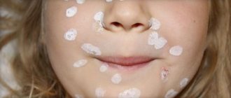
The disease begins with the appearance of lumps - tubercles ranging in size from a pinhead to a pea, brown-reddish in color with a yellowish-brown tint, soft, doughy consistency with a smooth, slightly shiny surface. Lupoms are poured out in a group, at first they are located in isolation, then they merge; along their periphery there is always a stagnant-hyperemic zone.
The “apple jelly” phenomenon (diaskopia, obelmus). If you press a glass slide onto the lesion, yellow-brown spots appear. This symptom is explained by the squeezing out of blood from dilated blood capillaries and the transmission through the epidermis of magnifying glass with a granulomatous structure
“Probe” symptom (Pospelov’s sign). If you press a button-shaped probe on the tubercle (lupu), then it is easily immersed into the depths of the tissue. In this case, light bleeding and minor pain occur. The symptom is more pronounced with fresh lupoma. This occurs due to caseous necrosis, accompanied by the death of collagen and elastic fibers.
There are several clinical forms of tuberculous lupus: macular (Lupus planus maculosus), tubercular (Lupus planus tuberculosus), tumorous lupus (Lupus tumidus), erythematous, squamous-hyperkeratotic, verrucous and ulcerative.
- In some cases, destructive ulcerative changes involve deep underlying tissues (cartilage, bones, joints) in the process with the formation of mutilations, fibrous, keloid scars and disfigurement of the nose, ears, fingers, eyelids, limbs (Lupus mutilans).
- When the mucous membranes are affected (Tuberculosis luposa mucosae), small bluish-red tubercles the size of a millet grain are formed, which, due to their close grouping, give the affected area a peculiar granular appearance. Diagnosis is facilitated by the simultaneous presence of manifestations on the skin. When the mucous membranes of the nose are damaged, a soft, lumpy, bluish infiltrate is formed, which disintegrates to form an easily bleeding ulcer.
- Pityriasiform lupus (Lupus vulgaris pityriasiformis) is characterized by pithiarisiform scaling, and lupus psoriasiformis (Lupus vulgaris psoriasiformis) is characterized by silvery-white scales.
- There are other types of the disease: serpiginating, exfoliative, rupioid, crustose, etc.
Tuberculous lupus can be complicated by erysipelas, lymphangitis, pyoderma, and the development of skin cancer (Lupus carcinoma).
Histologically, epitheloid and giant cells surrounded by lymphocytes are detected in the dermis. Giant Langhans cells are located in the central part of the tubercle, in the peripheral zone there are lymphocytes and plasma cells in large numbers; caseous necrosis is poorly expressed. Mycobacterium tuberculosis is detected with difficulty and in small quantities.
Diagnosis and differential diagnosis
Differential diagnosis is carried out with tubercular syphilis, tuberculoid form of leprosy, tuberculoid form of cutaneous leishmaniasis, actinomycosis, discoid form of lupus erythematosus.
Causes
Koch's bacillus has a shell that is resistant to acidic environments. It is able to survive freezing, drying, exposure to alkalis, and much more. etc. Characterized by the ability to form so-called L-forms with increased adaptability. The most pathogenic microbacteria for humans are:
- Mycobacterium tuberculosis humans;
- Mycobacterium bovis.
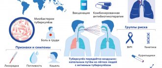
Microbacteria enter the body through contact, air, mixed and other routes. This is how the first inflammatory focus is formed. A child can also be infected during the mother's pregnancy - through the placenta or during childbirth, if it swallows amniotic fluid. Source: https://www.ncbi.nlm.nih.gov/pubmed/24548085 Marais BJ Tuberculosis in children J Paediatr Child Health . 2014 Oct;50(10):759-67. doi: 10.1111/jpc.12503. Epub 2014 Feb 19
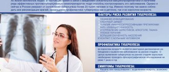
Children at risk include:
- not vaccinated with BCG;
- long-term treatment with antibiotics, hormonal or cytostatic agents;
- having HIV status;
- living in unfavorable conditions (social and/or sanitary);
- having diabetes mellitus;
- with weakened immunity;
- under 2 years of age;
- and etc.
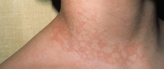
Rosacea-like Lewandowski tuberculosis
This form of the disease occurs as an allergic vasculitis. Erythema (redness) and telangiectasias (numerous dilated small vessels) appear on the skin of the face. Gradually the skin acquires a bluish-reddish tint. Rashes (tuberculids) are dense formations up to half a centimeter in diameter, without necrotic elements. During healing, atrophic scars form at the site of tuberculides.
Rice. 27. The photo shows Lewandowski’s rosacea-like tuberculosis.
Prices
| Name of service (price list incomplete) | Price |
| Appointment (examination, consultation) with a pulmonologist, primary, therapeutic and diagnostic, outpatient | 1750 rub. |
| Consultation with a candidate of medical sciences | 2500 rub. |
| Professor consultation | 4500 rub. |
| Consultation (interpretation) with analyzes from third parties | 2250 rub. |
| Prescription of treatment regimen (for up to 1 month) | 1800 rub. |
| Study of external respiratory function (RPF) with drug tests | 1800 rub. |
| X-ray of the chest organs (survey) | 1900 rub. |
| X-ray of the chest organs in 2 projections | 2900 rub. |
Ulcerative skin tuberculosis (Tuberculosis cutis ulcerosa)
The disease most often affects men who already had tuberculosis of internal organs. Tuberculosis bacilli enter the skin from the urine, feces or sputum of the patient himself, affecting areas of natural external openings: the skin around the anus, glans penis, nose, area around the mouth, mucous membrane of the tongue.
Symptoms of the disease
Merging, tuberculous tubercles form small yellowish nodules. Over time, the nodules suppurate and open with the formation of painful ulcers that make natural acts difficult.
The ulcers have soft, undermined edges. The color of the ulcers is pale red. The size of the ulcers is up to 1.5 cm, the bottom is granular. Javas often bleed. Sharply painful. As the disease progresses, tuberculous tubercles reappear at the bottom of the ulcers and around them. The tissues are destroyed, forming yellowish microabscesses, the so-called “Trel grains”, containing a large amount of MBT. The disease is difficult. Ulcers heal slowly. In their place, thin scars form, located below the skin level (atrophic scars).
Rice. 21. Ulcerative tuberculosis.
Papulonecrotic tuberculosis of the skin (tuberculosis cutis papulo-nectrotica)
Papulonecrotic tuberculosis of the skin is more often recorded in young people. The disease is manifested by the appearance of papules, the size of which does not exceed 3 cm in diameter. The skin of the buttocks, abdomen, extensor surfaces of the limbs and chest is affected. The skin over the papules becomes pinkish-bluish in color. Over time, the papules become ulcerated. At the site of the ulcers, a grayish-white crust appears, which is replaced by a whitish scar.
Rice. 25. Tuberculosis cutis papulo-nectroticа. Multiple bluish-purple infiltrates and nodes on the legs.
Rice. 26. Papulonecrotic tuberculosis of the skin of the legs. Multiple bluish-purple infiltrates and nodes on the legs.
Treatment
Chemotherapy, antibiotics
Bactericidal and bacteriostatic drugs are used to achieve complete recovery. In this case, the correct combination of drugs is important, taking into account the resistance of some bacteria to this type of therapy. First, treatment is aimed at suppressing the growth of bacteria and eliminating their resistance to medications. Residual infection in the cells is then eliminated. Duration of therapy is 6-12 months.
Surgery
Pulmonary resection is practiced as a radical technique. The operation is performed for bronchial stenosis, fibrous-cavernous lesions, pleural empyema, abscesses in the lungs, tuberculomas prone to progression, etc. In addition, decortication is used, that is, removal of fibrous layers, and cavernotomy - cleansing of the opened cavity.
DOTS
A treatment system consisting of several levels: bacterioscopic examination, chemotherapy, anti-tuberculosis treatment.
Sources:
- N.M. Koretskaya. Tuberculosis in children and adolescents in modern conditions // Siberian Medical Review, 2010.
- A.V. Mordyk, E.A. Tsygankova, L.V. Puzyreva, A.A. Turitsa. Tuberculosis in children of the Russian Federation at the present stage // Pediatric pharmacology, 2014, v. 11, no. 3, pp. 27-30.
- https://www.ncbi.nlm.nih.gov/pubmed/24548085 Marais BJ. Tuberculosis in children // J Paediatr Child Health. 2014 Oct;50(10):759-67. doi: 10.1111/jpc.12503. Epub 2014 Feb 19.
- V.N. Krivohizh. Modern methods of early detection of tuberculosis among children and adolescents // Health is the basis of human potential: problems and solutions, 2013, pp. 570-585.
The information in this article is provided for reference purposes and does not replace advice from a qualified professional. Don't self-medicate! At the first signs of illness, you should consult a doctor.
Etiology of the disease
- The causative agent of skin tuberculosis is Mycobacterium tuberculosis, which was first discovered by R. Koch in 1882.
- The tuberculosis bacillus belongs to the family of radiant fungi, the genus of mycobacteria, which, in addition to it, includes the causative agents of leprosy, scleroma and more than 150 species of atypical mycobacteria.
- Mycobacteria reproduce by division and budding. This process lasts 24 hours.
- MBT exhibit significant stability in the external environment. They cannot be frozen out. They remain viable in boiling water for up to 15 minutes. They live in manure for up to 15 years, and up to 1 year in wastewater. When dried, the pathogen remains viable for 3 years.
- Mycobacteria are resistant to phagocytosis (macrophages cannot destroy the mycobacterium, although they begin to fight it - incomplete phagocytosis).
- There are human, bovine and intermediate types of MBT.
- The pathogen has the appearance of an elongated rod of a rather complex structure: a three-layer cell wall and intracellular membrane contain polysaccharides, lipoprotein complexes and proteins. Proteins are responsible for antigenic properties (tuberculin). Polysaccharides play a role in antibody detection. Lipid fractions help MBT resist acids and alkalis.
Rice. 2. Mycobacterium tuberculosis.
Types and forms of pathology
According to the period of occurrence in medicine, stages are distinguished:
- infiltration;
- decay;
- contamination;
- resorption;
- seals;
- the appearance of scars;
- calcification.
| Form | Peculiarities |
| Chronic and early | It occurs against a background of body temperature up to 40℃, cough, pain in the lungs, uneven breathing, wheezing, decreased appetite, and loss of strength. |
| Respiratory damage | Includes pathologies:
|
| Other localizations | There may be lesions:
|
Ways of spreading skin tuberculosis
- Patients with tuberculosis spread the infection through sputum, urine, through fistulas and household items. The source of infection is also sick animals and food products contaminated with MBT from sick animals. During the formation of immunity, primary skin tuberculosis occurs (tuberculous chancre, lichenoid tuberculosis of the skin).
- There are a number of diseases that are associated with tuberculosis not directly, but indirectly. These are allergic vasculitis, resulting from allergic immune (“paraspecific”) inflammation in response to infection with mycobacteria (rosacea-like Lewandowski tuberculide).
Rice. 3. The photo shows tuberculosis of the skin of the face and neck.