Deep vein thrombosis (DVT) occurs when a blood clot (thrombus) forms in one or more deep veins in your body, usually in the legs. Deep vein thrombosis can cause leg pain or swelling, but may be asymptomatic.
- Symptoms of deep vein thrombosis
- Causes of deep vein thrombosis
- Risk factors
- Complications of DVT
- Prevention of thrombosis
DVT may be associated with diseases that affect the blood clotting process. A blood clot in your legs can also form if you have not moved for a long time, such as after surgery or an accident. But walking over extremely long distances can lead to the formation of blood clots.
Deep vein thrombosis is a serious condition because blood clots in your veins can travel through your bloodstream and become lodged in your lungs, blocking blood flow (pulmonary embolism). However, pulmonary embolism can occur without evidence of DVT.
When DVT and pulmonary embolism occur at the same time, it is called venous thromboembolism (VTE).
Symptoms
Signs and symptoms of DVT:
- Swelling of the affected leg. In rare cases, swelling appears in both legs.
- Leg pain. The pain often begins in the calf and may feel like cramping or soreness.
- Red or discolored skin on the leg.
- Feeling of warmth in the affected leg.
Deep vein thrombosis may occur without noticeable symptoms.
When to see a doctor
If you have signs or symptoms of DVT, contact your doctor.
If you develop signs or symptoms of pulmonary embolism (PE), a life-threatening complication of deep vein thrombosis, get emergency medical help.
Call 103
Warning signs and symptoms of pulmonary embolism include:
- Sudden shortness of breath
- Chest pain or discomfort that gets worse with deep breathing or coughing.
- Feeling dizzy or dizzy or faint
- Rapid pulse
- Rapid breathing
- Coughing up blood
Do you suspect deep vein thrombosis? Contact the professionals.
Thrombophlebitis of the superficial veins of the lower extremities
It is this form of thrombophlebitis of the legs that is considered the most common, and it is most often preceded by varicose veins. A blood clot is indicated by a seal on the vessel, which, as a result of varicose veins, has taken on a convex shape and is clearly visible under the skin. As the blood clot expands and lengthens, the vein becomes like a subcutaneous cord.
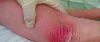
The tissue around the clot swells and the skin turns red. The process is accompanied by acute pain, which increases when you press on the area where the blood clot forms. Typically, thrombophlebitis of the veins of the lower extremities does not prevent the patient from moving the leg, but movement can be very painful. Body temperature does not exceed 37.5 degrees. Sometimes inflammation can spread to neighboring tissues - periphlebitis occurs.
The acute form mainly occurs due to infectious exposure. A blood clot forms at the site of infection, provoking, in turn, inflammation of the vessel wall. Acute superficial thrombophlebitis of the lower extremities causes the temperature to rise to 38o, and then it normalizes. Lumps are felt under the skin, the size of which depends on the diameter of the vein in which the blood clot has formed.
The chronic form is characterized by prolonged congestion in the lower limb, increased swelling and pain after exertion on the legs. In addition, pus may form in the affected tissues, and abscesses may form on the skin.
Risk factors
Many factors can increase your risk of developing DVT, which include:
- Age. At age 60, the risk of DVT increases, although it can happen at any age.
- Sitting for long periods of time, such as while driving or flying. When your legs remain motionless for several hours, your calf muscles do not contract. Muscle contractions promote blood circulation.
- Prolonged bed rest, such as during a long hospital stay or paralysis. Blood clots can form in the calves if the calf muscles are not used for a long time.
- Trauma or surgery. Injury to the veins or surgery may increase the risk of blood clots.
- Pregnancy. Pregnancy increases pressure in the veins of the pelvis and legs. Women with an inherited bleeding disorder are at particular risk. The risk of blood clots from pregnancy can last up to six weeks after the baby is born.
- Birth control pills (oral contraceptives) or hormone replacement therapy. Both factors can increase the blood's ability to clot.
- Exposure to drugs or chemicals. Certain drugs can cause blood clots. Consult your physician before use.
- Overweight or obese. Excess weight increases pressure in the veins of the pelvis and legs.
- Smoking. Smoking affects clotting and circulation, which may increase the risk of DVT.
- Cancer. Some forms of cancer increase levels of substances in the blood that cause blood clotting. Some forms of cancer treatment also increase the risk of blood clots.
- Heart failure. Increases the risk of developing deep vein thrombosis and pulmonary embolism. Because people with heart failure have limited heart and lung function, symptoms caused by even a small pulmonary embolism are more noticeable.
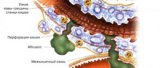
- Inflammatory bowel diseases. Bowel diseases such as Crohn's disease or ulcerative colitis increase the risk of DVT.
- Personal or family history of DVT or PE. If you or someone in your family has had one or both of these, you may be at greater risk of developing DVT.
- Genetics. Some people inherit genetic risk factors or disorders, such as factor V Leiden, that make their blood clot more easily. The hereditary disease itself may not cause blood clots unless it is combined with one or more other risk factors.
- Risk factor unknown. Sometimes a blood clot in a vein can occur without an obvious underlying risk factor. This is called unprovoked VTE.
Compression treatment
This method is used most often in the chronic course of the disease, after inflammation has subsided, and for prevention. In addition, it is effective only for thrombophlebitis of the superficial veins . It is not prescribed for deep vein pathology.
Medical knitwear is used: tights, stockings, knee socks of different lengths and with compression class 2 or 3. The use of special elastic bandages is effective. Bandaging is carried out at the beginning of the day, and the bandages are removed at night. This helps maintain limb tissue in good shape. Knitwear and bandages prevent the expansion of veins and the separation of a blood clot. After a few days it may completely resolve.
Also, in chronic and subacute forms, warm compresses are prescribed. They improve blood circulation and elasticity of tissues and blood vessels.
Complications
Complications of DVT may include:
- Pulmonary embolism (PE). PE is a potentially life-threatening complication associated with DVT. This occurs when a blood vessel in your lung is blocked by a blood clot that travels to the lung from another part of your body, usually your leg. If you have signs and symptoms of PE, it is important to seek medical help immediately. Sudden shortness of breath, chest pain when inhaling or coughing, rapid breathing, rapid pulse, feeling weak or faint, and coughing up blood may occur with PE.
- Postphlebitic syndrome. Damage to the veins by a blood clot reduces blood flow to the affected areas, causing leg pain and swelling, skin discoloration, and skin ulcers.
- Complications of treatment. Complications may arise from blood thinners used to treat DVT. Bleeding is a side effect of anticoagulants. It is important to have regular blood tests when taking these medications.
Thrombophlebitis of deep veins
The signs of this type of pathology largely depend on the location of the affected vessel. Among the acute forms, thrombophlebitis of the deep veins of the leg stands out in its frequency. It is characterized by the appearance of sudden pain, bursting the muscles of the lower leg. The pain may subside a little if you put your leg in a horizontal position. However, an almost painless manifestation is possible in the presence of other symptoms of thrombophlebitis: swelling, bluish color of the skin and its tension. When pressing on the skin, a dimple remains, which gradually smoothes out.
On the other hand, the disease can pass without any obvious signs at all. With thrombophlebitis of the femoral vein and deep veins of the pelvis, the presence of a blood clot may be indicated by slight swelling on the inner surface of the thigh and mild pain in the groin area. Thrombophlebitis of the iliofemoral segment of the main vein also has subtle signs. It manifests itself as mild pain in the lower back, near the sacrum, and in the lower abdomen. And in some cases, the only symptom may be thromboembolism - when the blood clot has already broken off.
Thrombophlebitis of the deep veins of the lower extremities also manifests itself as a general malaise. Body temperature can rise to 40o. And if the common femoral vein is affected, the patient may have a fever and there are signs of intoxication of the body.
The acute form in almost all cases is characterized by the appearance of acute pain at the site of blood flow disturbance. The swelling may be loose at first and then gradually harden. In the affected area, veins clearly appear on the surface of the skin, and the temperature rises significantly. When making a diagnosis, the doctor places a sphygmomanometer cuff near the affected area and monitors the increase in pressure that causes acute pain. In acute cases, a pressure of 45-60 mmHg is sufficient.
The chronic form by a long course, and the symptoms of thrombophlebitis can appear from time to time for 2-3 years without significant deterioration. The thrombus may be partially destroyed, the patency of the vessel temporarily improves, and the vein wall is overgrown with connective tissue. But since the outflow of blood from the lower extremities is impaired, the symptoms return:
- swelling becomes stable;
- a feeling of fullness and heaviness in the leg haunts when walking;
- severe muscle cramps occur at night;
- dense formations appear under the skin;
- the skin itself acquires ring-shaped pigmentation.
Since such symptoms of thrombophlebitis of the lower extremities are extremely likely to cause dermatitis, eczema, and trophic ulcers, it is necessary to start treatment as early as possible.
Prevention
Measures to prevent deep vein thrombosis include the following:
- Don't sit still. If you have had surgery or were on bed rest for other reasons, try to get back to work as soon as possible. If you sit for any period of time, do not cross your legs as this can block blood flow. If you are traveling long distances by car, stop every hour or so and take a walk. If you're on an airplane, stand or walk occasionally. If you can't do this, stretch your shins. Do some exercises. Try raising and lowering your heels while keeping your toes on the floor, then lifting your toes while pressing your heels into the floor.
- Do not smoke. Smoking increases the risk of developing DVT.
- Do exercises and control your weight. Obesity is a risk factor for DVT. Regular exercise reduces the risk of blood clots, which is especially important for people who sit a lot or travel frequently.
How to treat thrombophlebitis?
Depending on the form and severity, thrombophlebitis is treated at home or on an outpatient basis. Treatment can be carried out at home for damage to the superficial vessels of the hand, forearm, foot, and leg. In this case, the patient’s physical activity is limited, special ointments are applied locally to improve blood circulation, cold, and elastic bandages. Inside – anti-inflammatory drugs, antibiotics. Physiotherapeutic procedures are prescribed - magnetic therapy, pulsed currents, etc. Particular attention is paid to prevention.
Treatment for symptoms of thrombophlebitis of the lower extremities, characterized by damage to the deep veins , is carried out exclusively in a hospital, as there is a threat of embolism. Surgery may be necessary to remove a large blood clot, suppuration, gangrene, etc.
Drug treatment
Prescription of medications for thrombophlebitis should be comprehensive:
- anticoagulants reduce blood clotting;
- non-steroidal anti-inflammatory drugs and antibiotics act on the very cause of blood clots – infection;
- for severe pain, painkillers are used;
- special external ointments help dissolve blood clots; anti-inflammatory gels are also applicable;
- Enzyme therapy is carried out to dissolve the blood clot and eliminate swelling;
- the action of venotonics is aimed at strengthening the walls of blood vessels and increasing their elasticity;
- Angioprotectors may also be additionally prescribed.
For the treatment of thrombophlebitis, the use of rutin is important. It increases the tone of large vessels, strengthens their walls, reduces swelling and inflammation. Heparin contained in ointments is no less important. Their use allows you to avoid taking medications internally.
In the absence of severe complications, the effect of drug therapy is felt within a few days.
Physiotherapy
Physiotherapeutic treatment of thrombophlebitis of the lower extremities is effective for the chronic form of superficial thrombophlebitis and includes a number of measures:
- infrared rays;
- Sollux;
- magnetic therapy;
- electrophoresis;
- ultraviolet irradiation;
- pulse currents.
These methods are not used in the presence of trophic formations and during periods of exacerbation. A trip to a balneonological resort is possible only after consultation with a doctor and requires a special appointment.
Separately, it is worth noting hirudinotherapia - treatment with leeches. This method is applicable in the acute form and if the patient experiences intolerance to anticoagulant drugs (reducing blood viscosity). In this case, the role of these drugs is played by hirudin, contained in the glands of leeches and entering the patient’s blood. However, their use is strictly individual, has serious contraindications and requires mandatory consultation with the attending physician.
Diagnosis of thrombosis
Primary diagnosis is based on a detailed history and anthropometry (calf or thigh circumference).
The Wells scale is used to diagnose acute thrombosis and diagnose pulmonary embolism [8,9]. Instrumental diagnostics include compression or duplex scanning of veins, Doppler ultrasound with compression of veins, impedance plethysmography, pulmonary angiography, radiocontrast or MRI venography [6,9], CT and MRI angiography [7,9].
To diagnose arterial thrombosis, physical tests are used (6-minute walk test, treadmill test), determination of pulsation of superficial arteries (arteries of the dorsum of the foot), duplex scanning of the arteries of the extremities, angiography (X-ray of a vessel filled with a radiopaque substance) and measurement of transcutaneous oxygen tension [ 7].
Reasons for development
- Changes in blood composition due to increased viscosity. This happens, for example, with hereditary diseases, oncology, after surgery or serious injuries. Blood viscosity also increases during pregnancy or due to taking medications: hormonal contraceptives, drugs for treating oncology or stopping bleeding.
- Damage to the vessel wall. This happens with injury, burns, serious infection, or after long-term damage to the vascular wall by cholesterol plaques. Radiation or chemotherapy and even the installation of a catheter for administering intravenous drugs also affects.
- Change in blood flow. This can happen with heart rhythm disturbances. Arrhythmias cause turbulence in the blood flow and clots form. Another option is to slow down the blood flow. This happens when there is a lack of movement, for example, after a long flight.