Pneumonia (ancient Greek πνευμονία - “lung disease”, from ancient Greek πνεύμων - “lung”), pneumonia - inflammation of the lung tissue, usually of infectious origin, with predominant damage to the alveoli (the development of inflammatory exudation in them). Pneumonia caused by infections is a form of acute respiratory infection that affects the lungs. The main causative agents of pneumonia are bacteria and viruses, less commonly it is caused by mycoplasmas, fungi and parasites.
The term “pneumonia” unites a large group of diseases, each of which has its own etiology, pathogenesis, clinical picture, radiological signs, characteristic laboratory findings and treatment features. In general, pneumonia can be microbial (bacteria, viruses, protozoa), toxic, allergic, autoimmune, burn, or radiation etiology[7].
The main diagnostic methods are x-ray examination of the lungs and sputum examination, the main method of treatment is antibacterial therapy. Late diagnosis and delay in starting antibacterial therapy worsen the prognosis of the disease. In some cases, death can occur.
Pneumonia causes 15% of the deaths of children under 5 years of age worldwide. 808,694 children under 5 died from pneumonia in 2021. Every 64th person who falls ill with this dangerous disease dies from pneumonia. Pneumonia causes dangerous complications to various organs of the sick person.
Signs of lobar pneumonia in adults
With lobar pneumonia, symptoms appear acutely. Key ones:
- a sharp increase in temperature to 39 - 40 ° C;
- severe chills, weakness;
- chest pain when breathing;
- cough - at first dry, but quickly becomes wet, with rust-colored sputum;
- severe shortness of breath;
- wheezing that may be heard when breathing;
- rapid heartbeat, low blood pressure;
- pale skin, nausea, muscle weakness.
The general condition is serious, the patient needs the help of doctors in the hospital.
What is pneumonia?
Pneumonia, popularly called “pneumonia,” is damage to the lung tissue. Depending on the stage of the disease, it can affect the lining of the lung and various parts, including the diaphragmatic pleura. Pneumonia is a very dangerous disease, often leading to death, so if there are signs of the disease, doctors prescribe an x-ray.
Causes of the disease
In most cases, pneumonia occurs due to harmful microorganisms entering the organ: viruses, bacteria, fungi. Infection occurs by airborne droplets. Another type, eosinophilic pneumonia, is allergic in nature and requires urgent X-ray examination. Much less often, pneumonia is congestive, for example, when it occurs as a result of prolonged bed rest. It can also be a complication of other diseases. What pneumonia looks like on an X-ray largely depends on the nature and stage of the disease.
Signs of illness
Symptoms of pneumonia do not take long to appear, manifested by a dry or wet cough, with a temperature of up to 40 degrees. With a wet cough, the doctor pays attention to the nature of the sputum: “rusty”, bloody with pus, the presence or absence of odor. Other signs of pneumonia include pain in the chest, side, and even stomach—in this case, an x-ray is needed to confirm the location of the lesions in the lung.
Stages of lobar pneumonia in adults
Lobar pneumonia proceeds according to a special scenario, with a change of stages with typical changes in the lung tissue.
Stage one - high tide. A lot of blood flows to the lung tissue, which is why it turns red, and blood stagnates in small vessels. The stage lasts from 12 - 14 hours to 3 days.
Stage two – red liver. During this period, some of the fluid and red blood cells leak out of the small vessels of the lungs into the alveoli, which is why they work poorly. The affected part of the lung becomes dense, similar in appearance to liver tissue. Fibrin accumulates inside the lung sacs, a sticky substance that gives the tissue its density. This stage lasts up to 3 days.
Stage three – gray hepatization. Red blood cells stop entering the lung tissue, they are replaced by leukocytes and fibrin, epithelium. Therefore, the color of the lungs changes to gray and greenish. The stage lasts from 2 to 6 days.
Stage four – resolution. Gradually, fibrin dissolves, the alveoli are cleared, the lungs are restored and begin to breathe normally. This process is the longest, can last up to 2 - 3 weeks.
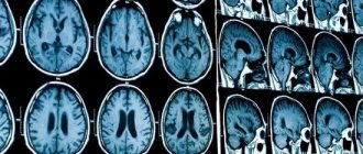
A thousand "photos"
— During the examination, an X-ray beam directed by a computer rotates around the patient, illuminating the body from different sides.
From hundreds, or even thousands of images, a single picture is formed - a tomogram. All this happens in a matter of seconds. Just like in regular X-rays, bones and structures that contain calcium appear in white, soft tissue (such as muscle) appears in shades of gray, and tissue close to the density of air appears in black.
The lungs are an excellent organ for tomography; they contain a lot of air, so areas with pathologies are more clearly visible: they are denser, which means they will differ in color. In photographs of patients with Covid, even an unprofessional eye can easily guess the areas affected by the inflammatory process - they are gray, have unclear outlines and are unevenly distributed in the lungs. This effect is called "frosted glass".
Lungs. CT scan.
Anna Zaikova.
Modern methods of treatment
To treat lobar pneumonia, your doctor will prescribe two antibiotics.
One drug is administered intravenously, the second intramuscularly. Usually these are drugs from the group of penicillins and cephalosporins. Stronger medications are required less often. Additionally, complex therapy is required:
- immunocorrection (blood plasma and immunoglobulins are administered);
- correction of blood clotting disorders (intravenous blood thinning solutions, heparin);
- normalization of the protein composition of the blood (intravenous albumin, retabolil);
- oxygen (supplied through a nasal catheter or mask);
- hormonal drugs (for severe cases, prednisone is used for a short course);
- antioxidant therapy (ascorbic acid, rutin);
- bronchodilators (Berodual, Eufillin, Atrovent);
- expectorants (ACC, Bromhexine, Ambrobene, Lazolvan);
- antipyretic drugs (Nurofen, Paracetamol, Ibuklin);
- physiotherapy, massage, exercise therapy.
As the patient’s condition improves, he undergoes a course of rehabilitation to completely eliminate foci of inflammation in the lungs.
Thin "slicing"
— The famous surgeon Nikolai Pirogov compiled the first atlas of topographic anatomy back in the century before last, cutting frozen corpses layer by layer. Thanks to CT, it is possible to “saw” a person transversely into thin layers intravitally and take pictures of each layer separately.
In one projection we see a person as if he is lying with his feet towards us, in another - frontal - as if he is facing us.
The tomograph step can be from centimeter to millimeter. The smaller the step, the clearer the tomogram. The obtained information can be converted into a three-dimensional image.
Interpreting an x-ray takes longer than the scanning procedure itself. Sometimes 10 minutes is enough to assess the patient’s condition and make a description, but in other cases you have to look in different projections and “rotate” the picture for an hour to give a clear conclusion.
Popular questions and answers
Pulmonologist Marina Samoilova told us why lobar pneumonia is so dangerous and how to treat it.
What complications can occur with lobar pneumonia?
If a person has a weakened immune system or treatment is started untimely or incorrectly, complications are possible in the form of:
- abscess (purulent melting of lung tissue); gangrene of the lung;
- purulent pericarditis (inflammation of the lining of the heart);
- purulent brain damage;
- blood poisoning;
- respiratory failure and even death.
Pneumococcal pneumonia, even in our time, is a fairly common cause of death, especially in older people, smokers, people suffering from alcoholism, with cancer and systemic diseases, and HIV-infected people.
How is an x-ray taken for pneumonia?
For pneumonia, an X-ray of the lungs is usually taken, regardless of the cause of the disease. Before the study, it is necessary to remove everything that could distort the results: metal objects, jewelry, long hair, etc. X-ray of the lungs
done in a vertical position, having first undressed to the waist and stood in front of the machine as the radiologist says. At the doctor’s command, the patient draws air into his lungs and holds his breath, while the device takes a picture.
Is it possible to treat lobar pneumonia with folk remedies?
As a rule, patients with lobar pneumonia should be hospitalized for constant monitoring by medical staff and emergency measures at the slightest sign of complications.
Often such patients experience nausea, vomiting, bowel movements, and confusion, so medications and solutions are administered intravenously. Thus, we cannot talk about treatment at home without the participation of qualified specialists. Fortunately, in our time, subject to a healthy lifestyle, such serious diseases as lobar pneumonia are becoming less and less common, and with modern treatment methods, in most cases it is possible to avoid serious complications and save the patient’s life.
Published on the portal kp.ru
Contraindications to X-rays
X-rays should not be taken for pregnant women at any stage, as well as for children under 15 years of age. In some cases, the patient's condition may be an obstacle to conducting the study. A patient in a supine position cannot undergo the study due to the inability to assume an upright position. In an inadequate condition, when the patient cannot stand still, signs of pneumonia on an x-ray cannot be recorded properly.
How often can an x-ray be taken for pneumonia?
A confirmed diagnosis is an indication for regular x-ray examinations to monitor the course of the disease. Whether the treatment prescribed by the doctor helps will show what pneumonia looks like on an x-ray. In this regard, many patients are concerned about how often this procedure can be performed without harm to health. After the start of treatment, depending on the type of disease, an x-ray for pneumonia must be done after 6-10 days. If positive dynamics are noticed, treatment continues as it was started. The patient, who is considered to be cured, is subsequently re-examined after 1, 3 and 6 months. With favorable treatment, the patient receives a small dose of radiation, far from acceptable levels. This is possible thanks to the development of medical technologies and protective measures during the procedure.
Incidental finds
— In addition to the lungs, the tomogram shows the mediastinum with large vessels, lymph nodes, bronchial tree, trachea, ribs and shoulder blades, fatty tissue of the chest, spinal cord, spine, etc. Therefore, it happens that we find other pathologies in “Covid” images.
Yesterday, for example, we did about 90 studies. Three people were diagnosed with lung cancer, and another had tuberculosis. In general, about 3-5% of patients at the Diagnostic Center receive news about an inflammatory process in the lungs and other serious pathologies.
It requires detailed research, so data on patients who have signs of cancer is sent daily to the regional oncology clinic, and data on tuberculosis is sent to the corresponding regional medical institution. There the tomograms are examined again in more detail.
The clinic at the place of residence will also learn about such preliminary diagnoses in order to conduct additional examinations, confirm the diagnosis and begin treatment as quickly as possible. On CT scans, tumors can be seen at very early stages, so Covid studies secretly help patients prolong their lives.
Lungs. CT scan.
Anna Zaikova.
Publications in the media
Pneumonia is a group of acute infectious (mainly bacterial) diseases, different in etiology, pathogenesis and morphological characteristics, characterized by focal damage to the respiratory parts of the lungs with the obligatory presence of intra-alveolar exudation. Bacterial pneumonia is pneumonia of bacterial etiology. Classification • According to the conditions in which the disease developed •• Community-acquired pneumonia - acquired outside a medical institution (synonyms: home, outpatient) •• Nosocomial pneumonia - acquired in a medical institution (synonyms: hospital, nosocomial) •• Aspiration pneumonia •• Pneumonia in individuals with severe immune defects (congenital immunodeficiency, HIV infection, iatrogenic immunosuppression, etc.) • Along the course •• Mild - does not require hospitalization •• Severe - hospitalization is required.
Incidence • 236.2 cases per 100,000 adolescents 15–17 years of age • 522.8 cases per 100,000 population under 14 years of age • Community-acquired pneumonia - 1,200 cases per 100,000 population per year • Hospital-acquired pneumonia - 800 cases per 100,000 hospitalizations per year . The predominant age is under 20 and over 60 years. Predominant gender - no significant differences were found by gender. Etiology • Streptococcus pneumoniae - most common (30-50%) • Haemophilus influenzae (10-20%) • Atypical pathogens - Chlamidophila pneumoniae, Mycoplasma pneumoniae, Legionella pneumophila (8-25%) • Typical but rare (3-5 %) include Staphylococcus aureus, Klebsiella pneumoniae (less commonly other enterobacteria) • Moraxella catarrhalis (Branhamella catarrhalis) • Most often, community-acquired pneumonia is caused by pneumococci (their sensitivity to penicillin in many countries is significantly reduced) • In very rare cases of community-acquired pneumonia - Pseudomonas aeruginosa (with cystic fibrosis, bronchiectasis), in persons with severe immunodeficiency - Pneumocystis carinii • Escherichia coli • Anaerobic microorganisms • Atypical pneumonia.
Risk factors • Recent acute respiratory viral infection • Renal failure • Cardiovascular diseases • COPD • Immunodeficiency conditions: diabetes, chronic alcoholism, AIDS, malignant neoplasms • Dysbiosis • Risk factors for hospital-acquired pneumonia •• Mechanical ventilation •• Early postoperative period •• Dysbacteriosis • Risk factors aspiration pneumonia •• Impaired consciousness •• Convulsive seizures •• Diseases of the central nervous system •• Anesthesia •• Reflux esophagitis. Pathogenesis . Main pathogenetic mechanisms • Aspiration of oropharyngeal secretions (the main route of infection) • Inhalation of an aerosol containing microorganisms • Hematogenous spread of pathogens from an extrapulmonary source of infection (for example, with endocarditis, with septic thrombophlebitis) • Direct spread of infection from neighboring affected organs (for example, with liver abscess ) or as a result of injury and infection of the chest organs. Pathomorphology . Segmental, lobar or multifocal peribronchial compaction with stages of red (intra-alveolar exudation and erythrocyte diapedesis) and then gray (fibrous organization of intra-alveolar exudate) hepatization.
Clinical picture. • Complaints •• Cough with mucopurulent (sometimes “rusty”) sputum •• Chest pain when breathing (with concomitant pleurisy) •• Shortness of breath •• Weakness, fatigue •• Night sweats. • Intoxication syndrome •• Fever •• Tachycardia •• Tachypnea •• Hyperhidrosis •• Myalgia •• Headaches •• Anorexia. • Objective examination data •• Cyanosis •• Percussion: dullness of percussion sound caused by infiltration or pleural effusion •• Auscultation ••• Moist fine rales and/or crepitus (heard on inspiration) ••• With extensive infiltration or pleural effusion, breathing is weakened • •• Pleural friction noise with dry pleurisy •• In severe cases, meningeal signs and disturbances of consciousness (for example, disorientation and anxiety) may appear •• In 20% of cases, objective signs may be mild or absent. Laboratory tests • Leukocytosis with a shift of the leukocyte formula to the left •• Leukocytosis more than 10-12109/l is characteristic of a bacterial infection •• Values more than 25109/l or leukopenia below 3109/l indicate a poor prognosis • Hyponatremia • Increased activity transaminases • Bacteriological blood test to identify the pathogen (positive result in 20–30% of patients with community-acquired pneumonia, especially before the start of antibacterial therapy) • At the height of active inflammation and intoxication, protein may appear in the urine • Bacteriological and bacterioscopic examination of sputum: microscopy of a stained smear according to Gram, and culture of sputum obtained during deep coughing • Bacteriological examination of material obtained during bronchoalveolar lavage and thoracentesis • In the presence of pleural effusion and pleural puncture - examination of pleural fluid: counting leukocytes with a leukocyte formula, determination of pH, protein content, LDH, microscopy of a Gram-stained smear, culture for aerobic, anaerobic bacteria and mycobacteria • Study of the immune status of persons with suspected immunodeficiency. Special studies • Chest X-ray is a mandatory research method for pneumonia, allowing to visualize areas of infiltration of lung tissue (take into account the shape, size and location), assess the dynamics of the process •• The prevalence of infiltration, the presence of pleural effusion and signs of destruction of lung tissue reflect the severity of the disease and significantly influence the nature of treatment • CT scan of the lungs is performed if destruction or neoplasm is suspected • Fiberoptic bronchoscopy with microbiological and cytological examination of the biopsy specimen - if tuberculosis and tumor diseases are suspected • FVD study - for differential diagnosis with respiratory distress syndrome • With extensive infiltration, massive pleural effusion, In the presence of COPD, it is advisable to evaluate arterial blood gases, which may be the basis for hospitalization and low-flow oxygenation • The study of capillary blood gases is not very informative.
Diagnostic tactics , diagnostic algorithms. The diagnosis of pneumonia is considered definite if the patient has radiologically confirmed infiltration of the lung tissue and at least two of the following signs: • acute febrile onset of the disease (body temperature more than 38 ° C); • cough with sputum; • listening to local crepitus, shortening of percussion sound; • leukocytosis more than 10109/l and/or shift of the leukocyte formula to the left more than 10%. Differential diagnosis • Pneumonia of non-bacterial etiology (viral, fungal, caused by protozoa) • Tuberculosis (examination of at least three sputum smears, Ziehl-Neelsen stained, sputum culture, PCR diagnosis) • Pulmonary infarction (PE) • Bronchiolitis obliterans • Pulmonary contusion • Pulmonary vasculitis • Acute sarcoidosis • Exogenous allergic alveolitis • Eosinophilic infiltrate • Lung tumors • Other conditions that can cause infiltration syndrome on chest x-ray.
TREATMENT Diet. A complete diet with sufficient protein and a high content of vitamins A, C, group B • Limiting carbohydrates to 200–250 g/day, table salt to 4–6 g/day and increasing the proportion of dairy products • Introducing a sufficient amount of fluid (1500–1700 ml/day) • Saturation of the diet with foods rich in vitamin P (aronia, rose hips, black currants, lemon) • Inclusion of foods rich in B vitamins (meat, fish, yeast, wheat bran decoction) prevents the suppression of intestinal microflora by antibiotics • Foods rich in nicotinic acid • Foods rich in vitamins A and -carotene (carrots, red vegetables and fruits) promote the regeneration of the epithelium of the respiratory tract. Fruit and vegetable juices are recommended • Food is given in crushed and liquid form, meals are taken 6-7 times a day • Energy value is from 1600 kcal/day, increasing as recovery progresses to 2800 kcal/day. Indications for hospitalization • Age under 16 or over 60 years • Concomitant diseases (for example, diseases of the bronchopulmonary system or cardiovascular system, circulatory failure IIa and higher, diabetes, thyrotoxicosis) • Physical signs: respiratory rate more than 30 per minute, diastolic blood pressure less than 90 mm Hg. Art., pulse more than 125 per minute, body temperature less than 35 °C or 40 °C or more, disturbances of consciousness • Laboratory data: the number of leukocytes in the peripheral blood is less than 4.0109/l or more than 30.0109/l , arterial blood oxygen saturation is less than 92% (according to pulse oximetry), paO2 is less than 60 mm Hg. and/or paCO2 more than 50 mm Hg. when breathing room air, serum creatinine content is higher than 176.7 µmol/l or urea nitrogen is higher than 7.0 mmol/l, Ht is less than 30% or Hb content is lower than 90 g/l • X-ray data: pneumonic infiltration of more than one lobe, the presence of decay cavities, pleural effusion, rapid progression of focal infiltrative changes in the lungs (increase in the size of infiltration by more than 50% within 2 days) • Extrapulmonary foci of infection (meningitis, septic arthritis, etc.) • Sepsis or multiple organ failure with metabolic acidosis (pH <7.35), coagulopathy • Impossibility of adequate care and fulfillment of all medical prescriptions at home • Lack of effect from outpatient treatment for 3 days, long-term persistence of intoxication syndrome • Preference of the patient and/or his family members.
Drug therapy. The basis of treatment is antibacterial therapy. It begins from the moment of diagnosis, but after collecting material for bacterioscopic and bacteriological examination of sputum. At home, as well as in a hospital, in the first days of the disease (before receiving the results of bacteriological studies), drugs are selected empirically. After receiving the results of a bacteriological study, treatment is carried out taking into account the sensitivity of the pathogen to drugs •• Treatment of community-acquired pneumonia in an outpatient setting ••• Persons under 60 years of age without concomitant diseases are prescribed amoxicillin (500 mg 3 times / day) or amoxicillin + clavulanic acid, or macrolides ( spiramycin, clarithromycin, azithromycin, midecamycin, etc.), or pneumotropic fluoroquinolones (levofloxacin) ••• For non-severe pneumonia in patients over 60 years of age and/or with concomitant diseases, treatment with amoxicillin/clavulanate or cefuroxime is started; levofloxacin is an alternative ••• If it is impossible to take drugs orally, parenteral administration of ceftriaxone is used ••• The effect of treatment is assessed after 48–72 hours, primarily by reducing body temperature. In this regard, NSAIDs should not be used during this period without special indications. If the temperature and intoxication syndrome do not decrease, a change in antibiotic is indicated with a re-assessment of the advisability of hospitalizing the patient •••• If amoxicillin was used, switch to macrolides; if amoxicillin + clavulanic acid or cefuroxime were used, switch to levofloxacin; if macrolides were used, switch to amoxicillin + clavulanic acid or levofloxacin ••• Antibacterial therapy is stopped with stable normalization of temperature on the 3-4th day •• Treatment in a hospital ••• For mild pneumonia, therapy begins with intravenous administration of ampicillin (or amoxicillin/clavulanate, cefuroxime, cefotaxime or ceftriaxone) with switching to oral administration of drugs of the same group (stepped therapy) ••• In severe pneumonia, treatment begins with intravenous administration of a macrolide (erythromycin, spiramycin, clarithromycin) in combination with an intravenous β-lactam drug (amoxicillin/clavulanate, cefotaxime, ceftriaxone) •• • An alternative may be the intravenous administration of a combination of third-generation cephalosporins with early fluoroquinolones (ciprofloxacin, ofloxacin) or the intravenous administration of new fluoroquinolones (levofloxacin, moxifloxacin) •• Treatment of nosocomial pneumonia in general wards: prescribe IV amoxicillin + clavulanic acid, ampicillin / sulbactam or cephalosporins of II–III generations, alternatives are new fluoroquinolones (levofloxacin, moxifloxacin), carbapenems, cefoperazone + sulbactam, cefepime; in intensive care units and wards - carbapenems, cefepime or cefoperazone + sulbactam, ticarcillin + clavulanic acid, piperacillin / tazobactam •• In the treatment of aspiration pneumonia, intravenous forms of cephalosporins of III-IV generations, levofloxacin, moxifloxacin, ticarcillin + clavulanic acid, piperacillin / taz are recommended obactam , amoxicillin+clavulanic acid, ampicillin/sulbactam. For persons with periodontal diseases or alcoholism - a combination of III-IV generation cephalosporins with metronidazole or lincomycin •• In the treatment of pneumonia in patients with AIDS, intravenous combinations of co-trimoxazole with amoxicillin and itraconazole (or fluconazole) are recommended •• For destructive pneumonia (or abscess formation) they are used IV amoxicillin + clavulanic acid, ampicillin / sulbactam, cefoperazone + sulbactam or carbapenems, ticarcillin + clavulanic acid, piperacillin + sulbactam, combination of lincomycin with an aminoglycoside; combination of benzylpenicillin + metronidazole with a transition to the combination of amoxicillin + metronidazole orally (stepped therapy). The duration of therapy is 3–4 weeks or more.
• Tactics of antibacterial therapy after obtaining the results of bacteriological studies, if the sensitivity of microorganisms to antibiotics has not been determined •• For pneumococcal damage - benzylpenicillin 1-2 million units intramuscularly every 4 hours, erythromycin 500 mg every 6 hours, roxithromycin 150 mg 2 times a day or azithromycin 500 mg 1 time / day. For resistant strains - cefotaxime, ceftriaxone, imipenem + cilastatin •• For H. influenzae infection - co-trimoxazole 2 tablets every 12 hours. Reserve drugs: cephalosporins of the II and III generations (cefuroxime 0.25-1 g IV every 12 h, cefaclor 0.5–1 g orally every 6 hours), chloramphenicol 0.5–1 g every 6 hours, amoxicillin + clavulanic acid •• For Staphylococcus aureus lesions - oxacillin 6–10 g/day, first generation cephalosporins or clindamycin 600–800 mg IV every 6–8 hours. For resistant strains - vancomycin •• For Klebsiella lesions - aminoglycosides, third generation cephalosporins (cefotaxime 2 g IV every 6 hours, ceftazidime 2 g IV every 8 hours; ceftriaxone 2 g IV every 12 hours), fluoroquinolone derivatives (ciprofloxacin 500–750 mg 2 times / day), imipenem + cilastatin 1 g 2 times / day •• For Escherichia coli lesions - aminoglycosides, cephalosporins II and III generations. Alternative drugs - fluoroquinolone derivatives, imipenem + cilastatin, chloramphenicol •• For Pseudomonas aeruginosa lesions - a combination of aminoglycoside and carbenicillin or ceftazidime, azlocillin or imipenem + cilastatin •• For Enterococci lesions - a combination of ampicillin and gentamicin •• For Moraxella catarrhalis lesions - cefa II generation losporins or amoxicillin + clavulanic acid, co-trimoxazole, clarithromycin •• If Acinetobacter is affected - imipenem + cilastatin or aminoglycosides, co-trimoxazole. • Expectorants •• Agents that stimulate expectoration ••• Direct-acting drugs, for example potassium iodide ••• Reflex-action drugs, for example, thermopsis herb infusion, licorice root preparations, etc. •• Mucolytic drugs, for example acetylcysteine, trypsin, bromhexine, ambroxol. • Oxygen therapy for patients with cyanosis, hypoxia, shortness of breath.
Surgical treatment of pneumonia is not performed. The question of surgical intervention arises when an abscess forms, especially a chronic one, or when pneumonia is complicated by pleural empyema. Monitoring the effectiveness of treatment • Clinical indicators •• Fever •• Shortness of breath •• Cough • X-ray dynamics (lags behind clinical) • Bacteriological examination of sputum - if treatment is ineffective. Complications • Pleural effusion (complicated and uncomplicated) • Destruction/abscessation of lung tissue • Acute respiratory distress syndrome • Acute respiratory failure • Infectious-septic shock • Pleural empyema. • Secondary bacteremia, sepsis, focus of hematogenous dissemination • Pericarditis, myocarditis • Nephritis. Prevention • Prevention of aspiration in bedridden patients • Rational use of antibiotics • Annual influenza vaccination for people at high risk • Polyvalent pneumococcal vaccine (currently not available in Russia) is recommended for people over 65 years of age and children over 2 years of age with the following risk factors •• Dysfunction spleen or asplenia •• Lymphogranulomatosis •• Multiple myeloma •• Liver cirrhosis •• Chronic alcoholism •• Renal failure •• Immunodeficiency. Age characteristics • Children •• Focal-confluent nature of the lesion predominates •• In the clinical picture - acute onset, severe intoxication against the background of a weak (or absence) pain syndrome, pronounced auscultation pattern •• Dynamics against the background of antibacterial treatment - rapid positive effect •• High mortality in children under 1 year of age • Elderly and old people: morbidity and mortality are increased over the age of 70 years, especially with concomitant pathology or the presence of risk factors. Features for pregnant and lactating women • During pregnancy, the use of β-lactam antibiotics, macrolides, metronidazole is permissible; fluoroquinolones, tetracyclines, aminoglycosides, lincosamides, co-trimoxazole are contraindicated • When breastfeeding, penicillins, cephalosporins, azithromycin are acceptable with caution; macrolides, fluoroquinolones, carbapenems, tetracyclines, lincosamides, co-trimoxazole are not recommended. Course and prognosis • Depend on the severity of the course, the pathogen, the patient’s age, the time of initiation of treatment, the adequacy of initial therapy, the state of the immune system, concomitant diseases • Mortality from community-acquired pneumonia: 1–3% - among young previously healthy people, 15–30% - in older age groups with concomitant diseases.
ICD-10 • J13 Pneumonia caused by Streptococcus pneumoniae • J14 Pneumonia caused by Haemophilus influenzae [Afanasyev-Pfeiffer bacillus] • J15 Bacterial pneumonia, not elsewhere classified • J17.0* Pneumonia in bacterial diseases classified elsewhere
Shine, doctor, shine
— Conrad Roentgen discovered rays in 1895, and they almost immediately began to be used in medicine. He opened them by accident because he “photographed” his finger. How the devices were invented is a whole story. The main thing is that the X-ray tube has only 2% efficiency, all the rest of the energy is heat. Imagine how hot it was.
I didn’t catch this time, but old radiologists say that they tried to cool the tube with water, but it boiled and literally exploded with steam, burning the patient. Of course, this doesn't happen now. But this does not mean that modern devices are completely safe.
Radiation exposure with CT is several times greater than with classical X-ray examination. If a person often undergoes a tomogram, the radiation doses will subsequently affect his health. However, for older people this danger is minimal, and there is an explanation for this.
The cells of the body are defenseless to ionizing radiation during the period of division and growth. In old age, when degenerative processes begin, cells divide less frequently and, accordingly, become resistant to X-rays. At the age of 55-60, you can no longer be afraid of radiation exposure, and children and nursing women are most at risk.
If there are no indications for a CT scan from a therapist, then you should not prescribe this study yourself.
Lungs. CT scan.
Anna Zaikova.
Fluorography, X-ray and CT
— Fluorography and radiography of the lungs are one and the same when it comes to digital devices. It’s just that in our country it has historically developed that an X-ray machine for examining the lungs is called a fluorograph. The quality of the images is the same.
And it is absolutely correct that patients with suspected Covid undergo a fluorogram. It is several times cheaper than CT and allows you to evaluate large lung lesions. In addition, every clinic has a fluorograph, and we only have about 30 tomographs in the entire region. If using a fluorogram we have identified a pathology, then there is no point in doing a CT scan.
Of course, CT is a more “sensitive” method if we are talking about Covid viral pneumonia. Sometimes it is visualized as very small and isolated areas with a barely noticeable dullness. If a patient has been sick for several days with a high fever, and the x-ray shows nothing, then he really needs a CT scan.
Thus, a kind of primary “sorting” of patients occurs. Already at the clinic, doctors see the extent of lung damage, can make a diagnosis and decide on hospitalization.
Lungs. CT scan.
Anna Zaikova.