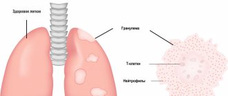
Lung cancer is a pathology that begins with damage to the parenchyma or bronchi.
The main symptoms of this disease are:
- Dyspnea;
- Cough with blood and sputum;
- Chest pain;
- Temperature;
- Thinness;
- Pulmonary hemorrhage;
- Pericarditis;
- Pleurisy.
To find out the exact diagnosis, you need to do a computed tomography of the lungs, radiography, bronchoscopy, study sputum and pleural exudate, and do a biopsy.
Treatment usually involves complex therapy: surgery, radiation therapy and chemotherapy.
This type of cancer is of epithelial origin. It develops from the mucous membrane of the bronchi and their glands or from the pulmonary alveoli. The mortality rate with it is quite high.
The development of the disease will be directly influenced by the histological structure of the tumor:
- Differentiated squamous cell carcinoma progresses slowly;
- Undifferentiated cancer progresses rapidly and metastasizes;
- Small cell cancer also develops rapidly, but unnoticed, and metastasizes.
It was revealed that cancer forms more often in the right lung than in the left. It also forms more often in the upper part of the lung than in the middle and lower parts due to the entry of more carcinogens into the upper part.
Metastasis occurs in three ways:
- Lymphogenic;
- Implantation;
- Hematogenous.
The lymphogenous route is the most common. Metastases go to various types of lymph nodes:
- Pulmonary (affected first);
- Bronchopulmonary (affected after them);
- Tracheobronchial;
- Paraesophageal;
- Bifurcation.
With hematogenous germination through the blood, metastases occur:
- Into the adjacent lung;
- Brain;
- Kidneys and liver;
- Spine.
Metastases
Germination of an implantation nature occurs along the pleura.
The main causes of lung cancer are:
- Tobacco smoking;
- Bad air;
- Radiation.
Based on histology, lung cancer is of four types:
- Glandular;
- Squamous;
- Small cell;
- Large cell.
Etiology and risk factors
In 8 out of 10 cases, the cause of lung cancer is smoking, including passive smoking.
But the disease is also found in non-smoking people. Risk factors include hereditary predisposition, regular exposure to volatile carcinogens, excessive chest radiation exposure, genetic disorders, certain viruses, alcohol abuse and malnutrition. At risk:
- smokers over 40 years of age;
- alcoholics;
- patients with chronic obstructive pulmonary diseases;
- patients with oncological diseases of the upper respiratory tract and lungs in anamnesis;
- people with a family history;
- people who regularly come into contact with asbestos, radon, arsenic, dust;
- people with low social status.
Factors independent of a person include age over 50 years, endocrine disorders, and chronic lung diseases.
Specialized clinics and treatment costs
Lung cancer treatment is carried out in the pulmonology departments of large university hospitals. This makes it possible to involve a multidisciplinary team of specialists in the therapeutic process and achieve successful treatment results. Here are some of these clinics:
- St. Vincent Clinic - Academic Hospital of the University of Freiburg, Department of Pulmonology
- University Hospital Heidelberg, Department of Cardiology, Angiology and Pulmonology
- University Hospital Aachen, Department of Cardiology, Pulmonology and Angiology
- University Clinic named after. Goethe Frankfurt am Main, Department of Gastroenterology, Hepatology, Pulmonology, Allergology, Endocrinology and Diabetology
- University Hospital Jena, Department of Cardiology, Angiology, Pulmonology and Intensive Care
Estimated cost of basic lung cancer treatment methods:
- Thoracotomy with segmental tumor resection for lung cancer – from 16,112 euros
- Video-assisted thoracoscopic (VATS) lobectomy – from 24,005 euros
- Lobectomy with da Vinci robot – from 28,510 euros
- Treatment of lung cancer using extended pneumonectomy – from 21,330 euros
- Treatment of lung cancer using embolization of tumor vessels – from 5,118 euros
- Treatment of lung cancer using radiation therapy – from 15,800 euros
- Treatment of lung cancer using proton therapy – from 44,325 euros
- Treatment of lung cancer with chemotherapy – from 2,265 euros
- Pulmonary rehabilitation – from 708 euros per day
If before starting treatment it is necessary to clarify the diagnosis or more accurately determine the stage of cancer, then an extended diagnosis is carried out.
Types of lung cancer
Lung cancer is classified according to various criteria. Based on the histological structure of the tumor, they are distinguished:
- adenocarcinoma;
- adenosquamous carcinoma;
- neuroendocrine carcinoma;
- diffuse idiopathic pulmonary hyperplasia;
- spindle cell carcinoma;
- large cell carcinoma;
- small cell carcinoma;
- squamous cell carcinoma.
Squamous cell lung cancer is detected in 8 cases out of 10. It spreads relatively slowly and can be treated surgically. In 17 patients out of 100, small cell lung cancer is detected - it grows rapidly, affects neighboring organs outside the lung tissue and metastasizes in the early stages. Responds to radiation and chemotherapy.
Based on location, central and peripheral lung cancer is distinguished. In direction of growth:
- endobronchial, or exophytic, - grows into the lumen of the bronchus;
- exobronchial, or endophytic – grows into the thickness of the lung tissue;
- branched with periorbital muff-like growth around the bronchi;
- mixed - grows in several directions with a predominance of one or another characteristic.
Principles of staging
The stages of lung cancer are determined based on three parameters:
- Main tumor
- Nearest metastatic foci
- Distant metastases
They are designated by the letters T, N and M, respectively. Below we will consider the main designations used in making the diagnosis and determining the stage of lung cancer.
Primary tumor
- Tx – tumor cells are detected, but the neoplasm itself is not detected, or its assessment is impossible.
- T0 – there is no primary tumor (the disease may well manifest itself with the appearance of metastatic tumors, while the primary tumor remains very small and invisible).
- Тis – cancer in situ (does not extend beyond the bronchus).
- T1 – a neoplasm in the lungs is no more than 3 cm in size, does not protrude beyond the pulmonary parenchyma and does not grow into the pleura, and according to bronchoscopy, does not spread beyond the lobar bronchus.
- T2 – the neoplasm is larger than 3 cm, the main bronchus is affected, and the tumor has spread to the visceral pleura. Atelectasis may be present - this is a collapse of the lung lobe when the alveoli do not fill with air and do not straighten.
- T3 – a neoplasm of any size extends to the chest, diaphragm, mediastinum, pleura or cardiac sac. T3 can also be determined with atelectasis of the entire lung or damage to the main bronchus, when the tumor is less than 2 centimeters from the carina of the trachea (the place of its division).
- T4 - diagnosed when the tumor grows into the esophagus, heart, main blood vessels, trachea, spine, distant lobes of the lung, or if metastatic pleurisy develops.
Regional metastases
- Nx – it is not known whether there are nearby metastases
- N0 – the patient has been examined and there are no metastases
- N1 – there is metastatic damage to the hilar or intrapulmonary lymph nodes
- N2 – the lower tracheobronchial lymph nodes or mediastinal nodes are affected, but only on the side of the localization of the primary neoplasm
- N 3 – metastases of lung cancer are present in the supraclavicular or hilar lymph nodes on any side relative to the main tumor
Distant metastases
- Mx – it is not known whether metastases are present in distant organs and tissues
- M0 – no metastatic foci
- M1 – distant metastases detected
Stages of lung cancer and their signs
- The first stage – the tumor is no more than 3 centimeters, there are no metastases.
- The second stage – metastases appear in the regional lymph nodes, the tumor increases to 6 centimeters.
- The third stage – metastases appear in the lymph nodes of the respiratory system, the tumor increases in volume.
- Stage four – the tumor extends beyond the boundaries of the lung, there are local and distant metastases.
The stages are divided into substages: A – initial, B – severe Source: Travis WD, et al. The 2015 World Health Organization Classification of Lung Tumors. J. Thorac. Oncol. 2015. Vol.10, No.9, P. 1243-1260..
How does lung cancer manifest?
Clinical picture
Consult your doctor at the first symptoms of the disease:
- prolonged increase in temperature;
- heavy sweating;
- increasing weakness;
- cough;
- chest pain when coughing or inhaling;
- bloody sputum;
- severe shortness of breath;
- weight loss;
- deterioration of general condition.
Characteristic signs of lung cancer are a whistling sound when breathing and a hoarse voice.
Symptoms depend on the location of the tumor. For example, peripheral lung cancer is asymptomatic and painless for a long time, since the tumor does not affect the nerve endings. And central lung cancer manifests itself in the early stages in the form of cough with sputum mixed with blood and pus, constant shortness of breath, voice changes, and general malaise. In peripheral apical lung cancer, one side of the face and shoulder girdle is affected. When metastasis occurs, the pain radiates to neighboring organs, and the face acquires a bluish tint. In addition to the listed symptoms, thrombophlebitis, muscle pathologies and neuritis, dermatoses, lipid metabolism disorders, rheumatoid conditions, bone lesions and pain in the joints are possible at different stages. With advanced lung cancer, complications develop - obstruction of the bronchi, respiratory failure, pulmonary hemorrhage, collapse (closure) of the lung, and general exhaustion.
In most clinical cases, the disease occurs with non-specific symptoms, and therefore requires accurate (hardware and laboratory) diagnosis.
Diagnosis of the disease
Initially, the doctor collects anamnesis; in other words, he listens to the person's complaints. Based on the typology of complaints, the specialist prescribes a comprehensive examination, consisting of an assessment of the person’s physical data, laboratory tests of urine and blood, lymph node biopsy, cytological analysis of sputum and bronchial lavage, pleural puncture, fibrobronchoscopy, X-ray and computed tomography of the lungs.
In order to see the full picture of the ongoing process, the specialist finds out the morphological nature of the tumor. In order to accurately determine the disease, differential diagnosis can be carried out. It helps to distinguish the symptoms of cancer from signs of other diseases (pneumonia, tuberculosis, lymphogranulomatosis, etc.).
If an accurate diagnosis cannot be made, a diagnostic thoracotomy is prescribed.
Diagnostics
To diagnose lung cancer, use:
- radiography (a number of tumors are detected during preventive fluorography);
- CT, MRI are the most informative methods for diagnosing pathology;
- bronchoscopy of the trachea and bronchi up to 6-7 orders with taking a biopsy for histological examination;
- PET-CT – to assess the extent of metastases and monitor the effectiveness of treatment;
- endoscopic or endobronchial ultrasound - to assess the condition of the lymph nodes and stage the process;
- bronchopulmonary electromagnetic sounding makes it possible to diagnose deep neoplasms in the early stages;
- thoracoscopy - examination using a video camera through a puncture in the chest;
- molecular genetic blood tests for specific tumor markers.
Recommended tumor marker combinations
- If adenocarcinoma or large cell cancer is suspected - CYFRA 21-1 and CEA.
- Small cell carcinoma (SCLC) – NSE and ProGRP, CYFRA 21-1.
- Squamous cell carcinoma – CYFRA 21-1, CEA and SCC.
- Carcinoma of unknown histological type - combinations of CEA, CYFRA 21-1, NSE and ProGRP.
- Neuroendocrine lung tumors (carcinoid, neuroendocrine large cell and small cell cancer) – chromogranin A, NSE.
The European Group on Tumor Markers recommends including at least three indicators in the examination (CYFRA 21-1, CEA and NSE), since lung tumors differ in histological structure.
Where are surgeries to remove lung tumors performed?
Removing a lung cancer tumor or metastases is a complex intervention that can only be performed by experienced surgeons in specialized clinics, public or private.
Large budget medical organizations have all the capabilities to carry out such operations, but the timing of their implementation and the conditions of stay in the hospital often leave much to be desired. For those who do not want to waste precious time and days of their lives waiting, we offer treatment at an oncological center. We have all the necessary doctors - true professionals with extensive experience in the field of diagnosing and fighting cancer, the most modern equipment and our own laboratory, which reduces the time required to obtain research results to a minimum. We do not create queues, quickly perform all procedures and provide not only surgical but also other oncology treatment, including chemotherapy, immunotherapy and targeted therapy using original drugs that give predictable results. Our specialists are fluent in the most advanced innovative techniques and perform any interventions, and the staff does everything to make the stay in the Center as comfortable as possible for every visitor.
Lung cancer treatment methods
Surgery, radiation and chemotherapy are traditionally used to treat lung cancer at different stages. Immunotherapy is one of the modern methods.
When the possibilities of antitumor treatment are exhausted or it is contraindicated, palliative therapy is used. Surgery is indicated if the location of the tumor allows it to be completely removed. During the intervention, regional lymph nodes must be removed. If possible, the procedure is carried out without incisions - using an endoscope and microsurgical instruments.
The amount of intervention depends on the stage of the process (1, 2, 3A Source: Masters GA, Temin S, Azzoli CG, et al. Systemic Therapy for Stage IV NonSmall Cell Lung Cancer. American Society of Clinical Oncology. Clinical Practice Guideline Update. J Clin Oncol 2015 ; 33:3488-3515):
- loboectomy – removal of one lobe of the lung;
- bilobectomy – removal of two lobes of an organ;
- pneumonectomy – complete removal of the lung.
Preference is given to the most organ-preserving techniques.
Radiation therapy is carried out as an independent treatment at stage 2 or in combination with chemotherapy at stage 3A. It may also be prescribed to patients with stage 1 cancer if surgery needs to be delayed (for example, due to a myocardial infarction). The method involves the destruction of tumor cells under the influence of a powerful stream of gamma rays. They affect the tumor itself and the area of regional metastasis.
Most patients at various stages are indicated for chemotherapy - treatment with drugs that destroy tumor tissue. It happens:
- neoadjuvant - prescribed before surgery to reduce the size of the tumor and the volume of intervention;
- adjuvant – prescribed after surgery to destroy remaining tumor cells and reduce the risk of relapse.
At stages 3B and 4, patients are treated only with chemotherapy.
A promising area of conservative cancer treatment is immunotherapy. With the help of medications, they specifically activate their own immune cells to destroy cancer cells.
Palliative treatment for incurable patients with stage 4 lung cancer is aimed at maximizing quality of life and symptomatic relief.
Survival prognosis:
- 1A – 50-70% – 5-10 years Source: Howlader N., et al. SEER Cancer Statistics Review, 1975-2013. Natl. CancerInst. 2021;
- 1B – 40-60% – 3-7 years;
- 2A – 55% – 3-4 years;
- 2B – 40% – 1.5-3 years;
- 3A – 20-25% – 14-23 months;
- 3B – 7-9% – 10-16 months;
- 4 – 2-13% – 6-18 months.
Life expectancy
Unfortunately, today's forecasts are unfavorable, because... The mortality rate for this lung pathology is excessively high. If there is no qualified treatment, the person will die within 2 years (probability – 90%).
Survival directly depends on the quality of treatment provided. The timeliness of the treatment started (i.e. the stage of the cancer) also plays a significant role. The survival percentage for each stage looks like this: for the 1st – 80%, for the 2nd – 40%, for the 3rd – 20%. The use of modern treatment techniques increases survival rate to 45%. In approximately 10% of cases, radiation and chemotherapy treatments provide 5-year survival.
Prevention
- eliminate smoking, alcohol abuse and systematic contact with carcinogens;
- lead a healthy lifestyle, balance your diet;
- promptly treat acute respiratory diseases;
- undergo regular screenings.
To consult with a specialized specialist in St. Petersburg, make an appointment using the form on the website.
Sources:
- World Cancer Report 2014. World Health Organization. 2014.
- Travis WD, et al. The 2015 World Health Organization Classification of Lung Tumors. J. Thorac. Oncol. 2015. Vol.10, No.9, P. 1243-1260.
- Masters GA, Temin S, Azzoli CG, et al. Systemic Therapy for Stage IV NonSmall0Cell Lung Cancer. American Society of Clinical Oncology. Clinical Practice Guideline Update. J Clin Oncol 2015; 33:3488-3515
- Howlader N., et al. SEER Cancer Statistics Review, 1975-2013. Natl. CancerInst. 2021.
The information in this article is provided for reference purposes and does not replace advice from a qualified professional. Don't self-medicate! At the first signs of illness, you should consult a doctor.