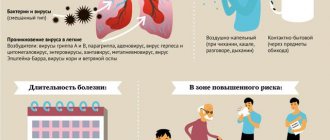
Right-sided lower lobe pneumonia develops more often than left-sided pneumonia. This is due to the peculiarities of the anatomical structure of the respiratory tract on the right. The right lower lobe bronchus has an oblique direction, so microorganisms penetrate it faster into the lung tissue. At the Yusupov Hospital, pulmonologists use modern examination methods to diagnose pneumonia and determine the source of inflammation. For treatment, effective drugs are used, registered in the Russian Federation, which have a minimal range of side effects.
Patients with mild right-sided lower lobe pneumonia are hospitalized in a therapy clinic. In life-threatening conditions, they are transferred to the intensive care unit. If indicated, they undergo artificial ventilation of the lungs using modern portable and stationary ventilators. All severe cases of right-sided lower lobe pneumonia are discussed at a meeting of the Expert Council, in which professors and doctors of the highest category take part.
Causes
The vast majority of right-sided pneumonia caused by microorganisms is an independent disease. Less common is pneumonia, which is a manifestation of an acute infectious disease. Right lower lobe pneumonia is predominantly caused by the following pathogens:
- pneumococci;
- staphylococci;
- hemophilus influenzae;
- mycoplasma.
Less commonly, right-sided lower lobe pneumonia develops under the influence of chlamydia and Klebsiella. In patients who are in the hospital, the cause of right-sided lobar pneumonia can be infection with gram-negative microorganisms (enterobacteria, Pseudomonas aeruginosa, acinetobacter), Staphylococcus aureus and anaerobes. In case of right lower lobe pneumonia in patients with immunodeficiency, in addition to gram-negative bacilli and pneumococci, pneumonia is often caused by cytomegaloviruses, fungi, and mycobacteria. The main cause of aspiration right-sided lower lobe pneumonia is the entry into the respiratory tract of microflora of the stomach or oropharynx (anaerobic bacteria, gram-negative microflora and Staphylococcus aureus).
The main causative agents of atypical inflammation of the lower lobe of the right lung are mycoplasma, chlamydia, and legionella. In viral-bacterial right-sided lower lobe pneumonia, respiratory viruses cause pneumonia only in the initial period of the disease. The main pathogens that determine the clinical picture, severity and outcome of the disease remain the bacterial flora.
Symptoms of right-sided pneumonia
Right-sided pneumonia of a bacterial nature is manifested by the following symptoms:
- cough;
- secretion of sputum;
- increased temperature;
- leukocytosis;
- blue skin in the area of the nasolabial triangle;
- rapid breathing and heart rate.
Viral right-sided pneumonia manifests itself:
- muscle weakness;
- rapid fatigue;
- fever;
- dry cough with little sputum production.
Signs
The following syndromes are characteristic of pneumonia of the lower lobe of the right lung:
- intoxication (pallor of the skin, loss of appetite, weakness, general weakness, muscle and headaches, shortness of breath, palpitations);
- syndrome of general inflammatory changes (fever, feeling hot, chills);
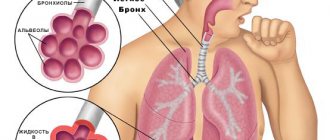
- syndrome of inflammatory changes in the lung tissue (cough with sputum production, increased vocal tremors and bronchophony, shortening of percussion sounds, changes in the frequency and nature of breathing, moist rales and crepitus);
- syndrome involving other organs and systems in the pathological process.
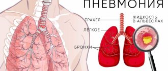
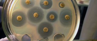
An X-ray sign of right-sided lower lobe pneumonia is the presence of large areas of clearing, involving the lower lobe of the right lung. At the Yusupov Hospital, patients undergo radiography in two projections or large-frame fluorography. Bacteriological examination of sputum or bronchial washings is carried out before prescribing antibiotics. It helps to detect the pathogen and determine its sensitivity to antibiotics.
To clarify the nature of pneumonia, immunological studies are used. Viral and viral-bacterial pneumonia are detected using virological and serological studies. Patients can undergo complex procedures at partner clinics.
A general blood test allows you to determine the severity of the inflammatory process in the right lobe of the lower lung. In patients with pneumonia, the white blood cell count and erythrocyte sedimentation rate increase. With severe intoxication syndrome, the number of red blood cells decreases and toxic granularity of neutrophils appears. C-reactive protein appears in the blood, fibrinogen content and the level of sialic acids increase.
Bacterial pneumonia is characterized by leukocytosis; with viral pneumonia, the number of leukocytes in the blood decreases. In mycoplasma and ornithosis inflammations of the lungs, leukopenia is combined with a very high erythrocyte sedimentation rate. For patients with a prolonged course of right-sided lower lobe pneumonia and complications of pneumonia, the immunological reactivity of the body is determined. In order to find out the degree of involvement of other organs in the pathological process, an electrocardiogram is recorded, echocardiography is performed, and indicators of external respiratory function are determined using spirometry.
Lobar pneumonia
Pneumonia has several types of classification that allow it to be considered according to causes, stages, development and other characteristics. According to the clinical and morphological course, pneumonia can be focal and lobar.
What is lobar pneumonia?
Lobar pneumonia is an acute infectious-allergic inflammatory process that affects one or more lobes of the lung, as well as the pleura. Pathology is diagnosed in most cases in adults. Children get sick with this form less often, but their lobar pneumonia is much more severe. Therefore, the initial manifestations of the development of this disease should be a reason for immediate contact with the clinic, this will avoid the development of life-threatening complications.
Kinds
According to the localization of the focus of the inflammatory process, lobar pneumonia can be divided into:
upper lobe;- lower lobe;
- middle lobe;
- interlobar;
- right-sided;
- left-handed;
- bilateral.
According to the nature of the course, the pathology is divided into acute and protracted forms.
Causes
In most cases, the causative agents of pneumonia are pneumococci; less commonly, the disease is caused by other microorganisms. Pathogenic flora enters the lungs through bronchogenic, less often lymphogenic or hematogenous routes.
Lobar pneumonia is associated with the persistence of the pathogen in the nasopharynx and sensitization of the body to antigens.
Risk factors:
- previous influenza or acute respiratory viral infection;
- hypothermia;
- immunosuppression;
- stress;
- injuries;
- excessive loads.
Background conditions that may contribute to the development of lobar pneumonia:
- tuberculosis;
- chronic obstructive pulmonary disease;
- cardiac ischemia;
- diabetes;
- chronic alcoholism.
Symptoms and signs
Lobar pneumonia begins acutely. Early symptoms include signs of intoxication, which include the following:
- temperature increase - may be above 39 ° C;
- chills;
- fever;
- intense headache;
- increased sweating;
- weakness.
There are also bronchopulmonary manifestations:
- pain in the pleural area;
- dyspnea;
- cough.
As the disease develops, the following symptoms appear:
- severe fatigue;
- cough with phlegm;
- chest pain when breathing;
- sleep disturbance;
- dizziness;
- pain in joints and muscles.
Signs of lobar pneumonia will vary depending on the location of the inflammation:
- Upper lobe inflammation - negative symptoms appear on the nervous and cardiovascular systems.
- Lower lobe inflammation - pathologies of the gastrointestinal tract appear.
- Middle lobe inflammation - symptomatic manifestations are weak.
Which doctor treats you?
Pneumonia is treated by a pulmonologist. Our clinic employs qualified pulmonologists with more than 10 years of experience. We also have our own modern diagnostic facilities - ultrasound, MRI. The patient, without leaving the walls of the clinic, can undergo all the necessary examinations, get an appointment with a competent doctor, who will make a diagnosis and prescribe treatment appropriate to the situation.
Treatment methods
Lobar pneumonia is treated with antibacterial drugs, which are prescribed even before bacteriological culture analysis. The duration of treatment and the regimen are prescribed only by the doctor. As a supplement, a specialist can prescribe antipyretic and antihistamines, mucolytics, and immunostimulants.
results
Provided timely diagnosis and proper treatment, the prognosis of the disease is favorable. However, lobar pneumonia has a negative effect on the immune system, so vaccination is recommended to prevent relapses.
Rehabilitation and lifestyle restoration
The general rules for rehabilitation after pneumonia include:
- adherence to a rest regime - you need a full night's sleep of 8 hours, and daytime sleep of 1-2 hours;
- proper nutrition - food should be rich in protein, calcium, vitamins A, B, C;
- regular ventilation of the room;
- inadmissibility of hypothermia and open drafts.
Physiotherapy indicated:
- medicinal electrophoresis;
- medicinal inhalations;
- chest massage;
- exercise therapy;
- UHF.
Lifestyle after lobar pneumonia
To prevent the recurrence of the disease, you need to:
- completely quit smoking and other bad habits;
- carry out hardening procedures;
- prevent emotional turmoil;
- exclude hypothermia of the body;
- to live an active lifestyle.
Lobar pneumonia greatly impairs the quality of life; the patient should consult a doctor as soon as possible for qualified help. Otherwise, severe and life-threatening complications may develop.
| Name of service | Price in rubles | Price until 19.12. |
| CT scan of the chest | 4 990 | 2 790 |
| CT lungs | 4 990 | 2 790 |
If you do not find a service in the price list, please call us and we will provide you with the necessary information.
Our doctors will help you:
Cardiologist, therapist, gastroenterologist
Shelekhova Violetta Valerievna
Right-sided pneumonia: treatment of the disease
After making a diagnosis, the doctor draws up an individual treatment plan using antibacterial drugs to prevent the spread of microorganisms. Self-medication in this situation is inappropriate, since independent choice of antibiotics and their incorrect dosage can contribute to a significant deterioration of the patient’s condition.
If the development of right-sided pneumonia was caused by a viral infection, the use of antibacterial therapy is inappropriate. In these situations, taking antiviral drugs and immunological stimulants that suppress negative symptoms is effective.
Drug therapy for right-sided pneumonia includes taking the following medications:
- antibacterial drugs (course duration is from seven to ten days);
- antihistamines;
- antiviral drugs;
- antipyretic drugs;
- expectorants and sputum thinners.
The patient is advised to stay in bed, eat fortified foods and drink plenty of fluids.
When choosing a treatment regimen, the doctor takes into account the characteristics of the patient’s body and the severity of pneumonia. Effective treatment can be considered when the patient’s general condition improves, blood counts normalize, and symptoms decrease.
Clinic
The main symptoms of pneumonia, which appear in both adults and children.
- Fever: occurs acutely, quickly reaches febrile levels
- Cough with copious discharge of purulent sputum; there may also be blood in the sputum (especially in the case of lobar pneumonia)
- Chest pain associated with breathing
- Shortness of breath, feeling of lack of air
- Manifestations of intoxication: severe weakness, fatigue, sweating, nausea, vomiting
- In the case of “atypical” pneumonia (Mycoplasma pneumoniae, Legionella pneumophila, Chlamydia pneumoniae, Pneumocystis jirovecii), there may be a gradual onset with a dry, unproductive cough, not without common symptoms - pain and sore throat, weakness, malaise, myalgia, headaches, pain in the abdomen - with minimal changes on the x-ray
- Elderly patients often have more severe general symptoms: drowsiness, confusion, anxiety, sleep disturbances, loss of appetite, nausea, vomiting, signs of exacerbation/decompensation of chronic diseases
Diagnostics
To identify pneumonia and its type, an integrated approach is used:
- anamnesis collection. The doctor finds out when the first symptoms of the disease appeared. The patient must tell in detail about his feelings, report the presence of chronic diseases, if any;
- physical examination. On auscultation, fine bubbling rales are heard. Bronchophony and weakened vesicular breathing are recorded. During percussion, dullness of the pulmonary sound is noted;
- radiography. In case of right-sided middle lobe pneumonia, the X-ray image shows a darkened area in the middle lobe of the right lung, the presence of single or multiple foci;
- general blood analysis. In patients with pneumonia, there is an increase in the number of leukocytes, an increase in neutrophils, an acceleration of ESR, a decrease in the level of hemoglobin and platelets;
- bacteriological analysis of sputum. Microscopy of the smear is performed and cultured on a special nutrient medium to determine the pathogen.
To diagnose the disease, specialists at the Yusupov Hospital use high-precision equipment, which allows them to reliably determine the type and nature of the disease. High-quality and reliable diagnosis is a key factor in prescribing the correct therapy.