Detailed description of the study
Rhinitis is an inflammation of the nasal mucosa, which can be either acute or chronic. It is customary to distinguish several types of process: catarrhal, atrophic, hypertrophic, allergic, neurovegetative. With each type, the nasal mucosa produces a large amount of secretion, the formation of which is commonly called a “runny nose.”
The walls of the nasal cavity are covered with ciliated epithelium with cilia, which vibrate under the influence of the secretion produced. Such vibrations are necessary for the movement of mucus along with various substances (microorganisms, dust) that enter the nasal cavity from the outside. When the immune system is functioning normally, they do not cause harm to a person. Otherwise, a decrease in local immunity can lead to the development of inflammation - acute rhinitis, which most often occurs when the body is hypothermic and reduced immunity in general. This is how catarrhal rhinitis develops in infectious diseases, usually with ARVI and influenza.
Allergic rhinitis has a slightly different mechanism of action. With this type of inflammation, the body interacts with an allergen - dust, wool, pollen, etc., which provokes the production of histamine and bradykinin. These substances contribute to the release of inflammatory mediators, dilation of the lumen of blood vessels, which leads to swelling of the nasal mucosa and increased secretion of mucus.
The cytological smear determines:
- lymphocytes;
- eosinophils;
- neutrophils;
- monocytes;
- ciliated and squamous epithelial cells;
- slime;
- mast cells;
- microflora.
Determining the number of eosinophils is a key point in choosing adequate therapy for rhinitis. Their number allows us to differentiate the most common rhinitis, allergic and infectious.
Rhinocytogram in the diagnosis of rhinitis.
Rhinocytogram is a microscopic examination of a smear of nasal discharge, stained using the Romanovsky-Giemsa method. With its help, you can determine the presence of an inflammatory process (rhinitis), as well as find out its nature - infectious or allergic.
Normally, all the walls of the nasal cavity are covered with a mucous membrane with a secretion that helps remove dust and germs. However, the nasal cavity of a healthy person contains a large number of microbes that do not cause harm to the body due to the normal immune response. If for some reason local (and/or general) immunity decreases, microbes and viruses can lead to inflammation of the nasal mucosa - rhinitis.
As a result of the immune system's response, the number of leukocytes (neutrophils or lymphocytes) increases in the nasal mucosa.
With allergies, the body is exposed to various allergens (plant pollen, animal dander, dust, etc.) to which an increased reaction of the immune system occurs. This reaction leads to the appearance of certain elements in the nasal mucosa that cause allergy symptoms. In the case of allergic rhinitis, these are eosinophils (a type of white blood cell).
The prevalence of allergic rhinitis has increased tenfold over the past century. In developed countries, allergic rhinitis affects 10 to 30% of the population.
There is vasomotor (neurovegetative) rhinitis, in which exposure to cold, physical or psycho-emotional factors causes acute swelling of the nasal mucosa and changes in the tone of the vessels of the nasal cavity.
Analysis of the rhinocytogram makes it possible to determine the nature of the inflammatory process in patients with nasal discharge, difficulty in nasal breathing and sense of smell, nasal congestion, itching in the nasal cavity, and also to evaluate the effectiveness of the treatment.
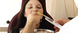
The material for the study is obtained by inserting a sterile cotton swab into each nostril to obtain a scraping of the nasal mucosa and applied to a glass slide.
A referral for research is issued and sent to the laboratory.
The collection of material for a rhinocytogram is carried out in the treatment room, or in the office of a doctor - otolaryngologist, immunologist - allergist.
Analysis time: 1 business day from delivery to the laboratory.
IMPORTANT!
The patient should avoid the use of nasal drops, sprays containing corticosteroids and antihistamines (anti-allergic) drugs for 24 hours before the test.
The interpretation of the obtained rhinocytogram results is carried out by the attending physician, comparing data from the anamnesis, symptoms and other types of studies (general blood count, total immunoglobulin IgE, rhinoscopy).
A significant increase in the content of eosinophils (more than 10% of the total number of leukocytes) indicates an allergic origin of the runny nose.
Timely diagnosed allergic rhinitis can quickly alleviate the patient’s condition, significantly reduce the severity of symptoms and the likelihood of relapse.
Elevated levels of neutrophils and lymphocytes are not specific for infection.
The absence of eosinophils, neutrophils and other types of leukocytes in the smear may indicate vasomotor rhinitis.
Nasal swab for eosinophils (rhinocytogram)
Examination of nasal discharge under a microscope is called a rhinocytogram. Its implementation makes it possible to determine the nature of the changes and conduct a differentiated diagnosis between allergic, infectious and other types of rhinitis (inflammation of the nasal mucosa).
The walls of the human nasal cavity are covered with mucous membranes that contain a specific secretion (liquid secreted by organs or tissues that contains biologically active substances). Its task is to neutralize the action of germs and dust that enter the nose. The ability to cope with them is due to the presence of ciliated epithelium with oscillating cilia, which, due to the movements performed, move mucus containing microbes and dust.
Normally, the nasal cavity is home to a large number of different microorganisms, including some varieties of streptococci, staphylococci, etc. Their presence does not create health problems if a person’s immune defense is in order. With a decrease in local protection, activation of conditionally pathological microflora occurs, resulting in the development of acute rhinitis - a disorder of nasal function, which is accompanied by inflammatory changes in the mucous membrane and the appearance of a runny nose.
One of the reasons for a decrease in local immunity and the appearance of rhinitis can be hypothermia of the body. Also, a runny nose can be caused by a slowdown in the movement of the ciliated epithelium.
The action of microbes activates the resistance of the body's immune system. This is manifested by an increase in the number of white blood cells - leukocytes - in the nasal mucosa. There are a number of types of white blood cells that perform different functions. For example, neutrophils are the main defenders during bacterial infections, lymphocytes - during viral diseases.
An allergic reaction of the body is caused by exposure to a certain substance - an allergen, which can be many of the most common components of the environment, for example, the fur or feathers of pets, dust, grass and tree pollen, etc. Increased sensitivity of the immune system to the allergen causes release of certain substances in the mucous membrane that cause the clinical manifestation of allergies. Such substances include, for example, histamine, bradykinin. An important factor in this process is the presence of one of the types of cells of the immune system - eosinophils. The development of an allergic reaction is accompanied by the accumulation of a large number of these cells in the blood, as well as in the nasal mucus.
Another type of rhinitis is vasomotor (neurovegetative), the causes of which can be:
- use of certain medications;
- hypothermia;
- other psycho-emotional or physical factors.
This type of rhinitis is characterized by acute swelling of the mucous membrane and changes in the tone of the vessels of the nasal cavity.
All forms of rhinitis have one common feature - the development of pathology is accompanied by the formation and secretion of a large amount of fluid, which is colloquially called a runny nose.
Allergic rhinitis is a very common pathology (according to some studies, symptoms of the disease are observed in approximately 38% of the population of the Russian Federation, allergic rhinitis has been identified in almost 40% of children). However, many cases of the disease remain undiagnosed. The purpose of a rhinocytogram is to identify eosinophils in the examined smear, the appearance of which may be a sign of the development of allergic rhinitis. Their presence in the material is determined by staining the smear using the Romanovsky-Giemsa method, after which they acquire a red color. This allows you to determine their quantity under a microscope.
Treatment methods for infectious and allergic rhinitis are fundamentally different, so in order to prescribe adequate treatment it is very important to identify the true cause of its development.
Rhinocytogram
A rhinocytogram is a comprehensive analysis of the microflora of the nasal sinuses. The test involves taking a nasal swab, which is most often prescribed to patients suffering from infectious diseases of the upper respiratory tract (especially if the disease does not respond to classical treatment or constant relapses occur).
Rhinocytogram refers to microbiological studies. This research method involves assessing the number of cells such as:
- lymphocytes;
- eosinophils;
- red blood cells;
- neutrophils;
- epithelium;
- macrophages;
- yeast mushrooms;
- microflora.
A rhinocytogram is an analysis of a smear taken from the nasal mucosa. The procedure is prescribed for adults and children. Often, such an analysis is prescribed for people who have a long-term persistent runny nose or frequent relapses of respiratory tract infections. High-risk groups include: children; people with weakened immune systems; have undergone organ transplantation; people with diabetes. This study is mandatory if the patient has one or several complaints, namely:
- inflammatory processes of the nasal mucosa;
- impaired nasal breathing;
- excessive nasal discharge;
- itching and sneezing;
- increased rhinitis.
This is a fairly simple analysis that does not cause any unpleasant sensations. Many patients wonder how to do a rhinocytogram and how to prepare for the study. To obtain accurate results, you must follow certain rules. Two days before the test, you need to stop using various ointments inside and outside the nose. It is prohibited to use drops and sprays containing antibacterial components and corticosteroids. 5 days before sampling, you must stop taking antibiotics. There is no need to rinse your sinuses the day before taking a smear.
A rhinocytogram in a child and an adult is performed at any time, but it is best to do this in the initial stages of the disease. The analysis is carried out in a fairly simple way. The procedure is completely painless, and even children do not need to be afraid of it. To collect material, the patient tilts his head back slightly, and the doctor or nurse uses a cotton swab to take the required amount of material for the study. The exact same procedure must be done with the other nostril.
A qualified doctor must describe the result obtained, since he knows how to decipher the analysis (rhinocytogram). The nasal mucosa consists of several cells, each of which performs a specific function. If bacteria or viruses enter the mucous membrane, the level of leukocytes increases to combat them. These cells are involved in the formation of immunity. An increase in leukocytes indicates the presence of infection in the body, and an excess of one of the leukocyte fractions indicates the cause of the disease. When allergens come into contact with the surface of the nasal mucosa, an allergic reaction occurs. It is caused by the formation of eosinophils on the surface of the mucosa. In addition, during the study, epithelium and bacteria can be detected.
When conducting research, it is imperative to know what the normal rhinocytogram is in children. Decoding allows you to determine the normal indicators of the components of discharge from the nasal sinuses for adults and children.
- Eosinophils
- Neutrophils (rods)
- Neutrophils (segment.)
- Lymphocytes
- Macrophages
The content of these cells is not constant; it can fluctuate throughout the day. In the morning or evening, this figure can decrease by about 20%, and at night it can increase by 30%. When deciphering a rhinocytogram, the norm for eosinophils in children under 4 years of age should be 0-7, and in children over 4 years of age, the indicators for adults are taken into account. The rest of the test indicators depend on the age of the child, so the doctor must interpret the results.
To determine the main cause of a prolonged, persistent runny nose, the quantitative ratio between cells is calculated during the study. When analyzing a rhinocytogram (deciphering), normally in children there should be up to several units of leukocytes in the field of view. During the course of the infectious process and inflammation, their number increases sharply. Normally, neutrophils should be 1-3%. In the presence of a bacterial type of runny nose, frontal sinusitis or sinusitis, their number increases sharply, in proportion to the intensity and complexity of the inflammatory process. This manifests itself in the appearance of thick yellow or green nasal discharge. Initially, a viral infection can be complicated by a bacterial one, which is why the number of neutrophils can increase simultaneously with lymphocytes. A sharp increase in the level of lymphocytes may indicate the presence of influenza, parainfluenza, adenoviral or any other viral infection. In addition, their number may increase with chronic runny nose. The presence of more than 10% eosinophils in the tests indicates the presence of hay fever, or allergic rhinitis, in the body. A significant amount of epithelium indicates an inflammatory process occurring in the body. When deciphering a rhinocytogram, children should not normally have red blood cells. If they are present, we can talk about the flu, since red blood cells in this case penetrate through thinned blood vessels into the nasal cavity. The microflora is normally negative, but if it is present, then the type of infectious agent is determined. Pathogenic microorganisms can occur with bacterial rhinitis or viral rhinitis. If the rhinocytogram is normal in children, and the runny nose does not go away for a long time, then this may be a sign of: vasomotor runny nose; drug-induced rhinitis; another type of rhinitis. Vasomotor rhinitis can occur as a result of vascular dysfunction. It is observed mainly under stress and under the influence of cold air. Rhinitis medicamentosa can begin as a result of prolonged use of vasoconstrictor drugs. In addition, a runny nose can occur as a result of disruption of the endocrine or nervous systems. All these disorders require additional research and comprehensive treatment.
Shapilova Natalya Vasilievna
Calling a laboratory technician to your home and taking blood at home
You don’t have to get up early and go somewhere to get tested. Especially if you have a small child, and, especially, a sick child. It is enough to call the laboratory team of Virilis Group of Companies by phone and experienced laboratory assistants will come to your home at a time agreed with you.
In this case, the child will not experience severe stress, as when taking tests in the clinic, and will not be at risk of becoming infected when interacting with other children in the clinic.
Laboratory diagnostics specialists at Virilis Group have undergone special psychological training and know how to take tests so that the child does not feel discomfort. We do all tests without pain or tears.
Which doctor prescribes a nasal swab and in what cases?
Pediatrician, general practitioner, therapist, ENT doctor, allergist, immunologist, endocrinologist, infectious disease specialist, as well as doctors of other specialties.
What is the purpose of a rhinocytogram?
- To find out the causes of chronic rhinitis and rhinosinusitis.
- Monitoring the condition of patients with chronic ENT diseases.
- Monitoring the effectiveness of treatment.
When is the study ordered?
With a long runny nose (several weeks or more) that is difficult to treat.
How is biological material collected for research?
To take biomaterial, the child is asked to blow his nose into wax paper. Mucus is extracted from wax paper using an applicator stick and spread onto two glass slides without smearing to avoid damaging the cells. In newborns or very young children or when there is a small amount of mucous discharge, a small nasopharyngeal swab is used to collect material. It is carefully inserted as far as possible into the nasal passage and scrolls around its axis. An imprint is made on a glass slide from the swab.
To increase the reliability of the results, it is recommended to repeat the examination after 1-2 weeks.