Aging of the body is an irreversible process, and it is very difficult to combat it. Age-related changes often primarily affect the musculoskeletal system. Statistics show that about 10% of the population, whose age has exceeded 40 years, suffers from arthrosis of the ankle joint.
Arthrosis of the ankle joint is a disease that can not only greatly reduce the patient’s quality of life, but also lead to disability!
Only timely diagnosis and treatment can protect against loss of ability to work.
What kind of disease is this?
The ankle joint is a rather complex joint in the human body. Participating in its education are:
- talus, femur and tibia;
- several articular ligaments;
- lateral and medial malleolus.
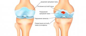
On the left is a schematic representation of the structure of the normal ankle joint,
On the right is destruction of the joint due to arthrosis.
Thanks to the correct functioning of this joint, a person has the opportunity to make many movements in the foot, soften the impulse from the plantar part of the legs when jumping, walking, and running. Also, this joint largely provides maneuverability of movements.
Like any joint in the human body, the ankle joint is equipped with cartilage layers. They reduce friction and provide shock-absorbing function. If an inflammatory process develops in the cartilage, accompanied by gradual degeneration and destruction of tissue, they speak of the development of arthrosis of the ankle joint.
The gradual thinning of cartilage tissue increases the load on the bones. Over time, when cartilage ceases to protect bone tissue, deformation of the joint affected by the disease begins.
Blood test for arthrosis
Since arthrosis has similar symptoms to other joint diseases, to distinguish it, for example, from infectious or rheumatoid arthritis, the following is prescribed:
- Clinical blood test. As a rule, arthrosis does not cause serious changes in blood counts, with the exception of a slight increase in ESR - up to a maximum of 25. With arthritis, ESR increases much more intensely - up to 40-80 units.
- Blood chemistry. The material is taken strictly on an empty stomach from a vein. With arthrosis, the indicators remain normal, but with arthritis, specific markers of inflammation appear - C-reactive protein, certain immunoglobulins, etc.
A blood test is needed to differentiate arthrosis from arthritis
Reasons for development
Arthrosis of the ankle joint is a disease that not all people experience during their lifetime. Doctors identify a number of factors that contribute to the development of pathology. The main roles in the gradual destruction of the ankle joint are played by 5 main factors.
- High loads
First of all, physical activity has an impact on the joint. If it exceeds the joint's ability to adapt, articular cartilage begins to gradually deteriorate. That is why athletes and people suffering from obesity most often suffer from the disease.
- Defect in juxtaposition of articular surfaces
If the articular surfaces involved in the formation of the ankle joint are not aligned correctly, this leads to the development of the disease. It is explained primarily by the uneven distribution of the load. Various injuries to the ankle area, arthritis, diabetes and some other diseases contribute to the formation of defects.
- Shoes
Women are primarily at risk. High-heeled shoes have a negative impact on the joint. However, men can also be affected by this factor if they wear uncomfortable shoes for a long time.
- It has been proven that hereditary predisposition also plays a role in the development of the disease.
- Fractures of the ankle joint.
Physiotherapy
In complex treatment, the patient should pay attention to performing active movements, after consulting with a physical therapy specialist. A set of exercises for deforming arthrosis includes:
- Rotate the foot alternately in one direction, then in the other. This exercise helps improve the elasticity of the ligaments.
- While lying on your back, turn your feet towards you, then away from you.
- While lying down, bend and straighten your knees. When extending, your legs should be parallel to the floor.
- Starting position: sitting on a chair. Make movements with your feet, imitating walking.
Exercises should be performed constantly, for a long time, gradually increasing the load. The purpose of such exercises is to prevent the development of muscle atrophy, strengthen ligaments and restore mobility in the joint. Physical activity when performing exercises on the ankle joint should be safe, dosed, but effective. Its volume is determined by the doctor individually based on the stage of the disease.
Types and degrees of pathology
Arthrosis of the ankle joint is a disease that does not develop immediately. The formation of the disease occurs gradually, in stages. There are 4 stages of the process in total.
- First stage . The doctor, conducting an objective examination, cannot detect any objective signs of changes in the joint. There are no changes on the radiographs.
- Second stage . Its development is most often triggered by trauma. The ankle area swells, movements become limited, and deformity is observed. The pain may gradually intensify. On an x-ray, the doctor will notice that the size of the joint space has significantly decreased. If we consider the lateral projections, we can note that the articular surface has become longer (bone growths), and the block of the talus has become flatter.
- Third stage . The third stage of the process is characterized by a clear change in the shape of the joint, which can be determined even without the help of radiographic examination. An atrophic change in the lower leg is noted. Movement is severely limited. The patient prefers to avoid movements in the damaged area.
- Fourth stage If treatment of the disease is not started on time, the process reaches a stage characterized by extreme narrowing and sometimes complete disappearance of the joint space. The bone contour can be deformed due to extensive growths. Subluxation is often observed.
Additionally, it is customary to divide the degenerative process in cartilage into primary and secondary. In the first case, initially intact tissues are damaged for some reason. In the second case, the joint is already subject to serious degenerative processes, and arthrosis only aggravates them. Some doctors understand secondary arthrosis as a post-traumatic form of the disease.
Symptoms of the disease
Ankle damage is considered quite insidious. In the early stages of the disease, symptoms may be completely absent. Naturally, a person who has no complaints does not go to the doctor. A patient visits the hospital for the first time only when signs of pathology begin to significantly reduce his quality of life.
Typical symptoms of the disease include:
- the onset of pain if the joint is forced to cope with significant loads;
- the appearance of sounds such as clicks, crunching, creaking when moving;
- gradual atrophy of muscles located in close proximity to the affected area;
- pain that occurs in the morning;
- rapid fatigue of the limb during exercise, including sometimes even simple walking;
- swelling of the affected area, sometimes a local increase in temperature;
- restriction of movement, which progresses over time to the point that the patient cannot move the foot;
- violation of the axis of the leg.
At the last stage of the disease, subluxations often occur. They are explained by the fact that the supporting function of muscles and tendons is disrupted, and large osteophytes appear.
Diagnostics
It is impossible to make a correct diagnosis at home without a doctor, so it is very important to choose a clinic where you can conduct a full examination:
- Laboratory diagnostics – blood tests that detect inflammatory, metabolic, and autoimmune disorders.
- Instrumental research methods:
- X-ray of the affected joint;
- magnetic resonance imaging (MRI) – if necessary to clarify the diagnosis in the initial stages of the disease;
- Ultrasound – to identify disorders in the surrounding soft tissues;
- arthroscopy with tissue biopsy – if necessary to clarify the nature of the pathological process.
Diagnosis methods
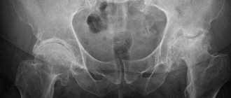
If you suspect the development of arthrosis of the ankle joint, it is recommended to immediately visit an orthopedic traumatologist. The doctor will conduct an examination and give recommendations on which diagnostic techniques are best to use. Radiography plays the most important role in diagnosing the disease. Using images, you can assess the condition of the joint axis and detect the bones and cartilage involved in the process. It is recommended to take the photo with a load on the leg. Radiography additionally makes it possible to understand whether neighboring joints are involved and, if so, how severe the process is. In some cases, based on radiography, the cause of the pathology can be assumed.
Additionally, computed tomography and MRI can be used.
A
b
a. on radiographs arthrosis of the ankle joint, b. CT scan of the ankle shows necrosis of the talus.
Basics of conservative treatment
First of all, it is important to eliminate the provoking factors, otherwise the treatment will not be effective. For some it’s excess weight, for others it’s high heels or standing for long periods of time. It will never be a bad idea to switch from flour, salted and smoked foods to fiber, fresh fruits and fermented milk products in order to stabilize the metabolism.
After such recommendations, the patient is prescribed medication according to indications:
- non-steroidal anti-inflammatory drugs and painkillers (tablets, ointments, injections);
- chondroprotectors containing glucosamine sulfate and chondroitin sulfate to give the cartilage layer density and elasticity (taken for a long time - more than six months).
Complex treatment may include:
- physiotherapy (regular procedures stop destruction in the joints);
- therapeutic massage - to strengthen the muscles of the foot and lower leg, as well as improve blood circulation;
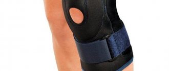
- wearing orthopedic shoes that reduce the load on the joint;
- hiking and swimming;
- sanatorium-resort treatment in specialized health resorts.
Best way to restore ankle mobility - swimming and walking
Principles of therapy
Treatment of ankle arthrosis should be carried out under the supervision of an experienced doctor, and it is best if the doctor guides his patient from the moment of diagnosis until the end of the course of treatment. If the doctor observes the patient from the very beginning and controls the entire treatment process, it will be possible to achieve significant results in therapy.
Dr. Petrosyan masters all approaches to the treatment of ankle arthrosis, using both conservative and surgical techniques in his practice.
Conservative treatment
Conservative therapy involves the use of medications, physiotherapy, physical therapy, massage and other techniques that are aimed at slowing down or completely stopping pathological changes in the joint.
First of all, drug therapy is selected for the patient, taking into account the patient’s personal characteristics and individual contraindications. The following groups of drugs are used for conservative therapy:
- NSAIDs designed to quickly relieve inflammatory and pain syndromes (for example, Ibuprofen, Naproxen, Diclofenac, etc.);
- chondroprotectors designed to protect the joint from further destruction (Aflutop, Artra, Terfalex, etc.);
- vasoprotectors, providing strengthening of vascular walls, local improvement of blood flow (Detralex, Trental, etc.);
- ointments with an analgesic effect (Deep Relief, Butadion, etc.).
The doctor will also select physiotherapeutic treatment for the patient, which not only has an analgesic effect, but also helps to dilate blood vessels and improve blood flow. But good blood flow improves tissue nutrition and promotes their restoration!
Conservative therapy will not do without nutritional correction. It is necessary to introduce more protein products into the diet, which contribute to the construction of new tissue. It is also worth adjusting the balance of micro- and macroelements and vitamins in food. A set of physical therapy exercises effectively complements therapy.
Surgical treatment
Unfortunately, a doctor can treat ankle arthrosis conservatively only if the disease has not reached the third stage. Later, when the joint is already destroyed, only surgery can help the patient. Three types of surgery are possible:
- Arthrodesis. In this case, the doctor, having removed the destroyed tissue, fixes the joint with various structures. The result is complete immobility in the joint, restoration of leg support, and relief from pain.
- Arthroplasty. During this procedure, the doctor completely preserves the joint, eliminating pathological changes in it. This is not a radical method and is rarely used today.
- Endoprosthetics. The operation is considered the most difficult and most progressive. During it, the patient’s own articulation (articular surfaces of the bones) is removed, and an implant made of artificial material is installed in its place.
Dr. Petrosyan has extensive experience in performing all three operations, and the clinic’s capabilities allow us to perform any of the interventions for which the patient has an indication. On the basis of the same hospital it is possible to undergo full preoperative preparation.
You can find out more detailed information in the ankle joint replacement section..
Synovial fluid examination
To perform the analysis, a joint puncture is performed. The main parameters of synovial fluid are subject to study:
- macroscopic indicators - color, volume, viscosity, turbidity and mucin clot;
- number of cells;
- cytology of the stained specimen;
- microscopy of the native preparation.
Normally, synovial fluid has a straw-yellow color and a transparent consistency. With arthrosis, its viscosity increases, the mucin clot is formed well, the number of cells is normal or slightly increased (maximum up to 5000/μl). Other indicators remain mostly normal. With reactive synovitis, the number of neutrophils is reduced by half.
A serious change is observed during inflammatory processes accompanying various forms of arthritis. One way or another, the interpretation of the results is carried out only by an experienced rheumatologist, taking into account the medical history, laboratory and instrumental studies.
Analysis of synovial fluid makes it possible to clearly distinguish arthrosis from arthritis
