Keratitis is an inflammatory disease that is localized in the cornea of the eye. This disease is characterized by unpleasant symptoms, but thanks to the achievements of modern medicine, it is highly treatable; the main thing is to seek qualified medical help in time.
The cornea is the most important part of the so-called optical system of the eye, responsible for visual effects. The cornea is the front transparent layer of the eye. The clarity of vision and its acuity largely depend on the condition of the cornea. There is a set of parameters of the cornea that ophthalmologists measure: surface curvature, structure, transparency, sphericity and others. Due to the inflammation that occurs with keratitis, changes may occur in the cornea. These changes cannot always be corrected by treatment - they can be irreversible. Therefore, it is important to consult a doctor in time and start therapy as early as possible so as not to worsen the problem.
At CELT you can get advice from an ophthalmologist.
- Initial consultation – 3,900
- Repeated consultation – 2,000
Make an appointment
Risk factors
The causes of keratitis are known, and there are also risk factors that significantly increase the likelihood of developing the disease.
Immediate causes include:
- Infections, parasitic infestations.
- Mechanical injuries, chemical, thermal damage.
- Allergy.
These causes lead to the occurrence of infectious (for example, herpetic), traumatic or allergic keratitis, respectively.
Among the risk factors for developing the disease, the following are most important:
- Presence of autoimmune diseases.
- Long-term wearing of contact lenses.
- Dry eye syndrome.
- Lack of vitamins.
- Various metabolic disorders.
- The presence of some systemic diseases: diabetes, gout, rheumatism.
Based on the list of risk factors, it is clear that keratitis is a fairly common pathology.
Causes of keratitis formation
External factors provoking the appearance of the disease:
- viruses (adenoviral infection, herpes simplex, parainfluenza, chicken pox, measles);
- fungi (aspergillus, candida, molds, fusaria);
- bacteria (pneumococci, staphylococci, streptococci);
- protozoan microorganisms (Toxoplasma, Leishmania, Acanthamoeba);
- the presence of an inflammatory focus in adjacent organs (ENT infections, dental problems).
Internal factors:
- allergic diseases (drug allergies, spring catarrh);
- neurological conditions (inflammation of the trigeminal nerve);
- chronic infectious pathologies (syphilis, tuberculosis);
- diseases associated with metabolic disorders (diabetes mellitus, rosacea, psoriasis);
- autoimmune diseases (polyarthritis nodosa, rheumatoid arthritis);
- mechanical thermal and radiation injuries to the eyes;
- glaucoma, chronic conjunctivitis, inflammation of the lacrimal sac, blepharitis;
- dystrophic pathologies of the cornea;
- incorrect wearing of lenses.
Complications and consequences of keratitis
Inflammation of the cornea of the eyes leads to clouding - this is a symptom that an ophthalmologist will immediately pay attention to during the initial examination. Cloudiness occurs due to the accumulation of leukocytes, lymphocytes and other components of the immune system in the tissues of the cornea. This is a natural response to inflammation. The accumulation of cellular elements has a special name - infiltrate. There may be one or there may be many. It can be located closer to the surface or in the depths (stroma) of the cornea - then it is called stromal.
Depending on the location and size of the infiltrate, keratitis can either go away on its own or develop into complications. Superficial infiltration may well resolve without a trace or with minimal consequences. But a deep lesion will result in scars on the cornea. They, in turn, lead to permanent visual impairment.
The clinical picture of this disease is also characterized by another unpleasant manifestation – vascularization of the cornea. This term refers to the ingrowth of blood vessels into any tissue. Vessels are needed for improved nutrition and accelerated resorption of the infiltrate. This is a protective reaction to inflammation. But it can also have adverse consequences - the transparency of the cornea, which is normally completely devoid of blood vessels, decreases. In the long term - problems with vision.
Viral keratitis
Keratitis caused by viruses is often accompanied by a blistering rash. The pathogenic agent can be almost any aggressive virus (measles, chickenpox, adenoviruses), but most often it is the herpes virus. As is known, over 95% of the population is infected with herpes infection, and in most cases, herpes occurs latently, in an asymptomatic form, and is activated only when immune resources are weakened, general exhaustion of the body and other unfavorable conditions. In this case, inflammation of the cornea is usually preceded by typical herpetic symptoms - rashes on the lips or other mucous membranes. With herpetic keratitis, as a rule, swelling and the appearance of fuzzy, vague infiltrates predominate.
Clinical picture
The clinical picture of keratitis is a series of manifestations specific to this disease, which have received a special name - corneal syndrome.
It includes symptoms:
- increased lacrimation;
- photophobia;
- narrowing of the palpebral fissure, it is impossible to open the eye completely;
- eye pain;
- sensation of a foreign object in the eye;
- redness of the eye.
In severe cases, the inflammatory process spreads to other parts of the eye and affects the sclera and iris. Another possible complication is ulceration at the site of inflammation. It can lead to perforation, in which infection enters the deep structures of the eye.
General information
The cornea is a relatively dense, transparent layer that protects the front, exposed surface of the eyeball. Both the shape of the cornea (it should be as flat, smooth, spherical as possible) and its transparency are of key importance, since any optical interference - turbidity, inhomogeneous inclusions, scars, etc. - distorts the normal refraction of light and/or reduces its intensity on the way to the retina inevitably affects the quality of the final visual image. Unlike some other inflammatory processes of the eye (blepharitis, conjunctivitis), which may not significantly affect visual acuity, with keratitis there is almost always a decrease in visual function. That is why it is extremely important to begin treatment as soon as possible, before irreversible scar changes form on the main optical axis of the eye.
Classification of keratitis
Depending on certain parameters of the disease, there are a number of classifications. So, based on the layer of the cornea that is affected by the pathology, it is customary to distinguish:
- Superficial keratitis - affects the upper layer, develops as a complication after conjunctivitis or dacryocystitis, is not accompanied by scars on the cornea;
- Deep keratitis - affects the internal layers, accompanied by scars on the cornea, which negatively affect visual acuity.
The table below shows the classification of keratitis depending on its etiology.
| Type of keratitis | What caused | Distinctive features |
| Spring (keratoconjunctivitis) | Intense irritation by allergens in spring |
|
| Bacterial | Bacteria Staphylococcus aureus or Pseudomonas aeruginosa | May develop due to injury or wearing contact lenses, characterized by:
|
| Amebic | Acanthamoeba amoebas that have penetrated the cornea of the eye | Most often develops in those who wear contact lenses, characterized by:
|
| Fungal | Parasitic fungi |
|
| Viral | Viruses, 70% - herpes virus |
|
| Purulent ulcer | Bacteria (penetrate the cornea due to constant irritation and microtrauma) |
|
| Neurogenic | Trigeminal nerve damage |
|
| Onchocerciasis | Prolonged irritation by allergen |
|
| Photokeratitis | Burn of the cornea and conjunctiva by UV rays due to prolonged exposure to the sun or use of a welding machine |
|
| Creeping corneal ulcer | Shallow corneal injuries due to small foreign bodies |
|
| Non-ulcerative | Gram-negative bacteria usually associated with lens wear |
|
Symptoms and signs
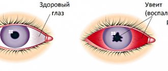
Almost all types and types of keratitis are characterized by the so-called. corneal syndrome, including increased lacrimation, increased sensitivity to light and uncontrolled closure of the eyelids (blepharospasm). As a rule, redness is pronounced, the vascular network becomes noticeable, and in severe cases, neovascularization (vascular neoplasms) occurs. The pain can be quite intense. The corneal layer swells, the surface loses its smoothness and often ulcerates (sometimes with a tendency to peel off), and infiltrates appear. Cloudiness of the cornea becomes one of the main symptoms and complaints with keratitis, since the opacity or translucency of the cornea has an extremely negative effect on the acuity and clarity of vision.
Keratitis is often accompanied by concomitant symptoms, the severity of which varies from mild to moderate discomfort (dry nasal and oral mucosa, headache, difficulty swallowing) to severe complications from the periodontium, gastrointestinal tract, etc. Various types of keratitis are characterized by their own specific features.
Keratitis: diagnosis
The effectiveness of treatment for keratitis directly depends on how correctly the diagnosis is made, since this disease is often mistaken for allergic conjunctivitis. Diagnostic methods include the following:
- A thorough history taking, identifying concomitant diseases and data on the presence of injuries;
- Carrying out an external examination of the eye area and eyeballs, palpation of certain areas (if possible);
- Examination of the fundus using special instruments to evaluate the retina, fundus vessels, and optic discs. With keratitis, accompanied by clouding of the cornea, a weakening of the fundus reflex is detected;
- Biomicroscopy - examination of ocular structures using a slit lamp, assessing their transparency, relief and depth of damage;
- Fluorescein test - as an additional diagnostic if ulceration is suspected;
- Microbiological laboratory tests - in the case of an infectious nature of the disease, aimed at identifying the causative agent.
Symptoms of keratitis
There are several characteristic symptoms by which doctors understand that treatment for keratitis is required. Here are the most typical ones:
- Pain in the eyes. This is the most common manifestation of the disease: a person feels pain, tingling, and there is a feeling that there is a foreign body in the eyes.
- Redness of the sclera. The eyes turn red and the cornea becomes dull.
- Photophobia. Bright light causes pain.
- Tearing.
- Involuntary closing of the eyelids (reflex blepharospasm).
- Reduced sensitivity of the cornea.
During examination, the ophthalmologist may notice traces of blood or lymph on the cornea. Due to these infiltrates, acute keratitis can cause tissue scarring: this will negatively affect vision and may lead to the need for surgery.
Keratitis: treatment
Treatment of keratitis is selected on an individual basis after conducting diagnostic studies and identifying the cause of the disease, the depth of the inflammatory processes and the severity of the pathology.
| Cause | Treatment |
| Bacterial infection | Taking antibiotics in the form of eye drops, ointments, injections into the tissue around the eyeball or under the conjunctiva of the eye. They prescribe Ofloxacin, Moxifloxacin, Levofloxacin, Oftaquix. |
| Viral infection | Taking antiviral drugs: Florenal, Acyclovir, Zovirax, others. |
| Fungal infection | Taking antifungal agents: Pimafucin, Amphotericin B. |
| Allergic reaction | Taking antihistamines: “Opatanol”, “Allergodil”. |
If the disease is accompanied by iridocyclitis, drops are additionally prescribed to dilate the pupils - mydriatics: Atropine, Cyclomed, Tropicamide and others. Their use eliminates the formation of posterior adhesions, which will eliminate a number of undesirable consequences for the patient.
For rapid healing of the cornea of the eye, restorative drops and eye ointments “Korneregel” or “Taurine” are prescribed. If opacities are detected in the patient, their resorption is stimulated by procedures such as phonophoresis and electrophoresis using proteolytic enzymes.
The prognosis depends on the form and volume of inflammation, the depth and how timely treatment is started. By contacting the ophthalmological service of the CELT clinic, you will receive high-quality treatment from attentive doctors and the highest chance of recovery.
What kind of disease is keratitis?
The cornea is the anterior avascular layer of the eye. Normally, it is a transparent, spherical, shiny and smooth tissue with high sensitivity. The cornea performs not only protective functions; it is the main refractive medium of the optical system of the eye. That is why her condition largely determines the sharpness and clarity of vision.
Eye keratitis is an inflammatory lesion of the cornea. Depending on the type of disease, it can be the result of a viral or bacterial infection, injury, allergic reaction, etc. The most common causes of inflammatory keratitis are the following:
- Damage to the eyes that leads to disruption of the integrity of the cornea (post-traumatic keratitis).
- Bacterial and fungal infections of the eyes. Because of them, ulcers or purulent infiltrates form on the surface of the cornea. The causative agents are strepto-, staphylo-, pneumococci, Escherichia coli, chlamydia and other bacteria. Among pathogenic fungi, the disease is most often provoked by aspergillus and candida.
- Conjunctivitis and its complications. The conjunctival cavity contains microflora, which, if the cornea is damaged, can cause an inflammatory process. If you touch your eyes with your hands during illness, there is a high risk of causing keratitis.
- Viral damage. The most popular causative agent of keratitis is herpes (60-70% of cases). It can also be caused by adenovirus, measles, smallpox and other viruses.
- Allergy.
- Ultraviolet radiation (photokeratitis).
- Vitamin deficiency (in particular, lack of vitamin A).
- Individual sensitivity to contact lenses or improper use of them.
- Some diseases (arthritis, Sjogren's disease, etc.).
This is what keratitis looks like in the photo.
Based on how deep the pathological process has penetrated into the tissue, superficial and deep keratitis are distinguished. The superficial one goes deep into no more than 1/3 of the cornea. Such keratitis responds well to treatment with drops and other methods, passes without consequences or leaves slight clouding of the cornea. Deep keratitis affects the entire cornea, requires complex therapy and can leave scarring that negatively affects vision.
Based on location, the lesion is divided into:
- central keratitis - damage to the cornea in the pupil area;
- paracentral keratitis - localized in the iris area;
- peripheral keratitis - affects areas outside the iris and pupils.
Keratitis: prevention
Prevention of this disease is quite simple and requires compliance with the following rules:
- Carrying out work in which the eyes may be injured, wearing special safety glasses;
- Strict adherence to hygiene rules when using contact lenses;
- Work with welding in a protective mask, avoiding prolonged exposure to the sun;
- Timely treatment of eye diseases such as: conjunctivitis, blepharitis, dacryocystitis;
- Timely treatment of common pathologies that stimulate the development of keratitis.
If you still notice the first signs of developing this disease, immediately seek professional medical help! Remember: the key to successful treatment is its timeliness!
Bacterial keratitis
Keratitis can be caused by a variety of pathogenic microorganisms, primarily cocci (Staphylococcus aureus, Streptococcus, Gonococcus) and Pseudomonas aeruginosa. A rare and very dangerous infection, even resulting in blindness, is acanthamoeba infection (named after the causative agent Acanthamoeba), which can exist, in particular, in the gap between the contact lens and the surface of the cornea. With the development of the so-called creeping corneal ulcers caused by gonococcal, tuberculosis, syphilitic and other bacterial infections, the risk of rapid and irreversible loss of vision is also very high.
Our services in ophthalmology:
The administration of CELT JSC regularly updates the price list posted on the clinic’s website. However, in order to avoid possible misunderstandings, we ask you to clarify the cost of services by phone: +7
| Service name | Price in rubles |
| Appointment with an ophthalmologist (primary) | 3 900 |
| Appointment with a surgical doctor (repeated, for complex programs) within 3 months after the initial one | 2 000 |
| Ultrasound scanning of the anterior segment of the eye | 1 000 |
| Removal of foreign bodies of the cornea, conjunctiva | 2 000 |
All services
Make an appointment through the application or by calling +7 +7 We work every day:
- Monday—Friday: 8.00—20.00
- Saturday: 8.00–18.00
- Sunday is a day off
The nearest metro and MCC stations to the clinic:
- Highway of Enthusiasts or Perovo
- Partisan
- Enthusiast Highway
Driving directions
Treatment of keratitis in adult patients
Treatment of the disease can be either conservative or surgical. The doctor determines how to treat keratitis. He may prescribe antibiotics, antihistamines, and antiviral drugs for oral administration. Locally - antibacterial, antiseptic, moisturizing drops, anti-inflammatory ointments and gels, agents that defeat the herpes virus. In addition, the specialist will select medications that promote regeneration.
If the process has gone too far, surgery may be indicated, which may involve excision of the affected area of the cornea, replacing it with a graft, and removing superficial scars and scars.
Prevention of keratitis of any etiology consists of maintaining immunity, timely treatment of general and eye diseases, eye hygiene and timely examinations by an ophthalmologist.
How is it diagnosed and treated?
For any eye injury, it is important to visit an ophthalmologist, even if nothing worries you. Early diagnosis of keratitis can prevent corneal clouding and preserve vision. During the examination, the doctor performs biomicroscopic diagnostics, which allows us to understand the extent and nature of tissue damage. Additionally, the following procedures can be performed: assessment of visual acuity, staining of the cornea with a fluorescein solution, algesimetry (testing the sensitivity of the cornea) and a number of other studies.
Treatment is carried out under the supervision of a physician and may include the use of eye drops and ointments with an antibacterial and disinfectant effect. Also, for keratitis, vitamin preparations are often prescribed, in particular B12.
MagazinLinz.ru team
Kinds
Keratitis in children and adults can be of the following types:
- Bacterial keratitis caused by pathogenic bacteria;
- Viral keratitis, in particular herpetic keratitis, caused by the herpes virus, is very common;
- Fungal keratitis is a very dangerous type, because. difficult to diagnose, occurs with constant relapses;
- Acanthamoeba keratitis - caused by the entry of protozoa - Acanthamoeba - into the eye;
- Allergic keratitis - develops with a pathological reaction of the immune system;
- Filamentous keratitis is a chronic process; against the background of reduced production of tear fluid, the cornea dries out, is easily injured and inflamed;
- Bullous keratitis – the appearance of blisters on the cornea due to a pronounced trophic disorder;
- Marginal keratitis is a lesion starting from the peripheral part of the cornea, which can lead to complete damage and the need for transplantation;
- Pigmentous keratitis is a chronic inflammation that leads to damage to corneal cells and their staining brown;
- Parenchymal keratitis is a chronic lesion of the middle parts of the cornea, which is most often caused by the causative agent of syphilis (treponema pallidum);
- Arborescent keratitis - the appearance on the cornea of a pattern resembling a tree (usually a consequence of herpetic keratitis);
- Ulcerative keratitis is the formation of a sufficiently deep epithelial defect on the cornea, which can lead to loss of vision;
- Pinpoint keratitis is an inflammatory lesion of the cornea, in which small ulcers like small dots appear on it.
Treatment
Therapy for the traumatic form of the disease depends on the subtype of pathology. If a foreign body gets in, you should immediately rinse with clean water and try to remove the foreign object; if unsuccessful, contact an ophthalmologist. For chemical burns, rinse eyes with running water for 15 minutes.
Treatment consists of the use of pain-relieving drops, antibiotic therapy and drugs that accelerate regeneration. When erosion occurs, medications are prescribed whose action is aimed at restoring trophic processes and epithelization.
The meibomian form is treated with antibiotics, antihistamines, massage, antiseptics and NSAIDs. Hormonal medications, eye wash solutions and drugs that restore the body's protective functions are prescribed. In case of ulceration, keratoplasty and laser coagulation are performed.
Causes
The main causes of the disease include:
- non-penetrating injury to the cornea;
- contact with the eye of chemicals - acids or alkalis that cause burns;
- contusional injury - blunt trauma with a non-sharp object (impact by a large object);
- entry of a foreign body;
- wearing contact lenses;
- eye diseases that increase the permeability of the cornea.
Post-traumatic keratitis is an inflammation of the cornea that can appear 4 days after damage to the visual organs during active treatment of injuries. It begins to develop due to the addition of a secondary infection.
Traumatic keratitis is predominantly the result of a work-related injury due to non-compliance with eye protection measures at work.