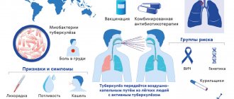
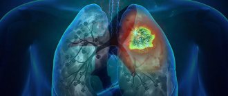
Tuberculosis is an infectious disease that occurs due to Koch bacilli entering the human body. The disease is dangerous because it affects the respiratory system. Less commonly susceptible to tuberculosis are the bones, skin, lymphatic, genitourinary, nervous, lymphatic systems, as well as other organs and systems.
The immune system (the body's natural defense against infection and disease) of most healthy people kills the bacteria without causing symptoms. Sometimes the immune system cannot kill bacteria, but can stop it from spreading in the body. In this case, no symptoms develop, but the bacteria remain in the body. This is called latent (hidden) tuberculosis.
Methods of transmission
- Airborne - through the sneezing, coughing of a patient with an open form of the disease, and even when drying out, the bacillus retains its pathogenicity.
- Alimentary – through the digestive tract. The infection enters the body due to poor hand hygiene or poorly washed and unprocessed food.
- Contact – the infection enters a person through the conjunctiva of the eyes, through kissing, sexual contact, through contact of contaminated objects with human blood, or the use of other people’s hygiene items.
Infiltrative TBC
This is a clinical form of the secondary tuberculosis process, which is characterized by the presence of inflammatory changes in the lung area. Most often, these changes are of an exudative nature with the presence of destruction of lung tissue and caseous necrosis in the middle. The infiltrate may have a round, cloud-like shadow, occupy a segment or lobe of the lung, and be located in interlobar fissures.
- Infiltrative tuberculosis accounts for up to 70% of all forms of tuberculosis of the respiratory system. It develops when a patient with a pre-existing infection is re-infected with mycobacterium, or it is a secondary outbreak and progression of focal tuberculosis.
- The size of the infiltrate varies from 2 cm to a lobe of the lung.
- During treatment, if the course is favorable, the infiltrate may resolve. If unfavorable, tuberculoma forms in its place or caseous pneumonia develops.
- 50% of cases of infiltrative tuberculosis begin acutely with general intoxication. In half of the sick, the disease does not show clear manifestations. Hemoptysis is a frequent accompaniment of this form of the disease.
During therapy, cough and hemoptysis, shortness of breath and chest pain first disappear, and then the temperature decreases. Weakness and sweating disappear. If the patient secretes Koch bacilli, this will stop within 3 months during therapy.
Types of tuberculosis
- Open form - the disease is clearly expressed, bacteria are easily detected in sputum and feces. The patient himself poses a danger to others, since the infection is transmitted by airborne droplets. Microbacteria can be found in sputum, urine, and feces.
- Closed form - not dangerous to others. Characterized by the difficulty of detecting infection in sputum. The most common tuberculosis is pulmonary tuberculosis, but this infection can also affect bones, joints, genitourinary system, intestines, peritoneum, meninges, central nervous system, peripheral lymph nodes, skin
Asymptomatic course of the disease
For a long time, pulmonary tuberculosis may be completely asymptomatic, or several mild symptoms may be present. Disseminated tuberculosis mainly manifests itself as a consequence of pneumonia, but most often, the disease is latent (latent form) and does not manifest itself. In this case, the disease can be detected by chance during standard annual examinations: X-rays, fluorography, Mantoux test.
The symptoms of a dangerous disease can often be confused with the usual symptoms of bronchitis, pneumonia or ARVI. In this case, the main sign that you should pay attention to is the presence of symptoms for a long time or the lack of improvement after treatment.
The first symptoms of tuberculosis
In the early stages, the disease is practically asymptomatic. As it develops, the patient's condition worsens, but no specific symptoms are observed. Increased fatigue, weakness, sudden weight loss for no apparent reason, a temperature of 37-38 ° C that does not subside for a long time, and night sweats appear. In children, the disease progresses faster than in adults.
The pulmonary form of tuberculosis is accompanied by a cough. Mild at first, it begins to progress over time. If your cough continues for more than three weeks, you should seek medical help immediately. The cough is initially dry, paroxysmal, especially at night and in the morning. Later, yellow-green sputum begins to be released, and at the cavity stage, hemoptysis is observed.
In the form of tuberculosis that affects the brain and its membranes, in addition to symptoms of general intoxication, sleep disturbances and headaches are observed, the intensity of which gradually increases.
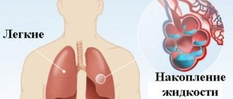
Pleurisy
Inflammation (primary or secondary) of the pleura surrounding the lungs can occur with the accumulation of fluid between them (exudative pleurisy). This option is the implementation of the allergic component of inflammation. It is also possible for tuberculous tubercles to form between the layers of the pleura and dry pleurisy to develop. In this case, the patient first addresses:
- complaints of shortness of breath
- stabbing pain in half of the chest
- dry cough
- weakness, fatigue
- temperature rises to 37.5-38.
Complications of pleurisy include purulent inflammation (pleural empyema), fusion of the pleural layers. Almost always, after pulmonary tuberculosis in the form of pleurisy, adhesions remain between the pleural layers.
How is tuberculosis diagnosed?
To diagnose the disease, you must first consult with your primary care physician, who, if tuberculosis is suspected, will refer you to a phthisiatrician (tuberculosis specialist) for further diagnosis and treatment. Diagnosis will depend on the type of disease.
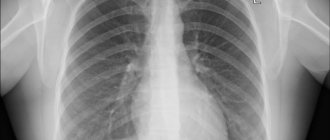
Diagnosis of pulmonary tuberculosis
An X-ray of the chest is required, which provides an image of the lungs. For pulmonary TB, the scan should show changes in the appearance of the lungs, such as scarring.
To confirm the diagnosis, samples of mucus and sputum are taken and analyzed under a microscope for the presence of tuberculosis bacteria.
Examination for extrapulmonary tuberculosis
- CT scan
- magnetic resonance imaging (MRI)
- ultrasound examination (ultrasound)
- blood tests
- Analysis of urine
- biopsy - a tissue sample is taken from the affected area to be tested for the presence of tuberculosis
Examination for latent tuberculosis
To prevent the disease, every person is required to undergo fluorography and an X-ray of the lungs at least once every two years.
Caseous pneumonia
Clinically, it resembles severe pneumonia with severe intoxication, pain in one of the halves of the chest, aggravated by breathing and coughing, an unproductive or dry cough, severe shortness of breath, indicating respiratory failure. This type of disease is the result of the spread of bacilli in the blood from the primary focus (caseous pneumonia is secondary). They may be complicated by a disseminated, infiltrative or fibrous-cavernous variant. Pneumonia is often complicated by bleeding from the lungs or pneumothorax.
How to treat tuberculosis?
Treatment of the pathology depends on its type, but most often a course of antibiotics is prescribed. Tuberculosis is a dangerous disease that requires immediate treatment. This allows the person to return to their normal lifestyle.
Antibacterial therapy is aimed at suppressing the proliferation of the tuberculosis pathogen.
Treatment takes place in 2 phases: in the first, several drugs are used at once to reduce the population of microbacteria, the second phase is maintenance therapy. Antibiotics stop the proliferation of bacteria and their release into the environment, the inflammatory process.
After taking such potent medications, a person needs additional supportive therapy that will strengthen the body and reduce the toxic effect. For this, immunostimulants (restore liver function), sorbents (remove toxic breakdown products of chemotherapy drugs) and vitamin complexes are prescribed.
After taking the drugs for two weeks, most people are no longer contagious and feel much better. However, it is very important to continue taking your medications as directed by your doctor and complete your entire course of antibiotics.
Survey plan
- Three-time (with a two-day break) examination of sputum for CD using microscopy. If the result is positive, consultation with a phthisiatrician and hospitalization.
- If the result is negative, a molecular genetic study of sputum is performed.
- Survey radiography of the chest organs.
- Diagnostic test with recombinant tuberculosis allergen.
- If the diagnosis is not confirmed or rejected, a spiral computed tomography is performed.
Other treatments for tuberculosis
Surgery
The goal of therapy is the elimination of tuberculosis foci in the lungs with ineffective treatment, elimination of the consequences of pulmonary tuberculosis, and elimination of organ damage. All this is necessary to prevent the recurrence of the disease and to exclude the occurrence of complications.
Indications for surgical intervention can be any form of respiratory tuberculosis, especially in the case of complications that threaten a person’s life.
Chemotherapy
used with an optimal combination of anti-tuberculosis drugs aimed at eliminating mycobacteria and suppressing their reproduction. The duration of such treatment can reach up to a year - it all depends on the form and stage of development of the pathology.
If chemotherapy is stopped early, exacerbation or complications of tuberculosis may occur. Therefore, it is important to follow all doctor's recommendations. And the doctor, for his part, must draw up a detailed treatment plan and make adjustments throughout the entire therapy.
A patient who is indicated for chemotherapy as a treatment for tuberculosis must be prepared for the negative consequences of such an aggressive method. Side effects on the action of medications often occur. There are 2 types of adverse reactions: toxic and allergic. Dysbacteriosis may also occur.
The doctor may prescribe outpatient treatment if the disease is detected at an early stage and there is no infection to others. In this case, you need to regularly visit your doctor and undergo diagnostics. Most often, the patient is transferred to outpatient treatment after observation in a hospital and undergoing an extensive course of therapy in a tuberculosis clinic. At this time, the patient is no longer contagious.
Manifestations of infection of the upper respiratory organs
Signs of infection may appear in different respiratory organs. To recognize the disease in time, you should know its symptoms. At the beginning of the development of tuberculosis, the patient develops dryness and soreness in the nose and oropharynx, and he cannot swallow food normally. In addition, there is severe shortness of breath and a hoarse cough.
After some time, severe chills appear, body temperature rises, and breathing becomes difficult. After another period, the voice changes and hemoptysis begins. At the same time, patients lose a lot of weight because they cannot eat properly due to pain.
Tuberculosis of the larynx is manifested by improper mobility of the vocal cords and leads to hoarseness, breathing problems, soreness, and changes in voice.
When the throat is affected, there is severe pain, a sensation of a foreign body in the throat, increased salivation, swelling, rashes and redness of the pharynx mucosa. If we are talking about nasal tuberculosis, then all the symptoms are very similar to ordinary sinusitis: itching, burning, crusts in the nose, frequent bleeding, congestion, pain, the presence of mucopurulent discharge.
Symptoms may be constant, come and go, or progress slowly (sluggish tuberculosis). Rapid progress is also common, when a person can die from the manifestations of the disease in a few weeks.
Incidence (per 100,000 people)
| Men | Women | |||||||||||||
| Age, years | 0-1 | 1-3 | 3-14 | 14-25 | 25-40 | 40-60 | 60 + | 0-1 | 1-3 | 3-14 | 14-25 | 25-40 | 40-60 | 60 + |
| Number of sick people | 12 | 13 | 19 | 90 | 90 | 90 | 90 | 12 | 13 | 19 | 90 | 90 | 90 | 90 |
Diagnostics
The following symptoms suggest abdominal tuberculosis: weight loss, fever, abdominal pain.
Even more suspicious is the presence of unclear formations or fluid in the abdominal cavity. Additional help can be obtained using: 1. X-ray examination of the intestine; 2. biopsy during surgery, laparoscopy of lymph nodes or peritoneum; 3. seeding of aspiration material obtained from the abdominal cavity.
Typically, the diagnosis of abdominal tuberculosis is made using clinical signs.
An anal fistula, or a leak formed in the anus, may be a complication of abdominal tuberculosis or its only objective sign. In countries with a high prevalence of tuberculosis, anal fistula is common. Anal fistula can also occur with ulcerative colitis and Crohn's disease.
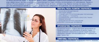
Prevention
Tuberculosis is one of the so-called social diseases, the occurrence of which is associated with the living conditions of the population.
The reasons for the epidemiological problem with tuberculosis in our country are the deterioration of socio-economic conditions, a decrease in the living standards of the population, an increase in the number of people without a fixed place of residence and occupation, and the intensification of migration processes. Men in all regions suffer from tuberculosis 3.2 times more often than women, while the growth rate of incidence in men is 2.5 times higher than in women. The most affected are persons aged 20 - 29 and 30 - 39 years.
The morbidity rate of contingents serving sentences in penal institutions of the Ministry of Internal Affairs of Russia is 42 times higher than the Russian average.
For the purpose of prevention, it is necessary to carry out the following measures: - carrying out preventive and anti-epidemic measures that are adequate to the current extremely unfavorable epidemiological situation regarding tuberculosis. — early identification of patients and allocation of funds for drug provision. This measure will also be able to reduce the incidence of illness among people who come into contact with sick people in outbreaks. — carrying out mandatory preliminary and periodic examinations upon entry to work on livestock farms affected by bovine tuberculosis. — increasing the allocated isolated living space for patients suffering from active tuberculosis and living in crowded apartments and dormitories. — more timely implementation (up to 30 days of life) of primary vaccination for newborn children.
Pathogenesis
Primary infection with Mycobacterium tuberculosis and latent course of tuberculosis infection.
Primary infection of humans with MBT usually occurs through the aerogenous route. Other routes of penetration - nutritional, contact and transplacental - are much less common.
The respiratory system is protected from the penetration of mycobacteria by mucociliary clearance (secretion of mucus by goblet cells of the respiratory tract, which glues incoming mycobacteria, and further elimination of mycobacteria using wave-like vibrations of the ciliated epithelium). Violation of mucociliary clearance during acute and chronic inflammation of the upper respiratory tract, trachea and large bronchi, as well as under the influence of toxic substances, makes it possible for mycobacteria to penetrate the bronchioles and alveoli, after which the likelihood of infection and tuberculosis increases significantly. The possibility of infection through the nutritional route is determined by the condition of the intestinal wall and its absorption function.
The causative agents of tuberculosis do not secrete any exotoxin that could stimulate phagocytosis. The possibilities for phagocytosis of mycobacteria at this stage are limited, so the presence of a small amount of the pathogen in the tissues does not appear immediately. Mycobacteria are outside the cells and multiply slowly, and the tissues retain their normal structure for some time. This condition is called "latent microbiism." Regardless of the initial localization, they enter the regional lymph nodes with the lymph flow, after which they spread lymphogenously throughout the body - primary (obligate) mycobacteremia occurs. Mycobacteria are retained in organs with the most developed microvasculature (lungs, lymph nodes, renal cortex, epiphyses and metaphyses of tubular bones, ampullar-fimbryonic sections of the fallopian tubes, uveal tract of the eye). Since the pathogen continues to multiply, and immunity has not yet formed, the population of the pathogen increases significantly. However, at the site of accumulation of a large number of mycobacteria, phagocytosis begins. First, the pathogens begin to phagocytose and destroy polynuclear leukocytes, but to no avail - they all die when they come into contact with the office due to their weak bactericidal potential.
Then macrophages are involved in the phagocytosis of MBT. However, MBT synthesize ATP-positive protons, sulfates and virulence factors (cord factors), as a result of which the function of macrophage lysosomes is disrupted. The formation of a phagolysosome becomes impossible, so the lysosomal enzymes of macrophages cannot act on the engulfed mycobacteria. MBTs are located intracellularly, continue to grow, multiply and increasingly damage the host cell. The macrophage gradually dies, and mycobacteria again enter the intercellular space. This process is called "incomplete phagocytosis".
Acquired cellular immunity Acquired cellular immunity is based on the effective interaction of macrophages and lymphocytes. Of particular importance is the contact of macrophages with T helper cells (CD4+) and T suppressor cells (CD8+). Macrophages that have absorbed MBT express mycobacterial antigens on their surface (in the form of peptides) and release interleukin-1 (IL-1) into the intercellular space, which activates T-lymphocytes (CD4+). In turn, T helper cells (CD4+) interact with macrophages and perceive information about the genetic structure of the pathogen. Sensitized T-lymphocytes (CD4+ and CD8+) secrete chemotaxins, gamma-interferon and interleukin-2 (IL-2), which activate the migration of macrophages towards the location of the office, increase the enzymatic and general bactericidal activity of macrophages. Activated macrophages intensively produce reactive oxygen species and hydrogen peroxide. This is the so-called oxygen explosion; it acts on the phagocytosed tuberculosis pathogen. With simultaneous exposure to L-arginine and tumor necrosis factor-alpha, nitric oxide NO is formed, which also has an antimicrobial effect. As a result of all these processes, the destructive effect of MBT on phagolysosomes is weakened, and the bacteria are destroyed by lysosomal enzymes. With an adequate immune response, each subsequent generation of macrophages becomes more and more immunocompetent. Mediators released by macrophages also activate B-lymphocytes, which are responsible for the synthesis of immunoglobulins, but their accumulation in the blood does not affect the body’s resistance to MBT. But the production of opsonizing antibodies by B lymphocytes, which envelop the mycobacteria and promote their adhesion, is useful for further phagocytosis.
An increase in the enzymatic activity of macrophages and their release of various mediators can lead to the appearance of delayed-type hypersensitivity cells (DSHT) to MBT antigens. Macrophages transform into epithelioid Langhans giant cells, which are involved in limiting the area of inflammation. An exudative-productive and productive tuberculous granuloma is formed, the formation of which indicates a good immune response to the infection and the body’s ability to localize mycobacterial aggression. At the height of the granulomatous reaction in the granuloma there are T-lymphocytes (prevail), B-lymphocytes, macrophages (carry out phagocytosis, perform affector and effector functions); macrophages gradually transform into epithelioid cells (carry out pinocytosis, synthesize hydrolytic enzymes). In the center of the granuloma, a small area of caseous necrosis may appear, which is formed from the bodies of macrophages that died upon contact with the office.
The PCI reaction appears 2-3 weeks after infection, and fairly pronounced cellular immunity is formed after 8 weeks. After this, the proliferation of mycobacteria slows down, their total number decreases, and the specific inflammatory reaction subsides. But complete elimination of the pathogen from the source of inflammation does not occur. Preserved MBTs are localized intracellularly (L-forms) and prevent the formation of phagolysosomes, therefore they are inaccessible to lysosomal enzymes. Such anti-tuberculosis immunity is called non-sterile. The MBT remaining in the body maintains the population of sensitized T-lymphocytes and provides a sufficient level of immunological activity. Thus, a person can retain MBT in his body for a long time and even throughout his life. When immunity is weakened, there is a threat of activation of the remaining MBT population and tuberculosis disease.
Acquired immunity to MBT decreases with AIDS, diabetes, peptic ulcers, alcohol abuse and long-term drug use, as well as with fasting, stressful situations, pregnancy, treatment with hormones or immunosuppressants.
In general, the risk of developing tuberculosis in a newly infected person is about 8% in the first 2 years after infection, gradually decreasing in subsequent years.
The emergence of clinically manifest tuberculosis In the case of insufficient activation of macrophages, phagocytosis is ineffective, the proliferation of MBT by macrophages is not controlled and therefore occurs in geometric progression. Phagocytic cells cannot cope with the amount of work and die en masse. At the same time, a large number of mediators and proteolytic enzymes enter the intercellular space, which damage adjacent tissues. A kind of “liquefaction” of tissues occurs, a special nutrient medium is formed that promotes the growth and reproduction of extracellularly located MBT.
A large MBT population upsets the balance in immune defense: the number of T-suppressor cells (CD8+) increases, the immunological activity of T-helper cells (CD4+) decreases. First, PCT to MBT antigens sharply increases and then weakens. The inflammatory reaction becomes widespread. The permeability of the vascular wall increases, plasma proteins, leukocytes and monocytes enter the tissue. Tuberculous granulomas are formed, in which caseous necrosis predominates. Infiltration of the outer layer with polynuclear leukocytes, macrophages and lymphoid cells increases. Individual granulomas merge, and the total volume of tuberculosis lesions increases. Primary infection transforms into clinically manifest tuberculosis.