Tuberculosis in Russia is so widespread that by the age of forty, 70-90% of the inhabitants of our country are infected with it. This does not mean that they are sick: after infection, most people's immunity copes with the bacteria. The probability of getting sick is on average 8% in the first two years after infection, then gradually decreases, and acquired cellular immunity is formed. Among adults, people who are weakened and live in poor conditions, with a lack of light and fresh air, are more likely to get sick. However, absolutely each of us can become infected. This is why it is so important to get regular TB screenings and have your children tested.
IMPORTANT! Information from the article cannot be used for self-diagnosis and self-medication! Only a doctor can prescribe the necessary examinations, establish a diagnosis and draw up a treatment plan during a consultation!
Why is BCG vaccination necessary?
WHO recommends mass vaccination of newborns against tuberculosis in all countries where the disease is common. Therefore, on days 3-5 after birth, while still in the maternity hospital, all newborns are given a free BCG vaccination. But this vaccine does not protect against tuberculosis infection. Her task is somewhat different. If an unvaccinated child under 2 years of age is infected with tuberculosis, he or she is extremely likely to develop tuberculous meningitis and generalized forms of tuberculosis, which very quickly lead to death. BCG quite reliably protects the child from these forms. It also protects children from pulmonary tuberculosis, but is less effective.
What other vaccinations do children under 2 years old need?
Mantoux test
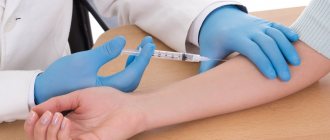
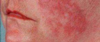
The Mantoux test is performed annually on all children under 7 years of age. This is not a vaccination (!), but an immunological test that shows whether the tuberculosis pathogen is in the body. In this case, tuberculin, a specific protein containing antigens of human and bovine tuberculosis, is injected intradermally. After 72 hours, the size of the papule is measured. The result of the Mantoux reaction can be affected by visiting a sauna, taking a long bath, swimming in a swimming pool, and scratching the injection site. Temporary contact with water does not in any way affect the results of the Mantoux test, therefore the opinion that Mantoux cannot be wetted is a myth.
Collection of material and preparation for research
To test urine for tuberculosis, a morning portion is used. It is necessary to toilet the external genitalia and not touch them to the reservoir for collecting material. The container must be sterile and dry. The collected biomaterial must be delivered to the laboratory as soon as possible, and must not be allowed to dry out or heat up. The day before the test, you need to give up foods such as blueberries, carrots, beets and other brightly colored vegetables and fruits. You should also limit your intake of diuretics, vitamins, and acetylsalicylic acid. Women should remember that urine tests are not taken during menstruation. A medium, intermediate portion of urine is given.
Blood donation occurs on an empty stomach, since food intake can distort the results of the study. It is worth consulting with your doctor about discontinuing certain medications. The day before you should not consume coffee, alcohol, or tobacco. Blood collection occurs under sterile conditions, in compliance with the rules for collecting material. Some studies require capillary blood, and some require venous blood.
Sputum is collected on an empty stomach, following the basic rules. The material must be coughed out so that mucus from the mouth or nasopharynx does not get into the container. A special spittoon made of dark glass with a tight lid is used. The sputum should not become chapped or dry out.
No special preparation is required for instrumental research methods. The exception is the use of a contrast agent. Renal function should be assessed before use. Most radiopaque substances are excreted by this organ, and the doctor must be sure that the excretory system can withstand this load.
Diaskintest test
It is carried out for children from 8 years old. Also a skin test. But if Mantoux uses antigens of both human and bovine tuberculosis, then Diaskintest uses only human tuberculosis antigens. Diaskintest is much more specific: a reaction to it occurs only if there are active tuberculosis bacteria in the body. Simply put, if the Mantoux test is positive, this does not mean anything: this test often gives false positive reactions. But after the Diaskintest, a reaction appeared - this is a more serious indicator.
The accuracy of Mantoux is 50-70%, Diaskintest - 90%.
Quantiferon test
Quantiferon test is one of the new methods for diagnosing tuberculosis using a blood test. It is based on the determination of interferon gamma in the blood, which is produced by cells in response to the introduction of the tuberculosis bacillus. Unlike Mantoux and Diaskintest, the Quantiferon test does not require the introduction of any antigens or bacteria into the body. Blood for research is taken from a vein. The result is ready in a few days and is much more accurate than skin tests. The test will only be positive if the person is truly sick.
Comparison of Mantoux and Diaskintest
Immunodiagnosis using Mantoux is carried out for children vaccinated against tuberculosis from 12 months of age until they reach the age of 18 years. An intradermal allergy test with tuberculin (Mantoux test) is performed once a year, regardless of the results of previous tests up to and including 7 years of age.
Diaskintest is performed once a year, regardless of the results of previous tests from 8 years of age to 17 years of age inclusive.
For those not vaccinated against tuberculosis, a Mantoux test is performed 2 times a year, starting from the age of 6 months.
Below is a comparative table of the Mantoux and Diaskintest tests.
| Mantoux | Diaskintest | |
| Place and method of drug administration | Forearm, intradermal | Forearm, intradermal |
| Compound | Proteins from killed mycobacteria extract | Recombinant protein obtained by genetic engineering |
| Indications | • Routine diagnosis of tuberculosis • Detection of the latent form of the disease • Disease risk assessment | • Identification of the causative agent of tuberculosis • Clarification of the diagnosis in case of doubtful Mantoux • Evaluation of treatment effectiveness |
| Contraindications | Eat | Eat |
| Side effects | Possible | Possible |
| Does it show a hidden form of the disease? | Yes | Yes |
| Is there a reaction to BCG? | Yes | No |
| Is there a reaction to other strains of mycobacteria? | Yes | No |
| Does it make it possible to determine the strength of immunity? | Yes | No |
| Test result | • Negative • Doubtful • Positive | • Negative • Doubtful • Positive |
| On what basis is the conclusion made? | Presence/absence, size of papules (seals, “bumps”) | Presence/absence, size of papules (seals, “bumps”) |
| Is it suitable for children | Yes (source: SanPin 3.1.2.3114-13 clause 5.1: “...from 12 months to 18 years”) | Yes (source: Order of the Ministry of Health of the Russian Federation No. 124n dated March 21, 2021: “use in children over 7 years of age and adolescents”, for ages under 7 years - according to indications) |
| Is it suitable for adults | According to indications | According to indications |
| Test frequency | For children – once a year annually. Adults - as needed | According to the doctor's indications |
| Risk of contracting tuberculosis | No | No |
If the result is positive or questionable, in both cases additional examination is required. This may be a laboratory test of blood, sputum or smear, chest x-ray, CT, MRI and other methods.
T.SPOT.TB (“tee-spot”)
This method of diagnosing tuberculosis by blood is similar to the quantiferon test. The difference is that the quantiferon test detects interferon gamma in the blood, produced in response to the introduction of the tuberculosis bacillus, and T-SPOT detects the T cells themselves, which produce interferon gamma in response to the presence of Mycobacterium tuberculosis. Both tests are equally sensitive and informative (up to 97%), they are more sensitive than Diaskintest and much more sensitive than the Mantoux reaction. “T-spot” practically eliminates false-positive and false-negative reactions, while the quantiferon test can still give a false-positive reaction if, for example, the person was sick with ARVI at the time of donating blood.
Modern methods of diagnosing tuberculosis
The following methods for diagnosing pathology are distinguished:
- bacteriological urine analysis;
- general urine analysis;
- biochemical blood test;
- T-SPOT.TB;
- microbiological diagnostic methods;
- study of the coagulation system;
- microscopic analysis of sputum;
- radiation diagnostic methods.
Go to analyzes
Let's look at these methods in more detail.
Bacteriological urine analysis
Indicated for extrapulmonary tuberculosis. This diagnostic method is extremely important, because tuberculosis of the genitourinary system often causes complications and does not cause symptoms for a long time. It is tuberculosis that can masquerade as an inflammatory process or urolithiasis for a long time. Therefore, long-term pathology of these organs, which is difficult to treat, is an indication for diagnosis. Also, a urine test is indicated for tuberculosis to exclude involvement of the genitourinary system in the process.
In order to determine the presence of a pathogen in the urine, bacterioscopy or culture on a medium is performed.
To detect mycobacteria, special staining is performed, in which the pathogen acquires a different shade, different from healthy cells.
Another method is to culture a urine sample onto a culture medium. If there is an increase in the culture of mycobacteria, this indicates the presence of a pathogen in the body.
General urine analysis
In a general urine test, characteristic changes in tuberculosis are observed. This is the appearance of drops of pus, the presence or traces of protein and a more acidic reaction. Modified leukocytes, bacteria, and red blood cells also appear.
Blood chemistry
At some stages of the disease, changes in biochemical blood tests are detected. With an inactive form, there are no changes in these tests, although they may appear with concomitant pathology.
The acute form of tuberculosis leads to a decrease in the albumin-globulin ratio. If the pathology leads to complications in the form of liver damage, the level of transaminases and different fractions of bilirubin increases. These indicators are included in the mandatory examination of a patient with tuberculosis. The patient takes this test over time to assess the condition.
Deterioration of kidney function may be reflected in an increase in creatinine and changes in glomerular filtration rate.
A biochemical blood test has no specific diagnostic value, but is an important component in assessing the quality of treatment and the patient’s condition.
Immunological diagnostic methods include the T-SPOT.TB technique
. The basis of the method is to study the reaction of T-lymphocytes. The procedure is highly sensitive and quite informative. False results are excluded even when other methods are insensitive to the result. The method is used in doubtful cases, for example, after vaccinations, in patients with autoimmune pathology, and in healthcare workers. This method allows you to quantitatively assess the presence of the pathogen, but does not provide information about the phase of the process.
This diagnosis occupies a special place in HIV carriers. The fact is that the virus infects lymphocytes. Despite this, diagnosis using the T-SPOT.TB method gives accurate results.
The high accuracy of the test is also explained by the fact that the test system is sensitive to the components of the pathogen, but specifically to those that are not found either in the BCG vaccine or in other microorganisms that have similar components. The essence of the method is the quantitative determination in the blood of effector T cells (CD4 and CD8) that produce IFN-ɣ (interferon gamma), which is produced in response to stimulation with specially selected antigens ESAT-6 and CFP10. We are talking about one of the fragments of the mycobacterium genome. It is noteworthy that high specificity is observed in both the latent and active phases.
No special preparation is required for the test; it is enough to limit your food intake two hours before the procedure. Venous blood is collected. This must be carried out under appropriate conditions, in compliance with the rules of asepsis and antiseptics.
A positive test result indicates that mycobacteria is present in the body, and a negative test result indicates the opposite.
Microbiological diagnostic methods
They are used for direct detection of the causative agent of tuberculosis in biological tissues of the body. Various techniques are used.
Ziehl-Neelsen staining
consists in the fact that a smear with the drug is treated with a specific dye. Mycobacteria acquire a characteristic hue, which indicates that a reaction has occurred. This way you can confirm or exclude the presence of a pathogen in the material. The method is economical and relatively fast. However, it is sensitive only when the concentration of microorganisms in the sample is high.
Fluorescence microscopy
gives higher resolution and improves coloring technique. The use of fluorochromes is required - specific substances that “illuminate” mycobacteria and make them more visible under a microscope.
Polymerase chain reaction
allows the reproduction of bacterial DNA from its fragments contained in tissues. This is a fast and informative diagnostic method that has high specificity and sensitivity.
Culture method
consists of growing a culture of mycobacteria. To do this, a fragment of biomaterial is taken and sown on a nutrient medium. Then the strain that grew on the medium is assessed and the diagnosis is confirmed or excluded. A specific medium is used that is more likely to grow the required medium. Grown colonies can be used to determine antibiotic sensitivity.
VASTES460
is a modern diagnostic method that uses a radiometric system and labeled CO2. The bacterium absorbs the indicator element and can be detected in the test material.
Study of the blood coagulation system
Coagulogram
often used in phthisiology, as patients with tuberculosis gradually develop hemoptysis or pulmonary hemorrhage. This leads to changes in hemostasis parameters. The analysis may change after surgical treatment of the disease.
Control of the coagulogram is based on the analysis of such indicators as aPTT, fibrinogen, bleeding time, coagulation time. These indicators can fluctuate in different directions, depending on the amount of blood loss. If it is insignificant, the coagulation system is stimulated and the indicators increase. The loss of a large volume of blood leads to a decrease in coagulation factors in the body and corresponding changes in the coagulogram.
Microscopic analysis of sputum
Sputum analysis
- mandatory testing for tuberculosis. With this pathology, sputum is released in small quantities, interspersed with blood, pus, and mucus. In the early stages, blood may not appear. The cavernous form of the disease leads to the appearance of so-called rice bodies in the material. Also, inclusions of crystals, elastic fibers, and cholesterol are observed. Protein in the general composition of sputum increases. If decay has occurred, the sputum contains calcium, fibers of various origins, cholesterol and, in fact, tuberculous mycobacteria.
Sputum bacteriology is also carried out. A specific staining method is used to determine the presence of mycobacteria. Cultures on nutrient media also have diagnostic value.
Bacterioscopy of sputum involves studying it under the necessary magnification of a microscope. At high concentrations of microorganisms, the technique is quite specific and sensitive. The sputum is examined 3 times in order to obtain the most accurate result.
Radiation diagnostic methods
Radiation methods
allow you to visualize changes in the body caused by mycobacteria. This is both a screening diagnostic method and a way to assess the severity and extent of pathology. Also, radiodiagnosis is a method of dynamic assessment of the patient. There are the following radiation techniques in the diagnosis of tuberculosis:
- fluorography;
- radiography;
- tomography;
- X-ray contrast studies.
Fluorography
used for screening diagnostics, which requires confirmation in case of suspicious results. A non-specific method that is used for preventive examination. The digital technique allows you to evaluate the image on the screen using magnification and zoom in on the picture. The method is simple, fast and economical.
Radiography
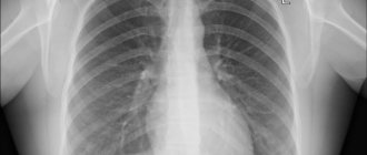
is a more accurate technique and allows you to fully examine the structures of the chest. The image can reveal signs of functional failure of various organs, trace the topography of the neoplasm, diagnose a cavity, atelectasis, abscess or violation of the integrity of the pleural cavity. Performed in two projections for more accurate results. Sometimes targeted diagnostics are used.
Tomography
is the most accurate technique for visualizing chest structures. This is the formation of layer-by-layer images of organs and structures. You can determine the spread and localization of the source of the disease, see what tissues it has affected and how deeply it has penetrated.
Techniques using contrast are used to obtain images of the bronchial tree (bronchography). Cavity changes, drainage dysfunction, structural changes and the presence of fistulas can be detected. Angiopulmonography is also used. contrast is injected into the vascular bed and allows assessment of pulmonary blood flow. This is especially important for hemoptysis and bleeding.
Radionuclide methods and scintigraphy are used. They are used to assess functional activity or impairment.
Ultrasound techniques are used to assess the activity of the heart, diagnose the condition of the pleural sinuses and lymph nodes.
Comparative characteristics of four tests for tuberculosis
| Mantoux | Diaskintest | Quantiferon test | T.SPOT.TB | |
| Accuracy | 50,00% | 90,00% | 97,00% | 97,00% |
| Data evaluation | Subjective | Subjective | Objective | Objective |
| False Positives | Often | Rarely | Rarely | No |
| With latent form of tuberculosis | Not reliable | Not reliable | Reliable | Reliable |
| Contraindications | A lot of | Eat | No | No |
| Adverse reactions | Eat | Rarely | No | No |
| For HIV and other immunodeficiencies | Not informative | Not informative | Informative | Informative |
| Research method | Skin test | Skin test | Blood analysis | Blood analysis |
What else is prescribed with this study?
Sputum analysis for Mycobacterium tuberculosis
16.15. Sputum 1 day
780 ₽ Add to cart
Diagnosis of tuberculosis infection using the T-SPOT method
20.136. Ven. blood 5 days
7,990 ₽ Add to cart
Mycobacteria, DNA (Mycobacterium tuberculosis complex, PCR) scraping, quality.
19.32.2. Scraping 1 day
450 ₽ Add to cart
Culture for tuberculosis (Mycobacterium tuberculosis)
148.0. Sputum, Urine 45 days
720 ₽ Add to cart
Fluorography
All adults over 15 years of age in Russia are recommended to undergo fluorography once a year. Children from 15 to 17 years old are allowed to undergo Fluorography, but this does not cancel the Diaskintest. A certificate with a recent photograph is required everywhere - even when visiting a child in the hospital. Fluorography is more of a “collective barrier” to tuberculosis than a method of choice for individual testing. Fluorography reveals only pulmonary forms of tuberculosis.
If you still have questions about tuberculosis diagnostic methods, call us: +7 812 331 00 00, we will answer them!
References
- Chernousova, L.N., Sevastyanova, E.V., Larionova, E.E. and others. Federal clinical recommendations for the organization and conduct of microbiological and molecular genetic diagnostics of tuberculosis, 2014. - 56 p.
- Yablonsky, P.K., Vasilyeva, I.A., Ergeshov, A.E. and others. Clinical recommendations for the diagnosis and treatment of respiratory tuberculosis in adults, 2013. - 51 p.
- WHO : The Stop TB Strategy : website / WHO - 2021, - URL: https://www.who.int/tb/strategy/stop_tb_strategy/en/.National
