Computed tomography (CT) of the lungs is considered the “gold standard” for diagnosing pneumonia, in particular pneumonia associated with COVID-19. Tomograms—multiple scans of the respiratory organ in three planes—visualize non-functional areas of compaction or infiltration of the lung tissue.
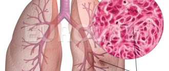
When they talk about lung damage during pneumonia, they mean that the alveoli - small bubble-shaped cavities of the lungs that are responsible for storing air and gas exchange, fill with liquid, mucus, fibrous tissue and “fail.”
In the early stages, pneumonia can be virtually asymptomatic or cause minor discomfort: cough, difficulty breathing, fever. However, it quickly turns into a more severe form and the person begins to feel a lack of air, a spasm in the chest caused by pulmonary edema, or acute respiratory distress syndrome - a widespread inflammatory process that causes complications in the heart and in some cases leads to death.
In this regard, it is very important to recognize pneumonia in time and begin treatment. Lung CT is the only diagnostic method that allows you to identify foci of infiltration and assess the degree of their severity, even if less than 5% of the lungs are affected.
After a computed tomography scan of the lungs, especially if there is suspicion of viral pneumonia, patients are primarily interested in the results and interpretation of the examinations. In this article we will talk about what CT1, CT2, CT3, CT4 mean in conclusion, and what you should pay attention to if pneumonia is still detected.
How does the procedure work?
No preliminary preparation is required before performing an x-ray examination.
The patient can continue to lead his usual lifestyle. Modern X-ray machines are high-tech and therefore capable of demonstrating any changes (from 1 mm in size). The result will be ready in a few minutes. All information will be immediately displayed on the screen.
The specialist who conducts the diagnosis will ask the patient to undress to the waist. It will be necessary to remove all metal objects; if they are present, the quality of the image may be spoiled. The procedure can be performed with the patient sitting, standing, or lying down. The doctor will ask you to hold your breath for a while.
The type of x-ray is determined by the purpose of the study. It can be overview or targeted. In the first case, the rays pass from two sides - directly and from the side, and in the second - the focus of the device is aimed at a certain area.
People need to undergo the described procedure to identify any diseases in the early stages.
What is the difference from fluorography
An X-ray of the lungs is also necessary for a healthy person. Thanks to this procedure, it is possible to diagnose diseases and dangerous conditions such as:
- tuberculosis;
- pneumothorax;
- various heart pathologies and chest injuries;
- the presence of objects of foreign origin;
- inflammatory diseases of the pleura and accumulation of fluid in it;
- tumors in the lungs, bronchi and trachea;
- helminthic infestation (presence of parasites);
- other diseases.
The principles of diagnostics using fluoro- and radiography methods are similar, but there are still differences:
- an x-ray image has a higher resolution, therefore, when enlarged, its quality will not be distorted;
- fluorographic diagnosis is more of a preventative method, since minor deviations in the image will be barely noticeable. In this case, an additional x-ray will be prescribed;
- with x-rays, the patient receives a lower dose of radiation;
- with fluorography, the image is obtained from a fluorescent screen, with x-rays - by exposing the film.
In many countries, fluorography is no longer used, as it is considered an outdated method. X-rays make it possible to detect pathologies at the earliest stages. Therefore, radiography is considered the best (in terms of information content) technique. Although it happens that in order to save time and consumables, fluorography is prescribed for all family members, employees of the organization and even residents of an entire locality.
Fluorography is a screening diagnostic method. In accordance with the regulations of the Ministry of Health of the Russian Federation, annual fluorography is mandatory for all adults. With its help, you can detect pronounced changes in the lungs, characteristic of tuberculosis and oncological processes.
About the dangers of radiography and fluorography
When using X-rays, the patient is exposed to radiation exposure, which ranges from 0.03-0.3 mSv per procedure, therefore, even when taking pictures in several projections, its total dose will not cause harm to health. In two weeks in normal life, a person receives approximately the same amount of radiation. That is, there is nothing wrong with an X-ray of the lungs, the main thing is to do it exclusively according to doctors’ indications and no more often than once every two months.
With fluorography, the radiation exposure is 0.9 mSv, which is 3-30 times higher than the above indicators, depending on the quality of the equipment used.
However, even this amount of radiation does not harm human health and is safe for it.
Radiography can only cause harm to a growing child’s body, due to its inhibitory effect on growing tissues, so such examination is not performed for children and pregnant women without special indications. When using this diagnostic method, it is important to pay attention to the equipment with which the study will be carried out.
The better and newer the X-ray machine, the less harm the examination will cause to the patient. You can also protect yourself from X-ray radiation with a protective shield. A “collar” for the neck, a “cap” for the head, an “apron” for the abdomen and a “skirt” for protecting the abdominal cavity and genitals can be used as such a screen. All of these protective screens have a lead layer.
At young childbearing ages, experts recommend protecting the genitals and abdominal area from radiation, since the greatest negative impact of the device affects the germ cells and blood.
Features when analyzing pulmonary fields
For the convenience of describing the localization of pathological shadows in the fields, they are usually divided into segments. In the description of the radiograph, the doctor indicates the serial number of the segment and the exact size of the formation.
Digital code
In the right lung it is customary to distinguish 10 segments, in the left, since its field is smaller due to the overlap with the cardiac shadow - 9. The principle of division into segments is based on the study of the branching of large bronchi. One segment is formed by one large bronchus.
[metasl >Chronic obstructive pulmonary disease is the main pathology that occurs in smokers. In the picture, COPD looks like obstructive bronchitis: the pulmonary pattern is enhanced, especially in the lower sections, and there are compensatory clearings - signs of emphysema.
At the same time, the intercostal spaces expand, the diaphragm lowers, the domes are smoothed, and the costophrenic angle turns from acute to straight. Clinically, with an increase in tissue volume, signs of hypoxia and insufficient ventilation are observed. Lung tissue loses its function.
Pathologies
Pathology in an image of the lungs can be determined by a combination of different numbers of shadows. This is how the doctor’s idea of the patient’s health condition is formed. Only after describing all the signs can the radiograph be analyzed.
White spots
Light spots on x-ray images can sometimes be a residual sign of previous diseases. But it is not always the case. As a rule, in patients they mean the presence of the following pathological conditions:
- development of occupational diseases;
- inflammatory processes;
- pleurisy.
When white spots are visible on a fluorography or x-ray, you should undergo some more research. In order to give the patient the most accurate diagnosis, the specialist will recommend that he take a repeat radiograph - in different projections or targeted.
It should be understood that white formations appear on the lungs at the initial stage of tuberculosis. This means that the structures of the lung fields are already infected with bacteria. A characteristic feature of tuberculosis will be the presence of a light path that leads to the root system from the source of inflammation.
Additional diagnostic methods should not be neglected, since if you recognize the disease at an early stage, it will be much easier to get rid of it.
Cavity
Another pathology revealed on an x-ray is a pulmonary cavity with a fibrous membrane, which is a cyst. In the image it will appear as a ring-shaped shadow. As a rule, you can notice it if you take a picture in a lateral projection. If there are thin walls, the cyst may not appear on the image. Thus, often such a formation simply goes unnoticed until it acquires more impressive dimensions.
Small lesions
There may also be minor lesions on the image. These are small spots, usually round in shape, and they are located in the lung tissue. Their number can vary: there are both single formations and entire clusters.
Foci in the respiratory organs are detected as some modifications in the tissues of the lungs. They may also occur when fluid accumulates. This may be sputum or blood discharge. Most experts consider the most important task to be establishing their origin.
After the examination, it is important to obtain an opinion from a doctor. If you suspect any pathology, you should undergo additional examination and, if necessary, begin treatment
Lung lesions CT2
CT2 means that more than three areas of ground-glass inflammation of the lungs with a diameter of no more than 5 cm have been detected. As in the case of CT1, this is community-acquired pneumonia, for which hospitalization is not required. The patient is treated at home, following the doctor's recommendations. A CT scan of the lungs will help answer the question of whether there is an active inflammatory process and a tendency toward consolidation of “ground glass” lesions. If treatment does not help and it gets worse, it is recommended to do a repeat CT scan of the lungs to assess the dynamics and adjust treatment. Since up to 50% of the lungs may be affected in a patient with moderate KT2 pneumonia, rehabilitation is necessary after primary treatment.
What is the difference between chest x-ray and fluorography?
A chest x-ray is an informative and detailed examination. It allows you to diagnose:
- tuberculosis;
- chest injuries;
- pneumothorax;
- pneumoconiosis;
- pathologies of the heart muscle;
- thromboembolism (PE);
- presence of foreign objects;
- diseases of the hematopoietic organs;
- inflammatory diseases of the pleura;
- parasitic diseases of the chest;
- accumulation of fluid in the pleural cavity;
- tumor and inflammatory processes in the lungs, bronchi, trachea.
The principle of obtaining an image using X-rays is based on the difference in the absorption of radioactive particles by body tissues. Thus, calcium-rich skeletal bones block the maximum amount of X-rays. As a result, bone tissue appears the brightest in the resulting image. Fat, muscle, fluids and connective tissue absorb less X-ray radiation. Therefore, they appear in grayscale in the image. Air allows a maximum of X-rays to pass through it. Because of this, the cavities filled with it look the darkest.
But what is the difference between classical fluorography and x-rays? It would seem that the diagnostic principle is the same and there should be no differences, but they exist and are due to differences in imaging technologies. First of all, every person going to the X-ray room should know that:
- During a fluorographic examination, the small size of the pathology is visible only as barely noticeable threads, so if the slightest suspicion arises, an x-ray is prescribed. Thus, today fluorography is more likely to be a preventive research method;
- X-rays allow you to obtain images with a resolution an order of magnitude higher, thanks to which they can be enlarged to enormous sizes;
- The radiation dose received from x-rays is several times less.
Traditional fluorography is considered an outdated method and is no longer used in many countries. While radiography is a more accurate diagnostic method, allowing not only to identify pathological processes in the early stages, but also to quickly monitor their changes. However, the price of x-rays is several times higher than classical fluorographic analysis.
Is it dangerous to do a CT scan of the lungs after an x-ray?
Ionizing (X-ray) radiation is not beneficial for humans, but in excess amounts causes radiation syndrome and can become a “trigger” for the development of cancer in patients predisposed to it. According to the current “Radiation Safety Standards”, up to 30-50 mV of radiation per year is permissible, but we should not forget about the natural radiation background. A CT scan of the lungs (about 2.5 mSv) after an X-ray (about 0.1 mSv) is safe, and such precision diagnosis can save the patient's life.
However, to avoid additional radiation exposure, it is most advisable to immediately do a CT scan of the lungs without resorting to x-rays.
What does a picture of healthy lungs say?
Before all this, you need to know the essence of any pulmonary disease. Even if it was one-sided pneumonia, after healing a scar often remains. From time to time, children are photographed and one/several are discovered, which frightens their parents. Such scars play the role of ordinary scars after injuries. That is why, after healing, the patient, even a child, may have back pain for some time or find it difficult to breathe. From time to time, scars disappear over time, from time to time ( a form of physical and mental processes, a condition for the possibility of change
) No. Here the answer is simple: it depends on the size of the outbreak and the scope of the infection itself.
X-rays of healthy lungs consistently show one list:
- pulmonary shadow from the heart;
- collarbone;
- the lung fields themselves on both sides;
- diaphragm.
When examining the image, specialists evaluate the homogeneity of tissues, bones, and the condition of the sinuses. But there is a certain range of disorders and configurations that will not be reflected on an x-ray:
- lesions less than 2 mm;
- wall compaction less than 1 mm;
- small dark spots on the lungs;
- small tissue seals;
- the presence of water in the sinus, if it is less than 250 ml.
This is a significant flaw in the study, when in ordinary people with the original form ( can mean: The shape of an object is the relative position of the boundaries (contours) of the object, object, as well as the relative position of the points of the line
) diseases may simply not see it. This is explained by the fact that the device itself has its own technical limitations. But radiologists have extensive practice, so they can intuitively suspect a deviation and recommend additional research. All advice is contained in the description of the result, which should be in the hands of the patient after the procedure.
By simplifying the definitions, we can outline what an x-ray looks like in the end. Finally, the dimensions of the lung sacs are included. The clarity of the contours and shape is also noted. A separate score is derived for the pulmonary outline.
X-ray image of healthy and clean lungs
Lots of pictures to show you what healthy lungs look like. See our article.
The X-ray method for the diagnosis and prevention of bronchopulmonary diseases is not only publicly available and simple, but also accurate. About 80% of all diseases and pathologies of the lung are diagnosed by radiography.
Radiography provides an image that reveals even the earliest stages of diseases of the respiratory system. That is why fluorography is an annual mandatory method for the preventive diagnosis of tuberculosis, tumor processes and other pathologies.
An X-ray of the lungs of a healthy person allows the doctor to evaluate the soft and bone tissues, the structures of the boundaries. In the absence of any negative changes in the lungs, a normal photograph of a healthy patient will look like the photo above. An image of a sick person will be distinguished by the presence of darkening, thickening, and lightening, which are not present in the image of a healthy person.
X-rays of healthy lungs will be interpreted taking into account the age and gender of the patient. The radiologist will tell you in detail what normal lungs look like:
- absence of visible focal, infiltrative shadows;
- normal root sizes;
- absence of altered contours of the diaphragm and phrenic-costal sinuses;
- heart shadow of normal shape;
- soft tissues unchanged.
Healthy lungs on x-ray and in pathology are distinguished by minor changes that the x-ray will show. Displacements, intensification of the pulmonary pattern, increase in shadows - all these are direct signs of the presence of lung diseases, the onset of pathological processes requiring medical intervention. Only a qualified specialist can give a correct assessment of the research results obtained. An ordinary person without medical education will not be able to distinguish a picture of healthy lungs from sick ones.
X-rays of a smoker's lungs are different from those of a healthy person. In a smoker, the pulmonary pattern thickens and bronchiectasis is observed (cavity formations in the bronchi).
A photo of the lungs will show:
- relative position of the organ among other structures of the body;
- number of pulmonary lobes;
- shapes, sizes, contours of the lungs;
- intensity of the pulmonary pattern.
Clean lungs do not have a number of signs that x-rays reveal. Fluorography can use these signs to determine the early stages of tuberculosis and other diseases.
Darkened areas may indicate pneumonia, tuberculosis, or neoplasms. Lung spots are classified by experts as follows:
- Partial darkening - pathologies, diseases affect part of the lung, pleurisy, pneumonia, atelectasis are diagnosed.
- Widespread darkening - the pulmonary field is almost completely changed, signs of pulmonary edema, effusion pleurisy, polysegmental pneumonia.
- Limited darkening is a small dark focus, a tumor and tuberculosis sign.
Tuberculosis on X-ray
Radiography is not able to assess the following changes:
- a small focus of inflammation less than 2 mm in diameter,
- a small area of enlightenment,
- small thickenings less than 1 mm in size,
- slight darkening with infiltrative changes in the bronchi,
- small focal thickening of lung tissue.
In general, radiography provides a reliable picture of the condition of the bronchopulmonary system. Accurate and professional interpretation of lung X-rays saves many lives, making it possible to diagnose the early stages of diseases and prescribe effective treatment in a timely manner.
Indications and contraindications for examination
To obtain high-quality information about the condition of the human respiratory system, doctors often prescribe x-rays of the lungs. Indications and symptoms for the use of this diagnosis are:
- suspected pneumonia;
- pleurisy;
- malignant tumors in the lungs;
- tuberculosis, bronchitis and other pulmonary abnormalities;
- prolonged cough;
- chest pain, shortness of breath;
- wheezing in the patient's audible lungs;
- monitoring the progress of therapy for diseases of the pulmonary parenchyma.
In many countries, lung examination is a mandatory preventive procedure every 2 years, and for some categories of employees and citizens, annual examination is strictly mandatory.
Such groups include people working in maternity hospitals, military personnel, carriers of HIV infection, people who have had tuberculosis, as well as those who often come into contact with such patients.
It is also important to undergo an annual X-ray examination of the lungs for those who suffer from bronchial asthma, diabetes mellitus, stomach ulcers and other patients with severe chronic diseases.
Children can undergo X-ray examination without indications only from the age of 15; before this age, x-rays can be prescribed by doctors in extreme cases necessary to clarify the picture of the course of the disease. X-ray examination is not recommended for pregnant women.
What healthy lungs look like on an X-ray
X-rays normally show the following structures:
Pulmonary fields. The lungs are projected on an x-ray as pulmonary fields. The organs of the mediastinum have their own anatomical features, which determines the difference between these fields: the right one is short and wide, the left one is longer and narrower, this is a normal case.
In a healthy person, these areas are transparent, as they are filled with air, which does not reflect radiation. The pulmonary fields are intersected by smooth and clear stripes - costal shadows, running obliquely.
Osteoarticular apparatus. In the spinal column, the bodies of the first three to four vertebrae are most clearly identified. At the same level their transverse processes are visible. In the upper part of the image, the apexes of the lungs are conventionally delimited by the shadows of the clavicles, which are located almost horizontally. The manubrium of the sternum may sometimes be visible on the radiograph.
Heart muscle is a dense tissue, so it reflects X-rays well. On a plain X-ray in direct projection, an intense, homogeneous cardiac shadow is localized in the central region, between the pulmonary fields, gravitating to the left side.
Important! The places of fixation of the ribs to the sternum are not shown, since this joint is represented by cartilage, which weakly reflects x-rays
Norm for other elements
In the superolateral part of the image, non-intense additional shadows of the pectoral muscles can be detected (mainly in men) of a triangular shape on both sides.
In women, the mammary glands are visible in the lower area of the image. Also, shadows of the mammary glands can be visible in older men.
Reference! The root of the lung is a complex of certain anatomical structures that passes through the hilum of the lung and connects the lung with organs located in the mediastinum.
This complex contains the pulmonary artery and vein, bronchi, lymph nodes and vessels, nerves, tissue and pleura. The main element in this shadow formation is the vascular-bronchial component, while other structures are covered by the mediastinum.
The shadows of the roots are organized asymmetrically: the left root is higher than the right one by one edge. They are heterogeneous, their width is no more than 1.5 cm.
Reference! Pulmonary pattern is a representation of the blood vessels of the pulmonary circulation. It looks like intertwining thin strands of shadows.
Towards the peripheral part of the lung, its visibility is weakened, as the number and diameter of the vessels decreases. The pulmonary fields are delimited below by the shadow of the upward-facing dome of the diaphragm.
Under the left dome you can see 1-2 clear spots - this is gas at the bottom of the stomach.
On the inferolateral sides, between the shadow of the diaphragmatic dome and the chest wall there are sharp angles - the cardiophrenic sinuses.