What is heart bypass surgery?
Coronary artery bypass surgery is an open-heart surgery in which a bypass path is created for cardiac blood flow, bypassing the affected arteries.
CABG surgery is performed using fragments of other vessels of the patient, which are most often taken from the lower extremities. Surgery is performed only in specialized clinics by highly qualified cardiac surgeons, with whom a transfusiologist works together to provide artificial blood circulation. It should be noted that the principle of this complex operation was developed by the Soviet doctor Vladimir Demikhov in the sixties of the last century.
Recovery after coronary artery bypass surgery
Immediately after surgery, the patient is on artificial ventilation for a certain time. And after natural breathing is restored, prevention of congestion is required. To do this, the patient is asked to inflate rubber balls several dozen times a day. As a result, the lungs expand.
Recovery from open-heart CABG involves caring for the surgical wound. The incision is made down the middle of the sternum. In addition, there is damage to the lower extremities - where the doctor took the material for bypass (the great saphenous vein). Skin wounds heal in 1-2 weeks. After this, the person is allowed to wash in the shower.
Banding after CABG is not mandatory, but is preferred to reduce the risk of sternal suture dehiscence. While skin heals in an average of 10 days, bone heals over several months. Patients often ask how long to wear a bandage after CABG. It is usually used for 4-6 months.
There should be no arrhythmia after CABG. If it occurs, it is not a consequence of the operation. There are a large number of arrhythmias. Some may be caused by myocardial ischemia, others by independent pathologies, and still others by previous myocardial infarction. Typically, the heart rhythm normalizes after surgery due to improved blood supply to the heart muscle.
The consequences of CABG surgery are assessed 2-3 months after it is performed. By this time, angina should no longer bother the patient. The person is given a stress test and IVF is removed to understand how much the blood supply to the myocardium has improved. If there is no pain after CABG during physical activity, the operation is considered successful.
Recovery after CABG can be carried out in a clinic, in a sanatorium or at home. Naturally, the most successful rehabilitation will be under the guidance of a specialist. But some patients undergo rehabilitation after CABG at home, which is usually associated with a desire to save money. If there is no money for quality restoration, you need to take care of yourself. Exercise after CABG surgery is very important. You need to start with walking - at least 1 kilometer a day. Then - gradually increase the load.
Patients often ask when they can sleep on their side after CABG. You can lie on your side almost immediately, if that’s comfortable for you. Most doctors do not prohibit this from happening to their patients, since sleeping on the side does not affect the results of the operation.
Find a specialized clinic
Varieties
Depending on which vessel is used for the bypass, there are two types of heart bypass:
- aortocoronary – CABG;
- mammarocoronary – MCS.
CABG, in turn, is divided into:
- autovenous, when the great saphenous vein of the leg is used;
- autoarterial - when using the radial artery (if the patient suffers from varicose veins).
For MCS, the internal mammary artery is used.
In addition, there is a division of bypass into:
- standard – using an artificial blood supply device (heart stopped);
- without the use of artificial blood supply to the heart (the heart does not stop), which requires the highest qualifications of the surgeon;
- hybrid, when different types of additional procedures are combined.
Options for aortofemoral bypass surgery
Aortofemoral bypass surgery can be performed in two options:
- Bilateral aortofemoral bypass surgery (aorto-bifemoral). This option involves restoring blood flow to both legs when both iliac arteries are blocked. The main branch of the prosthesis is sutured to the aorta, the branches of the prosthesis are sutured to the femoral arteries. 3 accesses are performed, two of them in both groin areas, one large on the left side.
- Unilateral aortofemoral bypass surgery is performed when one of the iliac arteries is blocked. Accordingly, only two accesses are required. One is carried out in the groin area on the affected leg, the other on the left side to the aorta.
For the operation to be successful, it is necessary to ensure good blood flow from the prosthesis; sometimes the arteries in the thigh are severely affected. In these cases, our clinic uses two-story bypass methods, when a connection is created in the groin area between the prosthesis and the most suitable artery, after which a shunt is launched further from the prosthesis from the great saphenous vein into the underlying arteries on the leg. Thus, the blood flow from the prosthesis is distributed throughout the leg and there is no stagnation of blood, leading to thrombosis and blockage of the prosthesis. If there is severe damage to the arteries in the groin, it is possible to perform femoral artery replacement. This made it possible to restore blood flow in poor condition of the peripheral vascular bed.
Indications for surgery
Bypass surgery of the affected heart vessels is performed for arterial stenosis causing ischemia. The most common cause is atherosclerosis, when the lumen of the arteries is blocked by atherosclerotic plaques, or thrombosis.
These pathologies are the main indications for surgical intervention. Additional examination is carried out when:
- retrosternal pain radiating to the left arm, shoulder, lower jaw, neck;
- hypertension;
- tachycardia; constant nausea, heartburn.
Coronary angiogram of the left coronary artery: critical stenosis of the left main artery with a good distal channel
One of the most important conditions for performing direct myocardial revascularization is the presence of a patent bed distal to the hemodynamically significant stenosis. It is customary to distinguish between good, satisfactory and poor distal bed. By good distal channel we mean a section of the vessel below the last hemodynamically significant stenosis that is passable to the end sections, without uneven contours, and of satisfactory diameter. Satisfactory distal flow is indicated in the presence of uneven contours or hemodynamically insignificant stenoses in the distal parts of the coronary artery. Poor distal flow is understood as sharp diffuse changes in the vessel along its entire length or lack of contrast in its distal sections.
Coronary angiogram: diffuse lesion of the coronary arteries with distal involvement
The most important factor for the success of the operation is considered to be preserved contractile function, the integral indicator of which is the ejection fraction (EF) of the left ventricle (LV), determined by echocardiography or radiopaque ventriculography. It is generally accepted that the normal value of EF is 60-70%. When EF decreases to less than 40%, the risk of surgery increases significantly. A decrease in EF can be the result of either scar damage or ischemic dysfunction. In the latter case, it is caused by “hibernation” of the myocardium, which is an adaptive mechanism in conditions of chronic deficiency of blood supply. When determining indications for CABG in this group of patients, the most important thing is to differentiate between irreversible scar and mixed scar-ischemic dysfunction. Stress echocardiography with dobutamine allows us to identify local contractility disorders in myocardial areas and their reversibility. Ischemic dysfunction is potentially reversible and can regress with successful revascularization, which gives grounds to recommend surgical treatment in these patients.
Contraindications
The following are traditionally considered to be considered for coronary artery bypass grafting: diffuse damage to all coronary arteries, a sharp decrease in left ventricular EF to 30% or less as a result of scar damage, clinical signs of congestive heart failure.
There are also general
contraindications in the form of severe concomitant diseases, in particular chronic nonspecific pulmonary diseases (CNLD), renal failure, and cancer. All these contraindications are relative. Advanced age is also not an absolute contraindication to myocardial revascularization, that is, it is more correct to talk not about contraindications to CABG, but about surgical risk factors.
Technique of myocardial revascularization surgery
CABG surgery involves creating a bypass for blood bypassing the affected (stenotic or occluded) proximal segment of the coronary artery.
There are two main methods of creating a bypass: mammarocoronary anastomosis and coronary artery bypass grafting with an autovenous (own vein) or autoarterial (own artery) graft (conduit).
Mammarocoronary bypass surgery.
Schematic illustration of a mammary-coronary anastomosis (a shunt between the internal mammary artery and the coronary artery)
In mammarocoronary bypass surgery, the internal mammary artery (IMA) is used; it is usually “switched” to the coronary bed by anastomosing with the coronary artery below the stenosis of the latter. The IAA fills naturally from the left subclavian artery from which it arises.
Coronary artery bypass surgery.
Schematic illustration of an aorto-coronary anastomosis (a shunt between the aorta and coronary artery)
In coronary artery bypass grafting, so-called “free” conduits are used (from the great saphenous vein, radial artery or SHA); the distal end is anastomosed with the coronary artery below the stenosis, and the proximal end with the ascending aorta.
First of all, it is important to emphasize that CABG is a microsurgical operation, since the surgeon works on arteries with a diameter of 1.5-2.5 mm. It was the awareness of this fact and the introduction of precision microsurgical technology that ensured the success that was achieved in the late 70s and early 80s. last century. The operation is performed using surgical binocular loupes (magnification x3-x6), and some surgeons operate using an operating microscope, which allows one to achieve magnification x10-x25. Special microsurgical instruments and the thinnest atraumatic threads (6/0 – 8/0) make it possible to form distal and proximal anastomoses with maximum precision.
The operation is performed under general multicomponent anesthesia
, and in some cases, especially when performing operations on a beating heart, high epidural anesthesia is additionally used.
Technique of coronary artery bypass grafting.
The operation is carried out in several stages: 1) access to the heart, usually performed by median sternotomy; 2) isolation of HAV; collection of autovenous grafts performed by another team of surgeons simultaneously with the sternotomy; 3) cannulation of the ascending aorta and vena cava and connection of IR; 4) clamping of the ascending aorta with cardioplegic cardiac arrest; 5) application of distal anastomoses with the coronary arteries; 6) removing the clamp from the ascending aorta; 7) prevention of air embolism; restoration of cardiac activity; 9) application of proximal anastomoses; 10) disabling IR; 11) decannulation; 12) suturing the sternotomy incision with drainage of the pericardial cavity.
+More details about the technique of coronary artery bypass grafting. Access to the heart is carried out through a complete median sternotomy. The IAV is isolated to its origin from the subclavian artery. At the same time, autovenous (great saphenous vein of the leg) and autoarterial (radial artery) conduits are collected. The pericardium is opened. Complete heparinization is carried out. The cardiopulmonary bypass apparatus (ACV) is connected according to the scheme: vena cava - ascending aorta. Artificial circulation (CPB) is performed under conditions of normothermia or moderate hypothermia (32-28˚C). To stop the heart and protect the myocardium, cardioplegia is used: the ascending aorta is clamped between the aortic cannula of the coronary artery and the ostia of the coronary arteries, after which a cardioplegic solution is injected into the aortic root below the clamp. Different surgeons use different compositions of cardioplegic solutions: pharmacocold crystalloid cardioplegia (St. Thomas solution cooled to 4˚C, Consol, Custodiol) or blood cardioplegia. In case of severe damage to the coronary bed, in addition to antegrade (to the aortic root), retrograde (to the coronary sinus) cardioplegia is also used to ensure uniform distribution of the solution and cooling of the heart. The left ventricle is drained through the right superior pulmonary vein or through the ascending aorta. Most surgeons first perform distal anastomoses of coronary artery bypass grafts. The heart is rotated to access the appropriate branch. The coronary artery is opened longitudinally in a relatively soft area below the atherosclerotic plaque. An end-to-side anastomosis is performed between the graft and the coronary artery. First, distal anastomoses of free conduits are formed, and lastly, a mammarocoronary anastomosis is formed. The internal diameter of the coronary arteries is usually 1.5-2.5 mm. Most often, three coronary arteries are bypassed: the anterior interventricular artery, the branch of the obtuse margin of the circumflex artery, and the right coronary artery. Approximately 20% of patients require four or more distal anastomoses (up to 8). Upon completion of the distal anastomoses after preventing air embolism, the clamp is removed from the ascending aorta. After removing the clamp, cardiac activity is restored independently or by electrical defibrillation. Then, proximal anastomoses of free conduits are formed on the parietally pressed ascending aorta. The patient is warmed up. After blood flow is turned on through all shunts, CPB is gradually completed. This is followed by decannulation, heparin reversal, hemostasis, drainage and wound suturing.
Numerous studies have convincingly proven that direct myocardial revascularization operations increase life expectancy, reduce the risk of myocardial infarction and improve quality of life compared to drug therapy, especially in groups of patients with prognostically unfavorable coronary lesions.
How to prepare for coronary bypass surgery?
Alshibaya M.M. "Clinical cardiology: diagnosis and treatment", 2011. edited by Bockeria L.A., Golukhova E.Z.
Contraindications
CABG surgery is not performed in the following cases:
- with renal failure;
- uncompensated diabetes mellitus;
- chronic nonspecific lung lesions;
- malignant neoplasms;
- post-stroke hypertension.
There are also relative contraindications for bypass surgery:
- heart failure;
- obesity;
- scarring of the heart, minimizing cardiac blood output below 30%;
- Menkenberg's arteriosclerosis - damage to all coronary arteries;
- elderly age.
AMI is considered as a contraindication at the discretion of the physician.
Diet after bypass surgery
The main factor that negatively affects the condition of the vascular wall and blood supply to the myocardium is excessive cholesterol in the blood. This is why it is so important to give up animal fats and diversify your diet with foods that remove cholesterol from the body and prevent its deposition on the walls of blood vessels.
The diet should contain sufficient quantities of vegetables, herbs, fish dishes, and chicken without fat. It is better to choose dairy products with a reduced fat content. It is recommended to consume vegetable oil as a source of fat - 2 tablespoons per day.
Preparing for the intervention
The basis of preparation for CABG surgery is coronary angiography, a procedure that thoroughly examines the relief of the coronary endothelium. To carry it out and subsequent detailed examination, the patient is hospitalized in a hospital. To perform coronary angiography, special catheters are inserted into the left and right coronary arteries, through which X-ray contrast is applied. X-rays then scan the endothelium. The procedure allows you to accurately determine the location and degree of narrowing of the vessel, while the patient receives a high dose of radiation. In addition, the manipulation cannot be performed if there is an allergy to iodine (x-ray contrast).
Therefore, in addition to coronary angiography, CT coronary angiography is used. It is more accurate, more expensive, but eliminates radiation exposure. True, contrast is still necessary, and a person weighing more than 120 kg cannot fit into the device.
If the examination reveals narrowing of the arteries by more than 75%, CABG is prescribed to minimize the risk of AMI or its recurrence. In addition to coronary angiography, the following are required: CBC, OAM, general biochemistry, coagulogram, lipidogram, ECG, EchoCG, ultrasound of the abdominal organs.
If the patient takes anticoagulants, their use is agreed with the doctor: usually a break is taken for two weeks before surgery. Other groups of pharmacological agents also require consultation with a doctor.
The day before coronary artery bypass surgery, the patient is examined by an anesthesiologist, correlating anesthesia with the height, weight, age of the person being operated on, and individual intolerance to drugs. On the eve of the intervention, the patient is sedated (taken sedatives). Necessarily:
- do not eat after 18:00;
- do not drink after midnight;
- prescribed medications are taken immediately after dinner;
- in the evening - shower.
Tests and diagnostics
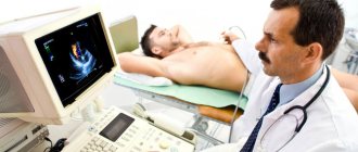
Complete information about the state of the vascular system of the heart is obtained after coronary angiography and multislice computed tomography. Both research methods make it possible to assess the degree of damage to the coronary vessels and determine further treatment tactics.
MRI of the heart and coronary vessels
Magnetic resonance imaging is considered a non-invasive diagnostic test, which is based on the nuclear magnetic resonance method. MSCT allows you to obtain a clear image of the heart and assess the condition of the coronary vessels. Absolute contraindications:
- the presence of clamps, staples and metal implants;
- the presence of an insulin pump, pacemaker, ferramagnetic implant and other electronic systems.
The price of the study ranges from 15 to 35 thousand rubles. Compared to the magnetic resonance imaging method, cardiac CT is more informative in terms of assessing the condition of the vascular system of the heart.
Additional examination methods:
- coagulogram;
- biochemical and general blood test;
- Ultrasound of the abdominal organs;
- lipid spectrum;
- chest x-ray;
- Ultrasound scanning of the lower extremities;
- ECG;
- EchoCG;
- Analysis of urine.
Progress of the operation
To perform a heart bypass, the doctor needs to cut the sternum, which then heals for a long time, which determines the length of the rehabilitation period. Depending on the type of CABG, a heart-lung machine is used or not. The heart is not stopped unless additional manipulations are required: removal of an aneurysm, replacement of valves. Bypass surgery on a beating heart has the following advantages: no complications from the immune system or blood; intervention time is shorter; rehabilitation is faster.
The essence of the operation is to create a workaround.
To do this: the surgeon opens access to the heart, takes a vessel for a shunt, if the heart stops, perform cardioplegia and turn on the artificial blood flow equipment. If the heart is beating, special devices are applied to the intervention area. The bypass itself involves stitching together blood vessels: one end of the bypass is connected to the aorta, the other to the coronary artery, which is located below the stenosis. After this, the heart is started again and the equipment is turned off. The sternum is secured with metal staples, and the skin on the chest is secured with regular sutures. The bypass lasts about four hours.
Features of the operation at the Innovative Vascular Center
Although aortobifemoral bypass surgery is one of the most common vascular operations and is performed in many vascular departments, our clinic uses certain approaches to improve the immediate and long-term results of the operation, especially in complex cases.
The main problem when performing ABBG remains the traumatic approach and associated early postoperative problems. In our clinic, a retroperitoneal approach is used to perform aortofemoral bypass surgery, without opening the abdominal cavity. This allows operations to be performed under epidural anesthesia without general anesthesia and ensures a comfortable postoperative period.
To perform repeated operations on the aorta in case of suppuration of vascular prostheses or thrombosis, our surgeons can use access to the thoracic aorta using an extended left-sided lateral approach. This approach made it possible to perform operations on patients who were refused by all other clinics.
Another important feature of surgical treatment in our clinic is the possibility of angiography during surgery. We definitely conduct a contrast study after aortofemoral bypass surgery in order to assess the hemodynamic correctness of the vascular reconstruction and identify possible problems. This approach increases the possibility of performing the operation and improves immediate results.
The use of intraoperative angiography makes it possible to operate on patients with severe calcification of the abdominal aorta, which does not allow the use of conventional methods of vessel clamping. To control bleeding, we use inflating a special balloon in the aorta, which allows us to block the blood flow while the vascular graft is sutured to the aorta. The balloon is passed through an access on the arm. The same technique allows us to successfully operate on ruptured abdominal aortic aneurysms.
The results of aortofemoral bypass surgery in our clinic are very good. Treatment success is achieved in 97% of patients with damage to the aortoiliac segment.
Complications
Often after surgery, the patient experiences a feeling of pain, heat, and discomfort in the chest. This is not a reason to panic; you need to inform your doctor about this, who will prescribe relief medications. The most common complications after cardiac bypass surgery are: pulmonary congestion, anemia, pericarditis and other inflammatory processes, phlebitis of veins adjacent to the shunt, immune disorders (during cardiac arrest), arrhythmias.
To prevent congestion in the lungs, it is recommended to inflate balloons up to 20 times a day. Anemia can be controlled with a special diet and, if necessary, with blood transfusions. Treatment of other complications is individual for each patient.
Symptoms
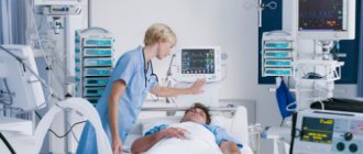
After surgery, patients are admitted to the intensive care unit, where urinary tract catheterization and artificial ventilation are performed. Painkillers are administered and antibiotics . Heart function is assessed on a monitor in the form of electrocardiography. After stabilization of the patient’s condition, the patient is transferred to independent feeding and breathing. The range of movements increases gradually, step by step.
At first, patients are bothered by chest pain, which is due to the specifics of the operation being performed, because it is open to the public. As the sternum grows together, the pain subsides. Also, discomfort is observed in the area where the vein was taken. All symptoms are temporary and the patient gradually returns to his usual lifestyle.
Rehabilitation
The recovery period is long. You cannot wash for two weeks after bypass surgery, as the wounds are extensive and there is a risk of secondary infection. Daily dressings, treatment with antiseptics. You will need to wear a chest bandage for six months to prevent the stitches on your sternum from coming apart.
Average rehabilitation is about three months. During this period, blood and blood flow will normalize, and the sternum will heal. An assessment test for the possibility of a full life is a stress test (for example, bicycle ergometry).
Cardiac rehabilitation results
The positive effects of cardiac rehabilitation at the EXPERT Clinic include:
- eliminating risk factors for coronary heart disease and reducing risk in general
- increasing physical activity
- smoking cessation
- normalization of blood pressure numbers
- weight loss
- improvement of lipid profile
- improving carbohydrate metabolism
- improvement of endothelial function
- slowing down the development of atherosclerosis and its clinical consequences
- improvement of psychophysical condition
- mobilization of patients to cooperate in the process of cardiac rehabilitation.
Dynamic (dispensary) observation is the main form of support for patients with chronic diseases in an outpatient setting. And this does not mean monitoring the natural development of the disease, as, unfortunately, often happens, but carrying out a set of secondary prevention and rehabilitation measures!
After surgical treatment, the patient is recommended to visit a cardiologist in the first days after discharge!
During the first year, visits to the doctor should be made every 4 months. Further, it is recommended to visit a cardiologist at least 2 times a year.
The goal of rehabilitation after cardiovascular accidents is to restore the patient’s physical and psychological health. Rehabilitation is indicated for all patients after surgery, regardless of the severity of the condition, both to gain self-confidence and strength, and to minimize the risk of re-exacerbation. Therefore, do not neglect your health and do not allow the disease to develop! Then you will get a chance to extend your life and improve its quality.
Which is better: a stent or a shunt?
If we compare two methods for correcting myocardial ischemia and hypoxia through the vessels supplying the muscle, the advantages and disadvantages of stenting and bypass surgery of the heart vessels become obvious:
| Evaluation criterion | Stenting | Bypass surgery |
| Scope of intervention | Minimal, intravascular | Technically complex intervention |
| Operation duration | From an hour to three | From three to nine or more |
| Heart failure | Need not | More than half of operations take place with the heart stopped |
| Incision | Deleted | The sternum is cut |
| Anesthesia | Local, less often - taking into account the individual threshold of pain sensitivity | Deep anesthesia |
| Rehabilitation | A few days | Up to six months |
| Acute cases | Relief of AMI is practiced | Not allowed due to the severity of the intervention |
| Capillaries | Correction of vessels with a diameter of 3 mm is possible | Cannot adjust small branches |
| Restoration of blood flow | For several years | Up to 10 years or more |
Treatment
Drug therapy is aimed at:
- maintaining normal blood pressure and pulse levels;
- prevention of thrombosis;
- lowering cholesterol , leveling the lipid spectrum;
- improvement of trophism and nutrition of the heart muscle.
Medicines
The main drugs prescribed after stenting and bypass surgery:
- Brilinta;
- Plavix;
- Bisoprolol;
- Lisinopril;
- ThromboASS;
- Atorvastatin;
- Preducted.
Lifestyle recommendations after surgery
Coronary bypass surgery is a reliable way to prevent heart attacks and angina attacks, since it eliminates ischemia for decades. However, the shunt can narrow; in every fifth patient this happens after a year, and after 10 years - in 100%. To minimize this possibility, you should adhere to seven rules:
- complete cessation of alcohol and cigarettes;
- anti-atherogenic nutritional profile (DASH diet included);
- movement: exercise therapy, walking, sports (swimming);
- minimizing stress;
- a balanced drinking diet (30 ml of water per 1 kg of weight);
- eight hours sleep;
- annual medical examination.
Life after CABG on the heart
After CABG, a person’s life changes only for the better. Full recovery takes about 6 months. During this time, the sternum grows together and tolerance to physical activity increases. Disability after CABG is not issued. Moreover, disability after CABG can be removed if you become able-bodied again. And if the operation is successful and there is no concomitant pathology, this will happen.
Several important questions that patients often ask regarding lifestyle after surgery:
- Showering - allowed 10 days after surgery (plus or minus 2 days) if the skin wound has healed
- A bath after cardiac CABG is allowed after 2-3 months, when you undergo a follow-up examination (because temperature changes can provoke spasm of the coronary arteries and an attack of angina)
- Alcohol after CABG is allowed in moderation after the early postoperative recovery period
- Smoking is prohibited because it leads to narrowing of the coronary vessels and increases the risk of atherosclerotic damage.
- Gymnastics after CABG - carried out under the guidance of a specialist in physical therapy, required to strengthen the heart muscle and improve tolerance to physical activity
- Diet after coronary artery bypass grafting is desirable, but not mandatory (you can reduce the consumption of animal fats to reduce the risk of atherosclerotic plaques in places where they do not yet exist)
Having surgery does not guarantee that you will not have angina or myocardial infarction in the future. Because CABG only allows you to bypass narrowed areas of blood vessels. But after a few years, new atherosclerotic plaques may appear. Therefore, it is important to adhere to a healthy lifestyle and take medications prescribed by your doctor.
Price
Since 2021, coronary artery bypass surgery is included in the system of state guarantees, that is, it is carried out under the compulsory medical insurance policy. The condition is a primary referral from a local doctor. Surgical intervention is performed in all government medical organizations at the appropriate level. If it is not possible to perform CABG at the regional level, referrals to federal medical centers are used.
If a patient wants to undergo coronary artery bypass surgery in a specific private clinic or abroad, he is not entitled to compensation for treatment. The average cost of an operation in Moscow is 120,000 rubles, St. Petersburg – 85,000 rubles, Kazan – 32,500 rubles.
Let us emphasize once again that bypass surgery is performed on an open heart, which requires special equipment, highly qualified doctors and a specialized hospital.
Causes
CABG improves coronary blood flow, which reduces the severity of pain and reduces the number of angina attacks. After surgery, patients can better tolerate physical activity, improve performance, and improve their psychological state. Surgeries to reconstruct the cardiac vasculature reduce the risk of myocardial infarction .
Indications for shunt installation:
- critical narrowing of the coronary arteries;
- angina pectoris grade 3 and 4 (attacks occur during normal physical activity and at rest);
- aneurysm due to coronary sclerosis ;
- inability to perform stenting;
- narrowing of the coronary arteries in combination with post-infarction aneurysm and structural defects of the heart .
Early postoperative period
After the operation, the patient lies in the intensive care unit for several days. During this period, vital signs are monitored, the seams are treated with antiseptic solutions, and the drainages are washed. Every day a blood test is performed, a cardiogram is recorded, and body temperature is measured. A slight fever and cough are normal at first. After the ventilator is turned off, the patient is taught breathing exercises to effectively remove fluid from the lungs and prevent congestive pneumonia. For the same purpose, the patient is often turned to the sides and X-rays of the OGK are performed several times. The patient receives the necessary medications.
If the condition is stable and the patient’s life is not in danger, he is transferred to the general ward to further observe and recover after heart bypass surgery. The motor mode is gradually expanded, starting with walking near the bed, along the corridor. Areas of postoperative wounds are treated. The patient wears elastic stockings to reduce swelling of the lower leg. Before discharge, the sutures from the chest are removed. The length of stay in the hospital ranges from a week or more.