The main cause of cerebrovascular accidents is atherosclerosis of the carotid arteries. Atherosclerotic plaques cause narrowing of the carotid arteries, which is an obstacle to normal blood circulation in the brain. Gradually, a complete blockage of the carotid artery develops, which is called occlusion. Impaired patency of the carotid artery is the main cause of ischemic stroke in the modern world. The risk of developing a stroke if the carotid artery is narrowed symptomatically by 70% or more is about 15% per year.
Many people die or become disabled from stroke every year, although modern vascular surgery can prevent it in most patients. Only regular diagnosis and trust in doctors will significantly reduce the risk of stroke. It is much easier to treat atherosclerosis of the carotid artery than ischemic stroke and its consequences.
Causes and risk factors
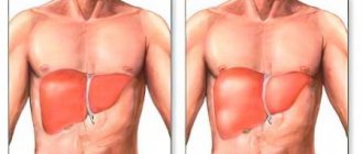
The carotid arteries are paired large arterial vessels that supply blood to the brain in those sections where the centers of thinking, speech, personality, sensory and motor function are located. The carotid arteries run through the neck and enter the brain through openings in the skull.
When fatty substances and cholesterol accumulate, an atherosclerotic plaque is formed, which narrows the carotid arteries. This reduces blood flow to the brain and increases the risk of ischemic stroke. A stroke occurs when blood flow does not reach any part of the brain. During a stroke, some brain functions are suddenly lost. If the lack of blood flow lasts more than three to six hours, then these disorders become irreversible.
Why does a stroke develop when the carotid artery narrows?
- Significant narrowing of the carotid artery reduces blood circulation in the brain, and with a sudden drop in pressure (suddenly getting out of bed, flying, overheating in the sun or major surgery), the blood flow suddenly stops, which leads to the death of nerve cells.
- Tearing off a piece of atherosclerotic plaque with its transfer by the bloodstream into small arteries of the brain, which leads to their blockage.
- Acute thrombosis (formation of a blood clot) due to narrowing of the carotid artery with complete cessation of blood flow in certain areas of the brain.
What manifestations are accompanied by the pathology?
The clinical manifestations of the disease depend on where the problem is located. Also, the nature of the onset of the disease, which can be sudden or gradual, plays an important role. It is worth remembering that in the first stages of the disease, compensatory mechanisms always turn on. For example, if the blood flow is partially disrupted, a system of collaterals develops, which will provide alternative routes for blood supply to the brain. The lack of oxygen supply is also compensated - the pulse and breathing become more frequent, intracellular processes change. For short-term conditions this is enough, but for serious pathologies it is not. In such cases, symptoms of the condition develop. Naturally, with an abrupt onset and significant stenosis, compensatory forces do not work and the clinical picture has a pronounced character, which indicates a stroke.
One of the first alarm signals is transient cerebral ischemia. These are short-term episodes of circulatory disturbance that can be compensated by collateral blood flow. The condition can be recognized by decreased sensitivity and motor activity, as well as visual disturbances. Patients usually complain of a feeling of numbness in the upper limb, half of the face, and loss of visual fields. Decreased muscle strength in the arm or forearm. The patient feels the movement of dark spots in front of the eyes, as well as a decrease in visual acuity. In some cases, the section of the artery that supplies the retina is affected. In some localizations, corresponding disorders occur - difficulty speaking, hearing, swallowing, facial asymmetry, severe pain. More common symptoms are dizziness, feeling unwell, weakness, and convulsions.
If stenosis develops gradually, the clinical picture also develops slowly. In some cases, it is so nonspecific that it is difficult to recognize the disease. For a long time, both the patient and the doctor may mistakenly believe that the cause of poor health is fatigue or increased stress. Also, the symptoms of occlusion overlap with the symptoms of a tumor process and dementia. This is indicated by increased irritability, depressive disorders, sudden changes in mood, increased drowsiness, decreased memory and mental abilities.
Nonspecific symptoms require a high level of responsibility from the patient and the doctor, since high-quality diagnosis is necessary. The diagnosis determines how correct the treatment of the pathology and the result will be.
Risk factors for carotid atherosclerosis
Risk factors for carotid artery disease are similar to those for other types of cardiovascular disease. They include:
- Age
- Smoking
- Hypertension (high blood pressure) is the most important risk factor for stroke
- High cholesterol
- Diabetes
- Obesity
- Sedentary lifestyle
- Family history of atherosclerosis
Men under 75 years of age have a greater risk of developing carotid artery stenosis than women in the same age group. In the group over 75 years of age, women have a greater risk of stroke. In patients suffering from coronary artery disease, narrowing of the carotid artery is often detected.
What else causes stenosis?
More rare cases are associated with a specific pathology. Narrowing of the arterial vessel is observed with a disease of the connective tissue that forms the stroma of the artery. This is called fibromuscular dysplasia - the vessels cease to maintain tone and collapse, stopping blood flow. Stenosis can accompany some inflammatory diseases of the vascular wall - arteritis, systemic vasculitis, Takayasu's disease, Horton's disease. There are rare pathologies, for example moyamoya disease, in which there is a slow progressive narrowing of the lumen of the arteries.
Stenosis can occur after an injury - a strong blow, a concussion. After some traumatic brain injuries, a special hematoma is observed - subintimal, which has the appropriate localization. A blood clot that forms due to increased blood clotting and rare types of anemia can block the lumen. There is antiphospholipid syndrome, in which young patients experience heart attacks and strokes precisely because of increased coagulation. Blood clots form when heart rhythm disturbances occur while taking certain medications.
The narrowing may be due to congenital structural features of the vessels - they can be tortuous and hypoplastic. Vascular pathology accompanies systemic diseases, diabetes, obesity and metabolic disorders.
Complaints and symptoms
Atherosclerosis of the carotid arteries can be asymptomatic or cause complaints associated with impaired cerebral blood flow. Most often, patients may complain of temporary impairment of brain function (transient ischemic attack) or persistent loss of brain function (ischemic stroke).
Transient ischemic attack (TIA)
A TIA occurs when blood flow to the brain is briefly interrupted. This is the initial phase of acute cerebrovascular accident, which is reversible. It has the same symptoms as a stroke, but these symptoms go away within minutes or hours.
A TIA requires emergency medical attention because it is impossible to predict whether it will progress to a stroke. Immediate treatment can save lives and increase the chances of a full recovery.
Modern research has shown that patients who have had a TIA are 10 times more likely to suffer a major stroke than a person who has not had a TIA.
Ischemic stroke has the following symptoms:
- Sudden loss of vision, blurred vision, difficulty in one or both eyes.
- Weakness, tingling, or numbness on one side of the face, one side of the body, or one arm or leg.
- Sudden difficulty walking, loss of balance, lack of coordination.
- Sudden dizziness.
- Difficulty speaking (aphasia).
- Sudden severe headache.
- Sudden memory problems
- Difficulty swallowing (dysphagia)
Ischemic stroke and transient ischemic attack begin in the same way, so any ischemic stroke can be called an ischemic attack if the symptoms completely resolve within 24 hours of the onset of the disease. The presence of a time interval between the onset of stroke symptoms and the death of parts of the brain allows for urgent surgery to restore cerebral blood flow.
Diagnostic program for the disease
First of all, a timely start to the diagnostic search is necessary. It is better to rule out the disease and continue to live peacefully than to postpone a visit to the doctor “for later.” For symptoms that cause concern, professional medical attention is necessary. The doctor begins making a diagnosis by interviewing the patient and general clinical examinations. If there are neurological symptoms, reflexes, sensitivity, motor activity, muscle and analyzer function are assessed. The following instrumental methods are used:
- Ultrasound of the arteries of the head and neck with Doppler sonography - triplex scanning. A safe, fast and inexpensive method that allows you to exclude or confirm the diagnosis;
- Functional tests during ultrasound examination;
- Computed tomography angiography - visualization of blood flow through vessels using a contrast agent;
- Magnetic resonance imaging of the brain.
To diagnose atherosclerosis, a laboratory method is used - the patient donates blood for a lipid profile. Also, it is necessary to evaluate the blood coagulation system and its coagulation properties. It is mandatory to measure pulse and blood pressure. To identify systemic vasculitis, specific laboratory tests and immunological studies are used.
MRI and CT are the “gold standard” in the diagnosis of ischemic disorders, including stroke. Depending on the location and extent of the lesion, treatment is prescribed.
Course of the disease
Once atherosclerotic plaques appear, they will no longer be able to resolve, but only gradually progress. The rate of growth of an athersclerotic plaque depends on many risk factors, including cholesterol levels. All people over 50 years of age are recommended to undergo an annual ultrasound of the carotid arteries in order to exclude the development of atherosclerotic plaques and the risk of ischemic stroke.
With the development of complications of atherosclerosis of the carotid arteries, discirculatory encephalopathy quickly progresses. Frequent TIAs, and even more so ischemic stroke, contribute to the death of part of the brain tissue and disruption of brain function. Patients with atherosclerosis of the carotid arteries often develop vascular dementia (dementia).
After restoration of the patency of the carotid artery, the phenomena of cerebrovascular insufficiency are stopped, and the likelihood of repeated cerebrovascular accidents is significantly reduced.
Function [edit]
Branches of the external carotid artery.
As it moves upward, the artery supplies:
- In the carotid triangle: [2] The superior thyroid artery, arising from its anterior surface
- Ascending pharyngeal artery - arising from the medial or deep side
- Lingual artery - arises from its anterior part
- Facial artery - arises from its anterior part
- Occipital artery - from its posterior part
The external carotid artery ends in two branches:
- Maxillary artery
- Superficial temporal artery
To remember the main branches of the external carotid artery, several mnemonics are usually used.
- Mnemonics - S OME
A natomists
L
IKE
F
reaking
O
ut
P
OOR
M
edical
S
tudents. - Mnemonics - S Ister
l ucy - y
Race
O
ften
P
owdered tr tracts
M edical
S
tudents
.
Forecast
Carotid atherosclerosis carries a significant risk of ischemic stroke. With asymptomatic narrowing of the internal carotid artery of more than 70%, the risk of ischemic stroke exceeds 5% per year. If the patient has had episodes of cerebrovascular accidents, then this risk is already 25% per year.
The risk of ischemic stroke in asymptomatic atherosclerotic plaques with a narrowing of less than 70% does not exceed that in patients without atherosclerosis.
After adequate restoration of blood circulation in the carotid arteries, the risk of ischemic stroke is reduced by more than 3 times.
Forecast and preventive recommendations
If the arterial lumen is blocked by half, the disease may occur without obvious signs. With such a picture, the risk of developing apoplexy is 10% of all cases within five years. A 75% vascular blockage leads to a 6% stroke rate in the first year. Complete thrombosis of the canal leads to inevitable neurological damage in 40% of incidents. Preventive surgical intervention with or without prosthetics makes it possible to reduce these risks to a minimum.
To prevent the closure of vascular walls, it is recommended to get rid of bad habits, balance your diet, and monitor weight gain. The lipid composition of the blood is determined by regular testing. If necessary, therapeutic recommendations help adjust the composition of the blood solution. If cardiac abnormalities are present, supportive treatment should be provided promptly. In case of congenital diseases of the cardiovascular system, it is also recommended to undergo regular examination as prescribed by a cardiologist.
Syndrome of blood flow insufficiency in the arteries of the vertebrobasilar system
Maksimova M.Yu., Piradov M.A. RMJ. 2021. No. 7. pp. 4-8 The article is devoted to the problem of blood flow insufficiency syndrome in the arteries of the vertebrobasilar system. Methods for diagnosing and treating vertebrobasilar insufficiency are presented, which should be aimed at preventing its progression, improving blood supply to the brain, and correcting certain syndromes and symptoms.
The independent clinical concept of “blood flow insufficiency syndrome in the arteries of the vertebrobasilar system” was formed in the 1950s, during the period of revision of views on the pathogenesis of ischemic cerebrovascular accidents (CVA) and the emergence of the concept of the leading role of cerebrovascular insufficiency in this case [1]. The peculiarities of the structure and functions of this arterial system, which provides nutrition to vital structures of the brain, and the uniqueness of clinical symptoms in case of disturbances of blood flow in it led to its identification in the latest version of the international classification into an independent symptom complex - “vertebrobasilar arterial system syndrome” within the framework of “transient transient cerebral ischemic attacks (attacks) and related syndromes” (International Classification of Diseases, 10th Revision, G45.0). Even earlier, a group of experts from the World Health Organization defined “vertebrobasilar insufficiency” as “a reversible impairment of brain function caused by a decrease in the blood supply to the area supplied by the vertebral and basilar arteries.” The ischemic nature and reversible nature of the disorders were emphasized, but the duration of neurological symptoms was not indicated, which previously did not allow them to be classified as transient ischemic attacks (TIA) and which has now become possible. Blood flow disturbances in the arteries of the vertebrobasilar system account for about 70% of all TIAs. Stroke with localization of focal changes in areas of the brain that receive blood through the arteries of this system develops 2.5 times less frequently than in regions belonging to the arterial basins of the carotid system [1].
Causes of blood flow insufficiency syndrome in the arteries of the vertebrobasilar system
The main causes of blood flow insufficiency syndrome in the arteries of the vertebrobasilar system, caused by arterial hypertension (AH) and atherosclerosis (AS), include [1, 2]:
- atherostenosis or atherobliteration of one of the vertebral arteries;
- characteristic arterial tortuosity, which in some cases can lead to kinking of the vertebral artery with the formation of septal stenosis and disruption of blood flow in it;
- congenital anomalies of the vertebral arteries (hypoplasia of one of the vertebral arteries, lateral displacement of the mouth of the vertebral artery), in which the insufficiency of blood flow in one of the vertebral arteries is compensated by another vertebral artery, but decompensation occurs against the background of AS and hypertension;
- compression of the vertebral artery by an osteophyte in the bone canal of the cervical spine, an articular process with instability of the cervical spine, an accessory cervical rib, a spasmodic neck muscle (posterior scalene muscle, longus colli muscle, inferior oblique muscle of the capitis), which is most often observed with congenital abnormally high entry vertebral artery into the spinal canal - at the level of 3–5 cervical vertebrae.
The syndrome of blood flow insufficiency in the arteries of the vertebrobasilar system can also be observed:
- with subclavian “steal syndrome”, in which, as a result of occlusion of the subclavian artery, blood flows not only to the entire vertebrobasilar system, but also to the arm through only one vertebral artery;
- with occlusion or severe atherostenosis of both internal carotid arteries
(ICA), since the vertebrobasilar system plays a significant role in the blood supply to the cerebral hemispheres and, under certain conditions, “steal syndrome” may occur; - with disturbances of general hemodynamics.
Subclavian “steal syndrome” is characterized by a phenomenon when a patient, during intensive work of the arm (retrogradely supplied with blood from the contralateral vertebral artery), experiences brainstem symptoms - most often dizziness. A certain contribution to the development of blood flow deficiency syndrome in the vertebrobasilar system can be made by changes in the rheological properties of blood (increased levels of fibrinogen, blood viscosity, platelet aggregation and hematocrit, increased rigidity of erythrocytes), leading to deterioration of microcirculation.
Diagnosis of blood flow insufficiency in the arteries of the vertebrobasilar system
Subjective data
The diagnosis of insufficiency of blood flow in the arteries of the vertebrobasilar system is based on a characteristic symptom complex that combines several groups of clinical symptoms found in patients with AS and hypertension.
These are visual and oculomotor disorders, disorders of statics and coordination of movements, vestibular disorders. In this case, the presumptive diagnosis is determined on the basis of at least two of these symptoms. They are short-lived and often go away on their own, although they are a sign of impaired blood flow in the arteries of this system, which requires clinical and instrumental examination. A thorough medical history is especially necessary to clarify the circumstances of the occurrence of certain symptoms [1, 2]. Visual disturbances
include a feeling of blurred vision, photopsia, scotoma, changes in visual fields, decreased visual acuity and are associated with transient ischemia of the occipital lobes of the brain.
Blurred vision in the form of a veil before the eyes and blurred vision often occurs at the height of a headache. Photopsia appear in the form of flashes of colored dots, most often red or green, black, with a light halo, as well as spots, fiery lightning, lines, rings, zigzags. Photopsia differ from the rainbow circles characteristic of glaucoma in that their appearance is not associated with an external light source; they also occur with closed eyes. Changes in visual fields are usually observed in the form of their concentric narrowing. Decreased visual acuity often develops after the onset of headache and progresses; vision deteriorates noticeably during headache attacks and after them. Oculomotor disorders
manifest themselves in the form of transient diplopia with mild paresis of the eye muscles and impaired convergence.
In most patients, these disorders are among the initial manifestations of the disease, and in a quarter of them they serve as one of the main complaints with vertebrobasilar insufficiency. Static and dynamic ataxia are also among the permanent symptoms that are manifested by patient complaints of instability and staggering when walking and standing. Coordination of movements is significantly less impaired; a persistent change occurs, as a rule, with cerebellar infarctions. Vestibular disorders
manifest themselves in the form of sudden dizziness - systemic, which is characterized by a feeling of “rotation of objects”, “an inverted room”, and non-systemic with a feeling of “motion sickness”, nausea, and less often vomiting.
Spontaneous nystagmus is also detected, sometimes only after special tests with turning the head to the side and fixing it in these positions (De Klein test). The development of dizziness is associated with ischemia of either the vestibulocochlear organ or the vestibular nuclei and their connections. The vestibular nuclei are most sensitive to ischemia and hypoxia. In this case, dizziness as a monosymptom can be regarded as a sign of impaired blood flow in the arteries of the vertebrobasilar system only in combination with other signs of its impairment in patients with a relatively persistent otoneurological symptom complex. Less known, although not uncommon, are optic-vestibular disorders. These include symptoms of “shading shadow” and “convergent vertigo,” in which patients experience dizziness or unsteadiness when flashing light and shadow or when looking downward. Characteristic symptoms are attacks of sudden falling
without loss of consciousness (“drop attacks”), usually occurring during sudden turns or throwing back the head.
Syncopal vertebral Unterharnscheidt syndrome has been described, in which loss of consciousness and muscle hypotonia are observed in the absence of evidence of epilepsy and other paroxysmal conditions. Manifestations of diencephalic
disorders include severe general weakness, irresistible drowsiness, disturbances in the rhythm of sleep and wakefulness, as well as various autonomic-visceral disorders, a sudden increase in blood pressure (BP), and heart rhythm disturbances.
These disorders are associated with ischemia of the structures of the reticular formation of the brain stem. The described symptom complex has now been supplemented by other signs, which, in combination with them, also make it possible to judge the insufficiency of blood flow in the arteries of the vertebrobasilar system. At various stages of vertebrobasilar insufficiency, patients often complain of decreased memory
(“forgetfulness”), concentration disorders and instability of active attention. Most often, memory for names, numbers, and recently occurring events decreases. The ability to memorize new material decreases, it becomes more difficult to retain what has been read in memory, what is planned for implementation is forgotten, and the need to write it down arises. It becomes difficult for patients to comprehend a large amount of information, which leads to a certain decrease in performance and limitation of creative possibilities in people engaged in mental work. At the same time, professional memory and memory of past events are preserved. This applies more to RAM than to logical memory. Often, a decrease in memory and performance is regarded by others as a result of overwork, and not as a manifestation of cerebral vascular insufficiency. During neuropsychological research, the preservation of the level of generalization, the correspondence of judgments to the general educational and cultural level, and the preservation of the stock of ideas and skills are noted. Impaired cognitive functions significantly reduce the quality of life and also affect the progression of cerebrovascular insufficiency. Decreased memory for current events in patients with vertebrobasilar insufficiency is associated with chronic ischemia of the medial parts of the temporal lobes, primarily the hippocampus and mammillary bodies. With vertebrobasilar insufficiency, attacks of transient global ischemia are also observed, during which working memory (the ability to remember new information) is impaired for several hours. The patient looks absent-minded, he is disoriented in space and time, sometimes excited, persistently tries to find out from those around him where he is, how he got here, but being unable to remember the answers, he constantly asks the same questions. With the return of the ability to remember, orientation is also restored, only the episode itself is amnesic. Acute amnesia can also be caused by acute cerebrovascular accident in the basins of both posterior cerebral arteries. In this case, amnesia may be accompanied by limitation of visual fields (unilateral or bilateral hemianopia), visual agnosia, alexia, amnestic aphasia, and sensory impairment. The combination of a number of characteristic symptoms makes it possible to diagnose the syndrome of blood flow insufficiency in the arteries of the vertebrobasilar system, although in this case only the ischemic nature of the cerebrovascular accident and the localization of the source of ischemia are determined, and not the reasons that determined this nature.
Objective data
The most accessible and safest methods for determining insufficiency of blood flow in the arteries of the vertebrobasilar system are neurological examination and ultrasound methods of studying the vascular system of the brain.
Among the objective signs revealed during a neurological examination
, one should first of all mention nystagmus, static and dynamic ataxia.
In the Romberg test, the patient deviates to the side. Walking with eyes closed reveals unsteadiness and persistent deviation to one side in a patient with insufficient blood flow in the vertebrobasilar system. When performing the Unterberger test, the patient is asked to march in one place with his eyes closed for 1–3 minutes. Normally, it remains in place or moves slightly relative to the starting point or rotates slightly around its axis. A forward shift of more than 1 m and a rotation of more than 40–60° (after 50 steps in place) are considered pathological. The results of the Babinski-Weil test (“star test”) are interpreted in a similar way. With eyes closed, the patient is asked to take two steps forward, turn 180° and take two steps back. Any deviations to the side or rotation indicate dysfunction of the vestibular labyrinth. If the patient is asked to walk in the forward and reverse directions several times, then as a result of a deviation to one side, the trajectory of his movement resembles the outline of a star (hence the name of the test). It is also necessary to measure blood pressure in both arms in a sitting and lying position. Objective signs of the syndrome include differences in pulse and blood pressure in the arms and noise in the supraclavicular region. With a significant decrease in systolic blood pressure (more than 20 mm Hg) in an upright position, symptoms reminiscent of insufficiency of blood flow in the vertebrobasilar system should be attributed to orthostatic hypotension. Subclavian “steal syndrome” is characterized by a phenomenon when a patient, against the background of intense hand work, develops brainstem symptoms—usually dizziness. Doppler ultrasound
allows you to obtain data on blood flow in the vertebral arteries, linear speed and direction of blood flow in them.
Compression-functional tests make it possible to assess the condition and resources of collateral circulation, blood flow in the carotid, temporal, supratrochlear and other arteries. Duplex scanning allows you to determine the condition of the artery wall, the structure and surface of atherosclerotic plaques that stenose these arteries. Transcranial Doppler ultrasound with pharmacological tests is important for determining cerebral hemodynamic reserve. Data on the condition of the great arteries of the head (MAG) and intracerebral arteries obtained from CT and MRI angiography are extremely informative. X-rays
of the cervical spine can provide information about the condition of the structures around the vertebral arteries and the effect of these structures on the vertebral arteries and the blood flow in them;
functional tests are used in this case. A special place among instrumental methods is occupied by otoneurological research
, especially if it is supported by electronystagmographic and electrophysiological data on auditory evoked potentials characterizing the state of brain stem structures, as well as MRI of these structures. The algorithm for using the listed instrumental research methods is determined by the logic of constructing a clinical diagnosis.
Treatment of vertebrobasilar insufficiency
Treatment of vertebrobasilar insufficiency is aimed at preventing its progression, improving blood supply to the brain, and correcting individual syndromes and symptoms.
The most effective measures in this direction are the elimination or correction of the main risk factors
for the development of vertebrobasilar insufficiency, which include smoking, hyperlipidemia, AS of the cerebral arteries, hypertension, diabetes mellitus, obesity, heart disease, disorders of the rheological properties of the blood, psycho-emotional stress, alcohol abuse [3 ].
A large place in the prevention of the progression of vertebrobasilar insufficiency is occupied by recreational activities, climate therapy at local resorts, in low-altitude conditions, at sea resorts, balneotherapy (radon, brine, carbon dioxide, sulfide, iodine-bromine baths). Moderate physical activity (therapeutic exercises, walking, swimming) and regular mental exercise are needed. The diet
should not be burdensome for the patient (do not overeat, limit the consumption of animal fats, easily digestible carbohydrates and foods rich in cholesterol, reduce the total calorie content of food, introduce fresh vegetables and fruits, wholemeal products, fish products into the diet).
Smoking is excluded and alcohol consumption is limited. When treating vertebrobasilar insufficiency, the following measures should be taken: early detection; determination of the severity of clinical symptoms; exclusion or correction of the main risk factors for the development of cardiovascular diseases; dynamic observation; timely initiation of treatment; its duration and continuity; treatment of concomitant somatic, neurological and mental disorders; medical, professional and social rehabilitation. Methods of drug treatment
of chronic musculoskeletal disorders include: antihypertensive therapy, the use of lipid-lowering drugs, improving blood supply to the brain using antithrombotic drugs, neuroprotective therapy [3, 4].
One of the most promising neuroprotective drugs from the perspective of evidence-based medicine is citicoline
[4, 5].
Citicoline, a natural endogenous compound also known as cytidine-5'-diphosphocholine (CDP-choline), is a mononucleotide consisting of ribose, cytosine, pyrophosphate and choline. When taken orally, citicoline is rapidly absorbed and hydrolyzed into choline and cytidine in the intestinal wall and liver. These substances enter the systemic circulation, pass through the blood-brain barrier and recombine to form citicoline within the central nervous system [6]. Phosphatidylcholine in brain cell membranes is broken down into fatty acids and free radicals by phospholipases under ischemic conditions. By restoring the activity of Na+/K+-ATPase of the cell membrane, reducing the activity of phospholipase A2 and participating in the synthesis of phosphatidylcholine, the membrane-stabilizing effect of citicoline is realized. In addition, citicoline affects the formation of free fatty acids, the synthesis of acetylcholine and an increase in the content of norepinephrine and dopamine in nervous tissue. Citicoline is also able to inhibit glutamate-induced apoptosis and enhance neuroplasticity mechanisms [7]. The first studies of citicoline, conducted at the end of the twentieth century, concerned patients with vascular dementia. Thus, R. Lozano et al. (1986) observed 2067 elderly patients treated in geriatric psychiatry departments and found a positive effect of a 2-month course of citicoline therapy on the severity of neuropsychological symptoms [8]. In a study by B. Chandra (1992), assessing the effectiveness of the drug in 146 patients with vascular dementia, it was demonstrated that therapy with citicoline at a dose of 750 mg/day IV for 2 months. led to a significant improvement in cognitive function scores (assessed on the MMSE scale) compared to placebo. Moreover, the effect of therapy was maintained after 10 months. after completion of treatment [9]. In 2005, a Cochrane review of the effectiveness of citicoline in the treatment of cognitive and behavioral impairment due to chronic cerebrovascular insufficiency in elderly patients was published [10]. The review included the results of 14 randomized placebo-controlled trials involving 1336 patients. The average dose of citicoline in these studies was 1000 mg/day, the duration of treatment was 3 months. The effectiveness of treatment was assessed using tests for memory, attention, and behavior. The review demonstrated the positive effect of citicoline on behavioral disorders, as well as improving memory. The only significant limitation of this review was the short duration of the included clinical studies. In subsequent years, researchers focused on studying the drug's effectiveness in patients with mild cognitive impairment (MCI) and post-stroke cognitive impairment. Thus, according to M. V. Putilina (2009), already at the initial stages of manifestations of cognitive impairment in patients with chronic cerebrovascular insufficiency, the use of citicoline (at a dosage of 1000 mg IM or IV for 10 days followed by oral administration in the form of a solution for oral administration for 3 months) contributes to the regression of these disorders. In addition, the drug has a positive effect on concomitant emotional, affective and behavioral disorders in this group of patients [11]. In 2013, the results of two controlled studies evaluating the effect of the drug on cognitive function in patients with chronic cerebrovascular diseases were published. In a placebo-controlled study, L. Alvarez-Sabin et al. (2013) took part in 347 elderly patients (mean age 67.2±11.3 years) who had suffered a stroke and had cognitive impairment. In the active treatment group (172 patients), citicoline was prescribed at a dose of 2000 mg/day per os for 6 months, then 1000 mg/day for another 6 months. The criteria for the effectiveness of treatment were the results of a neuropsychological examination (a battery of tests for memory, attention, executive (regulatory) functions, time orientation), as well as an assessment of clinical outcomes using the modified Rankin Scale after 6 and 12 months. after starting treatment. Long-term therapy with citicoline resulted in a slowdown in the progression of cognitive impairment and better functional recovery (compared to placebo) due to improved attention, regulatory functions, and time orientation [12]. The IDEALE study assessed the effectiveness of citicoline in the long-term treatment of vascular MCI in elderly patients. 349 patients with MCI of predominantly vascular origin were prescribed citicoline (265 patients) at a dose of 1000 mg/day per os for 9 months. or placebo (84 patients). Treatment with citicoline had no effect on measures of functional daily activities compared with placebo. At the same time, during treatment with citicoline, there was a positive dynamics of cognitive functions when assessed on the MMSE scale (improvement after 9 months by an average of 0.5 points); in the placebo group, progression of cognitive impairment was observed (after 9 months - worsening by an average of 1.9 points) (p=0.0001). Thus, long-term therapy with citicoline is associated with a decrease in the rate of progression of cognitive impairment in patients with vascular MCI [13]. Recently, new generic dosage forms have been widely introduced into practice. Among them is the domestic drug Neypilept
. The drug is produced from the Japanese substance of the KYOWA company in the form of a solution of 125 and 250 mg/ml for IV and IM administration, as well as two oral forms - an oral solution of 100 mg/ml in bottles of 30 ml and 100 ml. The volume of the 100 ml bottle corresponds to the sachet form of the original drug. As part of post-registration multicenter randomized studies, Neypilept was compared with the original drug in 152 patients in the acute period of ischemic stroke in the carotid system (RCT No. 396 of June 24, 2013) [14] and the effectiveness and safety of its oral form was studied in 128 patients with cognitive impairment (RCT No. 145 dated March 26, 2015) [15]. The results of the studies demonstrated the tolerability and effectiveness of Neypilept in these conditions comparable to the original drug [16, 17].
Conclusion
It should be emphasized that timely and systematic treatment can prevent the progression of cerebrovascular insufficiency and significantly improve the quality of life of patients. The adequacy and effectiveness of taking citicoline (Neipilept) is of particular importance. Adequacy of therapy implies a course of taking the drug, as well as cooperation between the patient and the attending physician in prescribing and carrying out treatment, the goals of which are to preserve the ability to work and maintain the patient’s quality of life. The following areas for assessing the effectiveness of treatment for vertebrobasilar insufficiency can be recommended (as early as 6–12 months from the start of treatment): reduction or disappearance of cerebral complaints, improvement of cognitive functions (primarily memory).
The original article was published on the RMJ website (Russian Medical Journal): https://www.rmj.ru/articles/nevrologiya/Sindrom_nedostatochnosti_krovotoka_varteriyah_vertebrobazilyarnoy_sistemy/#ixzz5NyXkC9vV
Appointment form...
Treatment methods at the Innovative Vascular Center
The vascular surgeons of our clinic have significant experience in unique operations on the carotid arteries with pathological tortuosity. The main problem for surgical treatment is determining clear indications for surgical treatment. Our clinic has developed a clear diagnostic protocol that allows us to determine the clinical significance of a particular tortuosity and the degree of its effect on cerebral blood flow. The experience of successful operations in our clinic for pathological tortuosity exceeds 200 cases.
Therapeutic exercise to relieve the syndrome
Specific exercises are prescribed only by a specialist with a medical education. If you detect the slightest signs of deterioration, you should immediately stop exercising and consult your doctor again.
- Head tilts. You should start with a small amplitude; as the condition improves, the number of inclinations increases and the amplitude increases. The sensation of a slight painless cracking sensation during bending is not considered a reason to stop exercising.
Head tilts to the sides - Shrug . The head should always be in a strictly vertical position. The shoulders are raised to the maximum amount. The number of repetitions is not limited, the exercise does not have a direct effect on pathological areas, it only helps to improve blood flow in nearby areas.
- Head turns. During this exercise, cracking sounds may also be heard in the cervical vertebrae. Such crackling sounds indicate the presence of changes in bone tissue. If they are painful, then the amplitude should be reduced. In most cases, the cracking stops after a few days of exercise, which indicates an improvement in the condition of the spine.
Head turns - Tilts the head forward/backward with increasing effort . A more complex exercise, you need to counteract the tilt with your hands. A very effective exercise for improving the condition of the muscle tissue of the cervical region. Strong muscles are able to maintain the correct position of the head, due to which the load on the vertebrae is significantly reduced, they occupy a natural physiological position and free up pinched or curved arteries.
- Tilts of the head to the sides with increasing effort . The exercise is similar to the one described above, the only difference is in the direction of the inclinations.
Head tilts forward and backward
Signs of arterial stenosis
Long-term disruption of the blood supply to the brain causes the following phenomena.
- Numbness of facial muscles. If there is a one-sided pathology, then the face may become distorted. When it is bilateral, facial expressions become more complicated, speech becomes unclear, and the face becomes white.
Numbness of facial muscles - Dizziness, inability to hold a stable position . Such symptoms may indicate an approaching fainting state. The patient should be immediately laid down and the head should be placed in the most comfortable position.
Long-term painful conditions have a negative impact on the psyche of patients and serve as an impetus for the onset of depression. The earlier the diagnosis is made and treatment measures taken, the less likely there are negative consequences. In the most complex forms, pathologies cause strokes.
Diagnostics
Diagnosis of vertebral artery syndrome presents certain difficulties and both overdiagnosis and underdiagnosis of vertebral artery syndrome often occur. Overdiagnosis of the syndrome is often due to insufficient examination of patients, especially in the presence of vestibulo-atactic and/or cochlear syndrome, when the doctor fails to diagnose diseases of the labyrinth.
To establish a diagnosis of vertebral artery syndrome, 3 criteria must be present.
- The presence in the clinic of symptoms of one of 9 clinical options or a combination of options
- Visualization of morphological changes in the cervical spine using MRI or MSCT, which may be the main reasons for the development of this syndrome.
- The presence of changes in blood flow during ultrasound examination when performing functional tests with flexion - extension of the head and rotation of the head.