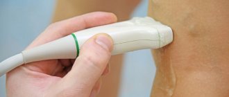
- home
- Services and prices
- Phlebology
Varicose veins (VV) is a fairly common disease that affects both men and women. It can affect the lower limbs of a person, as well as deep veins, leading to the development of thrombosis and post-thrombophlebitis disease.
Spider veins that appear on a person’s legs as a result of the development of the disease become the reason that he begins to feel unattractive. In addition to external ones, there are also internal manifestations of explosives, which are expressed in the appearance of discomfort and pain in the calf muscles of the leg. The development of the disease increases the risk of other pathologies of the circulatory system.
The key to success in the fight against pathology lies in timely diagnosis and competent treatment. A big mistake on the part of a person is the independent use of various ointments and creams, which in most cases do not bring the desired effect. As a result, time was lost that could have been spent on proper and effective therapy.
In order to get rid of varicose veins, you need to solve the following problems:
- Elimination of symptoms.
- Removal of varicose veins.
- Prevention of the development and reappearance of explosives.
Only a highly qualified specialist with sufficient experience in the treatment and prevention of pathologies of this kind can successfully cope with each of the above tasks. These are the specialists who work at the Yuzhny Medical Center. The high level of qualifications of doctors in combination with the latest equipment will make it possible to make the correct diagnosis and carry out competent therapy, guaranteeing the absence of progression or recurrence of varicose veins of the lower extremities.
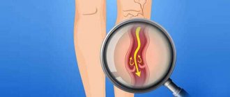
What are varicose veins?
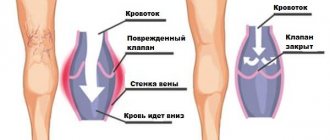
Varicose veins (in common parlance - varicose veins ) are overstretched, irregularly shaped, tortuous blood vessels that have lost their elasticity. They are increased in length and width and look like thick, convoluted blue strands that are visible under the skin. Veins become this way when the venous valves are missing or for some reason cannot perform their functions. If the valves do not work properly, blood flows through the veins in the opposite direction, downwards, accumulating in the lower sections of the veins and bursting their walls. As a result, the veins lose their natural shape, and a pathological chain of various complications begins.
Vessels: what you need to know?
22.10.2019
There are three main types of blood vessels in the human body: arteries , veins , and lymphatic vessels . They all look like a rubber pipe with many branches and different passages. pink arteries the veins are bluish and soft. Blood vessels are yellowish.
Story
Ancient anatomists connected arteries and veins with various organs. In the Middle Ages, scientists misunderstood the system of arteries ; they thought that they were not all connected to each other. This theory was refuted by the Italian physician Jacopo Berengario da Carpi at the end of the 15th century. He noticed that an artery is connected to every artery . In the 16th century, anatomists tried to answer the question of how blood gets from veins to arteries . This was clarified in the second half of the 17th century by William Harvey, who discovered blood circulation.
Under a magnifying glass
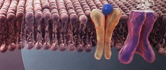
The wall of the vessel consists of three layers. Its inner part consists of a lining of flat cells (the so-called endothelium; the entire inner layer has other parts and is called intima). The middle layer consists of round and spirally oriented smooth muscle cells. The outer layer of the vessel is a compound that forms a flexible membrane (adventitia). The adventitia connects blood vessels and nerves to nourish and control vascular . Arteries have a thicker layer of muscle than veins . The capillary wall consists of a single layer of cells - the endothelium. In the veins, the endothelium forms a small pocket, a flap, in certain places, preventing blood flow. Blood vessels have a similar structure to veins .
Comparative anatomy of blood vessels
As is known, humans have a closed blood circulation. For example, mollusks, snails, arthropods and jellyfish have open blood circulation. Their “blood” or hemolymph, if present, is located directly between the organs of the body. Insects are less developed, they have only a simple tubular heart .
Anatomy
Blood vessels extend from the heart to all organs and cells. The aorta originates in the left ventricle, descends into the abdomen and flows into the pelvis, where it divides into two arteries that carry blood to the lower extremities. arteries (emanating from the aorta) also function blood to the head, upper limbs, all internal organs and skin. The arteries branch into smaller ones and eventually become capillaries. Arterial capillaries pass into venous capillaries, which converge further into veins . The superior vena cava carries blood to the heart from the lower extremities, abdomen and torso, and it also carries blood from the head and upper extremities. Blood vessels form blindly between cells flowing into stronger trunks and entering veins .
Functions
Blood vessels are used to transport blood in the body. A person has a so-called closed circulation. This means that blood flows only in blood vessels and does not accidentally “wash” certain organs. Arteries carry blood from the heart . Most of this blood is oxygenated, except for the blood that passes through the pulmonary artery from the right ventricle to the lungs . Veins carry blood to the heart . Except for the blood in the pulmonary veins , which is oxidized and flows into the left ventricle.
In the human body there are two circles of blood connected by the heart . The "small" or pulmonary circulation is where oxygenated blood is drawn through the pulmonary artery from the right ventricle into the lungs , where the pulmonary veins lead to the left ventricle. And the “large” circulation, where oxygenated blood from the left ventricle moves to the right atrium, and from there to other arteries and organs. There the blood loses oxygen and the veins return it to the heart . Blood vessels collect saliva in the intercellular spaces and transport it to the veins .
Published in Cardiology Premium Clinic
How common are varicose veins?
Varicose veins are one of the most common diseases of the vascular system. According to some statistical estimates, from varicose veins . The number of people who have varicose veins increases with age, and women are affected much more often than men. According to statistics, in the age group under 25 years only 8% of women suffer from varicose veins, and in the older age group - 55 years and older - 64% of women are affected by varicose veins.
How can you recognize varicose veins in yourself?
The most common sign of varicose veins is fatigue, dull pain, a feeling of heaviness and fullness in the legs after sitting or standing for a long time. Often these symptoms appear or worsen in the evening. However, it is usually impossible to determine exactly where it hurts. And if these unpleasant symptoms - fatigue, heaviness, pain - go away after resting with your legs elevated, then they are really caused by varicose veins (unless some other cause is reliably identified).
However, do not rush to blame everything on varicose veins, especially if there are no external signs in the form of dilated veins. Some other painful conditions may also exhibit the same symptoms.
Leg cramps
With varicose veins, painful nighttime cramps in the leg muscles can actually occur (in other words, “leg cramps”). Most often, cramps appear in the calf muscles and can sometimes be so painful that the patient wakes up. Moreover, night cramps usually occur after a hard day, when the patient had to stand or sit a lot.
Symptoms of varicose veins
Symptoms of a disease are signs that clearly indicate its development. They are divided into:
- Subjective: Mild and aching pain in the calf muscles.
- A burning sensation and itching along the veins affected by varicose veins.
- Heaviness in the legs, increasing towards the end of the day.
- Skin hyperpigmentation.
- Increased fatigue of the lower extremities.
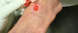
- Trophic venous ulcer of the leg.
- Pain in the calf muscles, aggravated by walking.
- The appearance of swelling in the lower legs and feet.
- Varicose veins of the saphenous veins, which are clearly visible even without the use of special equipment.
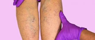
What veins look like
The very first warning sign of problems with the veins is swelling of the lower extremities at the end of the day. Swelling is especially pronounced if a person spends most of the day standing on his feet. It can disappear in the morning after a night spent resting.
However, if you do not pay due attention to this problem, the condition can worsen significantly. Intradermal veins in the legs with varicose veins become dark blue, protruding above the surface of the skin of the legs and feet. Outwardly, they look like bunches of red grapes that are overripe. Such external manifestations of pathology are accompanied by pain in the calves, a feeling of heat in the legs, swelling and cramps in the calf muscles. Over time, these symptoms are accompanied by a change in the appearance of the skin.
Is varicose veins inherited?
It is now known that varicose veins are hereditary. Scientists even believe that they were able to isolate a separate gene responsible for the development of varicose veins. It is not yet clear whether this gene causes malformations of the venous valves or malformations of the vein walls themselves. But there is no doubt that these studies will help develop a gene therapy technique - perhaps the most promising way to prevent and treat varicose veins. Unfortunately, this is still a matter of the rather distant future, and gene therapy is not yet available to patients with varicose veins.
Classification and stages
Like any disease, VV has several stages, differing from each other in the degree of spread of the pathology and symptoms. Among them, the following stages are distinguished:
- Initial (or compensation).
- Second (or subcompensation).
- Third (or decompensation).
A detailed description of each stage can be found here.
It is worth noting that complications can occur at any of the above stages, but their greatest likelihood is inherent in the last two. VV can serve as an impetus for the development of diseases such as:
- Thrombophlebitis.
- Erysipelas.
- Deep vein thrombosis.
- Trophic eczema.
A visit to a specialist, made at the first signs of the disease, will help reduce the risks of worsening the situation and begin removing varicose veins. You should not ignore even minor symptoms, because this can lead to undesirable and extremely negative consequences.
Varicose veins during pregnancy
Pregnancy does not cause varicose veins, but it is often a trigger for the appearance of varicose veins in those women who are predisposed to it. For example, in people with congenital insufficiency or even absence of venous valves. This fact has already been established quite definitely, because many pregnant women do not develop any varicose veins. Sometimes varicose veins appear only during the fourth, fifth or tenth pregnancy.
And in some women, they appear during pregnancy and disappear immediately after the birth of the child. Pregnancy acts as a triggering factor for varicose veins due to the fact that during pregnancy the content of sex hormones - estrogen and progesterone - in a woman’s blood increases sharply. These hormones, in high concentrations, help soften the venous walls, the veins stretch, and the valves cannot close normally because of this.
Physiology/hemodynamics
Normally, oxygenated blood leaves the left side of the heart through very large arteries. Which then branch into smaller and smaller arteries, then into arterioles and capillaries, which penetrate all organs and tissues of the human body. They are visible only under a microscope. The capillary bed connects the smallest arteries (arterioles) with the smallest veins (venules). Capillaries are very small vessels with very thin walls, thanks to which oxygen and nutrients easily flow from the blood into the tissues of the body. The tissues, in turn, release carbon dioxide and various waste products into the blood, which returns through the veins to the heart (Figure 1).
Picture 1.
Blood from the heart enters the large arteries (indicated in red), then it flows into smaller arteries and arterioles of the upper and lower extremities, as well as other organs and systems of the human body. Then the blood enters a network of tiny vessels - capillaries, which penetrate all human tissues and organs. The blood releases oxygen and various nutrients, and then returns through the veins (highlighted in blue) to the heart.
The venous system of the lower extremities consists of superficial and deep veins. Deep veins are large vessels through which the bulk of the blood moves due to the work of muscles. Superficial veins are smaller vessels that collect blood from the skin and subcutaneous tissue, and, due to the work of venous valves, move it upward, back to the heart. The superficial and deep veins communicate with each other through communicating or perforating veins, which are also equipped with valves. It is due to the operation of the valves that blood moves from the bottom up and from the superficial veins to the deep ones; such one-way movement is the key to the proper functioning of the veins.
Regulation of blood flow through the vessels is carried out by the nervous, endocrine system, as well as local vasoactive substances produced in tissues. This complex regulation allows blood flow to increase or decrease depending on the body's needs, for example, increased blood flow in the muscles during exercise, and decreased at rest. By changing the tone of skin vessels, body temperature is regulated. When it is cold, the blood vessels in the skin constrict, blood moves closer to the center of the body, due to this mechanism the body retains heat. On the contrary, when it is hot, the blood vessels in the skin dilate and the body gives off more heat. Various body injuries and injuries trigger processes that can cause blood flow to increase or decrease, for example, in the area of a skin burn or in a sprained area.
The walls of the veins are very thin and pliable, so the venous system can change its capacity to accommodate different amounts of blood. Blood volume is proportional to the pressure inside the veins. When the amount of blood in the veins decreases or its pressure on the vein walls decreases, the veins collapse like an empty balloon. When the volume of blood or its pressure on the walls of the vein increases, the veins expand, like an inflated balloon. If the pressure in the veins becomes very high, the venous wall stretches, its permeability increases and the vein allows fluid to pass through, which rushes into the tissue. This is how swelling occurs.
To maintain normal blood circulation in the body, the following 4 components are very important:
- Normal functioning of the heart, which, when contracting, works like a pump
- Pressure gradient between areas of low and high venous pressure
- “Muscular-venous pump” - the muscles of the lower extremities contract and work like a pump, pushing blood to the heart
- Normal, non-dilated, completely patent veins, with functioning venous valves
(1) The heart is the main pump of the human circulatory system. Blood moves through the arteries due to rhythmic heart contractions. It is important to understand that venous blood, after returning to the heart, must be pumped to the lungs, where it is enriched with oxygen. If the vein does not perform this function, as happens with heart failure, then venous blood stagnates and edema can appear even with absolutely normal veins.
(2) According to the laws of physics, any liquid moves from a high pressure zone to a lower pressure zone. The pressure difference between different zones is called gradient. There are such zones in the human body, thanks to which blood can move against the force of gravity. For example, the pressure in the veins of the lower extremities is higher than in the veins of the pelvis and abdominal cavity, and in the right parts of the heart it is even lower and may even be negative, which is why venous blood moves towards the heart. With some diseases of the lungs and heart, the pressure in the right parts of the heart may be increased, which can also lead to edema.
(3) “Muscular-venous pump” - like the heart, which pumps blood through the arteries, the muscles of the lower extremities contract to pump blood through the veins. The most important muscles performing the pumping function are the gastrocnemius muscles. There is also a venous network in the foot area; the muscles and ligaments of the foot also act as an additional pump. With each step, the muscles of the foot and lower leg contract rhythmically, pushing blood through the veins towards the heart, overcoming the effects of gravity. If a person leads a sedentary lifestyle and there is not enough activity in his life, the muscular-venous pump ceases to function normally and edema may appear. It can also occur as a result of injury or after a stroke. Sometimes the gait of older people changes, it becomes shuffling, the old people seem to shift from one foot to another. In this case, the muscular-venous pump also stops working and swelling may appear.
(4) Most veins in the human body are equipped with valves that allow blood to flow in only one direction. For the normal functioning of the venous system, the valves must be intact, that is, not damaged, and functioning correctly. As a result of contraction of the leg muscles, a portion of blood moves up through the veins, the valves allow blood to pass upward, and immediately close. They work like the rungs of a ladder, allowing blood to move forward towards the heart.
Figure 2.
The work of the “muscular-venous pump” is similar to a pump that pumps blood from the lower extremities to the heart. Due to the many valves with which the veins of the lower extremities are equipped, blood moves only in one direction: from the more superficial layers to the deep ones and from bottom to top, towards the heart. (a) When muscles contract, blood is forced out of the veins and moves upward. (b) When the muscles relax, the valves close, preventing blood from flowing back.
Each venous valve consists of two thin elastic flaps located opposite each other, opening and closing synchronously. If the vein dilates more than normal, as happens with varicose veins, the valve flaps cannot close and block the lumen of the vein, as a result, the blood moves in the opposite direction - this is called reflux. The flow of blood through a vein can also be disrupted if a blood clot forms that blocks the lumen of the vein. In both cases, venous pressure increases significantly, the vein wall becomes thinner as it stretches, and the liquid part of the blood seeps into the tissue, causing swelling. Edema is one of the main signs (symptoms) of improper functioning of the venous system.
If venous outflow is disrupted for a short time, for example after an air flight or prolonged static exercise, the main manifestation is swelling, which completely disappears overnight. If venous edema persists for a long time, for months, the skin and subcutaneous tissue begin to change, thickening and darkening may appear in the lower leg area, and subsequently infection, erysipelas, and microbial eczema may occur. All this can lead to the formation of long-term non-healing trophic ulcers.
Other causes of varicose veins
Such a widespread prevalence of varicose veins in highly developed Western countries is probably associated with the lifestyle of the population. For example, we spend a lot of time sitting on chairs. From kindergarten until graduation, a person sits for at least 40 hours a week (counting approximately 5 hours during the day in class, 3 hours in the evening doing homework, watching TV, and so on 5 days a week). Now let's multiply these hours by 10 months a year, and so on - up to 17 years. Then - work in some institution where you have to sit even longer. When a person sits in a chair, the veins running along the back of the thighs are compressed, and the calf muscles (the rhythmic contractions of which help move venous blood to the heart) do not work.
Another important factor is nutrition. In Western countries, people prefer a low fiber diet. With such a diet, fecal matter becomes denser, and constipation often occurs. When straining to move hard stool, the abdominal muscles tense and the pressure in the abdominal cavity increases significantly. High pressure spreads to the veins running along the back of the abdominal cavity and to the veins of the legs, which dilate, causing the venous valves to leak.
Varicose veins in older people
Why varicose veins more common in older people, and especially common in women?
1. To answer briefly - because their vascular system wears out with age and, sooner or later, fails. However, there are still many objective reasons why older women suffer from varicose veins more often than younger men and women. Firstly, since women generally live somewhat longer than men, there are correspondingly more elderly women than elderly men, and their veins have been working harder for a longer period of time.
2. Men don't get pregnant. Even if varicose veins that appeared in a woman during pregnancy disappear soon after the birth of the child, these veins were still abnormally enlarged within a few months. And with age, all the muscles of the human body, including the smooth muscles of the vascular walls, become less elastic than in youth. And the veins, which had already expanded once, during pregnancy, in old age again become a little wider than normal.
3. Nowadays, many women over the age of 30 are resorting to hormone replacement therapy, which was originally intended to relieve the unpleasant symptoms of menopause. There is no doubt that hormone replacement therapy helps women look younger, feel better, and generally cope with the menopause years more easily. Doctors' observations also confirm that hormone replacement therapy to some extent reduces the frequency of angina attacks and prevents a decrease in bone strength due to osteoporosis.
However, hormonal supplements at the same time soften the vein walls in the same way as increased levels of estrogen and progesterone during pregnancy. This side effect of hormonal pills is all the more dangerous because the walls of the veins are already becoming weaker - due to natural age-related changes in the muscle layer. So, additional clinical studies are needed to definitively clarify this issue.
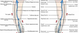
Veins of the lower extremities
Vienna
- these are vessels that ensure the outflow of blood from organs and tissues to the heart. The wall of the veins consists of three layers: internal (endothelium), middle (muscular) and external (adventitia). Unlike arteries, the walls of veins are thinner and contain few elastic fibers. Therefore, the veins are less elastic and collapse easily. In this case, the diameter of the veins is larger than that of the arteries. The peculiarity of the veins is that their diameter depends on many factors: body position, blood pressure, blood flow speed, condition of the valves and breathing phase.
The peculiarity of the veins in the legs is that they have valves. Venous valves are folds of the inner lining; they allow blood to flow towards the heart and prevent it from flowing back.
The flow of blood is carried out due to respiratory movements, the existence of constant muscle tone of the venous wall, constant support of blood from the arterial end of the capillary bed, and the suction action of the right parts of the heart. The main role in moving blood is played by the so-called “muscular-venous pump”. The deep veins that run in the legs are surrounded on all sides by muscles. When walking and physical activity on the legs, the muscles contract and squeeze blood upward.
If the valves are malfunctioning, during the operation of the muscle pump, there is no drop in pressure in the deep veins during muscle contraction. Venous blood is retained in the sinuses and venules, which leads to changes in capillary exchange parameters and the development of edema, pigmentation, itching and other symptoms of venous insufficiency.
The outflow of blood from the lower extremities is provided by three interconnected and clearly interacting systems: superficial veins, deep veins and communicating veins (perforators) connecting them.
Superficial veins
and their tributaries form venous networks under the skin. They can be felt and are quite visible. This network is especially clearly visible on the dorsum of the foot. From the superficial veins of the leg, it is customary to distinguish the large and small saphenous veins of the leg. Both saphenous veins receive other superficial veins along their path. Varicose veins on the legs concern the superficial veins.
Deep veins
. These veins are located between the muscles and connective tissue. The main outflow of blood (85-90%) is through the deep veins. These veins have valves that prevent blood from flowing back.
Superficial and deep veins are connected to each other by communicating veins
. The reason for the existence of these veins is to equalize the pressure between them. Damage to the valves of the communicating (perforating) veins leads to blood flowing from the deep veins to the superficial ones. Normally, the valves of these veins allow blood to flow in only one direction - from superficial to deep.
Types of varicose veins
Varicose veins are divided into two main groups:
- The first group includes primary varicose veins, caused by a hereditary predisposition to varicose veins.
- The second group includes varicose veins that appear after damage to the venous walls as a result of injury with the formation of blood clots in the veins or thrombosis.
When a clot or thrombus passes through a vein, the integrity of the venous valves is disrupted and secondary varicose veins are formed.
Varicose veins
Varicose veins are bundles of thin, purple or red veins that appear around the knees or ankles. (Sometimes such vascular “webs” can appear on the face, near the nose.) These vessels cannot be called varicose veins, since, by definition, varicose veins are veins that are increased in length and in diameter. In fact, these are slightly dilated venules (vessels connecting capillaries to the veins themselves), which are located close to the surface of the skin.
Such dilated venules appear due to increased levels of female sex hormones in the blood and are often found in women taking oral contraceptives. But venules can expand even in the presence of varicose veins of larger veins that do not appear externally. However, women with varicose veins often experience symptoms very similar to those of varicose veins.
Treatment of varicose veins
Treatment depends on the severity of the disease. If the disease does not manifest itself too strongly, then conservative treatment is best:
- regular rest with your feet up,
- elastic bandaging (or special elastic stockings),
- physical exercises for leg muscles.
If these measures are not enough, the veins affected by varicose veins must be surgically removed at the Phlebology Center. Or using new, experimental methods - elastic strengthening of the venous walls is carried out surgically. That is, a special elastic plastic cover is placed on the outer surface of the affected veins in places of varicose veins, where incompetent venous valves are located. And finally, to treat dilated venules or varicose veins of small veins remaining after surgery, sclerotherapy - that is, the introduction of sclerosing substances into the areas of dilation, which causes clogging of the pathological vein. Blood returns to the heart through normal venous vessels.
Introduction:
The circulatory system is responsible for moving blood throughout the body. The main components of this system are the heart and blood vessels. The heart is the central organ of the circulatory system. With each heartbeat, blood is pumped through the arteries and delivers oxygen and nutrients to various organs and tissues, after which the blood returns back to the heart through other blood vessels - veins.
There are three types of blood vessels that play different roles. The two main types are arteries and veins. Arteries carry oxygenated and nutrient-rich blood away from the heart, and veins return waste blood back to the heart. Lymphatic vessels are the third component; they filter and “purify” the liquid part of the blood - plasma, before returning it to the general bloodstream.
Complications during treatment
The main danger with conservative treatment (elastic stockings, exercise and resting with legs elevated) is its possible ineffectiveness.
Surgical treatment of varicose veins of the lower extremities should currently be performed by experienced vascular surgeons and phlebologists . Often complications and relapses after surgical treatment are caused by the fact that the operation was not performed by a specialist from the phlebology center.
With sclerotherapy, the main nuisance is small dark spots that can remain at the injection sites for several months, or in some cases forever.
Dilated veins after surgical treatment
If varicose veins have been removed, varicose veins will no longer appear in their place. However, sometimes varicose veins are found after surgery - in veins that were not previously affected, or in small veins that were not identified during the preoperative examination. Varicose veins after surgery appear because the blood is forced to find new outflow paths. At the same time, a larger volume of blood is redistributed to the remaining veins than before, and if there were any defects in the valves or walls, then new problems arise. New varicose veins, as a rule, bring cosmetic inconvenience and can be easily eliminated by a phlebologist using modern sclerotherapy techniques.
Treatment methods
Modern methods of treating varicose veins are aimed at reducing the degree of disability and trauma, which contributes to a faster recovery of the patient. Main therapeutic techniques include:
- Sclerotherapy. This method involves introducing a special medication into the lumen of varicose veins of the legs, causing a chemical burn of the internal venous wall. This leads to their gluing and cessation of pathological blood flow through them. Can be used alone or in combination with other types of manipulation. It is carried out without prior anesthesia with skin punctures using a thin needle. The duration depends on the scale of the lesion.
- Foam sclerotherapy, which involves the preparation by a specialist of a special medication of foam that can use an impressive area of the internal walls of the affected venous vessels. Used to treat large diameter veins.
- Endovenous laser coagulation, which is performed using a laser device on the main trunks of the leg veins and allows you to stop the pathological flow of blood through the affected veins due to the burn of their inner walls and their subsequent gluing. Laser treatment for varicose veins is available at the Yuzhny Medical Center.
- Miniphlebectomy, aimed at eliminating subcutaneous nodes and tributaries enlarged by varicose veins through punctures of the skin. It has excellent cosmetic effects and is used alone or in combination with other therapeutic methods under local anesthesia.
- Elimination of incompetent perforating veins, performed for the prevention of venous insufficiency and treatment of trophic disorders, including ulcers.
- Combined phlebectomy, which is a combination of some methods of treating veins, based on the indications and nature of venous pathologies.
Yuzhny Medical Center offers its clients modern approaches to the treatment of varicose veins. Qualified specialists work here, ready to conduct a competent examination in order to make a diagnosis and carry out effective treatment, more about which you can read here.