Chronic venous insufficiency (CVI) is a pathology that develops due to a violation of the outflow of blood from the veins of the extremities. Frequent symptoms are changes in skin color, cramps, swelling, pain. The most common is chronic venous insufficiency of the lower extremities.
Often, patients ignore the first symptoms of CVI and turn to a phlebologist with deteriorated health and the condition of the lower extremities. At the earliest stages, the disease is treated conservatively; when pronounced signs of impaired venous circulation appear, complex therapy is required, usually including treatment of varicose veins, nutritional correction, physical activity and wearing compression garments (1, English).
From the article you will learn the causes of chronic venous insufficiency and features of the development of the disease. Classic symptoms and method of diagnosis according to the international classification. Diagnostics and therapy are discussed in detail.
CVI – causes of chronic venous insufficiency
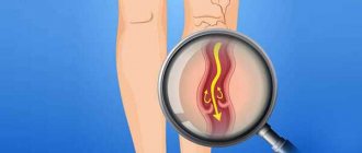
CVI is a symptom complex in which there are problems with the lumen of the veins. Usually the vessels have normal lumen. If there are disturbances, the capacity of the veins is significantly reduced. Overload of the venous system leads to impaired blood return, which is manifested by the appearance of symptoms of chronic venous insufficiency.
Impaired venous blood return leads to symptoms of CVI
Is CVI dangerous? Wikipedia states that CVI actively develops and progresses in women 30-50 years old, especially those with a hereditary predisposition. Often, symptoms of CVI appear during pregnancy, when body weight increases significantly.
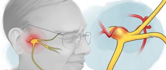
Among the factors leading to CVI is weak physical activity.
The appearance of CVI is provoked by:
- travel in transport without changing position;
- long static position at the workplace;
- excess weight;
- tendency to constipation;
- staying in the heat for a long time;
- straining when lifting weights.
In addition, CVI can occur with systematic exercise and taking hormonal contraceptives; CVI often occurs during pregnancy.
Diagnosis and treatment of CVI
At an appointment with a phlebologist at the Ambulatory Surgery Center, the patient is diagnosed based on the medical history, the patient’s complaints, and the results of the study. The main method of instrumental examination is duplex ultrasound scanning. In our clinic, functional diagnostic doctors during the study not only confirm the presence of CVI, but also help surgeons determine the extent of damage to the venous system and the choice of tactics for further action. In some cases, the attending physician may prescribe a duplex angioscan or X-ray contrast study (phlebography).
Treatment of chronic venous insufficiency is a set of measures, the purpose of which is to restore the functionality of the venous and lymphatic systems, eliminate the pathologies that arise from them and prevent relapses. The method of therapy is selected individually for each patient, taking into account the degree of pathological changes, medical history, age and concomitant diseases.
Conservative treatment
A course of treatment is prescribed, lasting on average 2 - 3 months, then after a certain period it is resumed. The course usually includes:
- medicines (phlebotrobic drugs);
- local use of antiseptic ointments;
- corticosteroid drugs;
- elastic compression (bandages and compression hosiery);
- treatment of associated secondary infections;
- elevated position of the legs when lying down.
In some cases, antibiotics and diuretics are prescribed to reduce swelling. The greatest role in the routine treatment of CVI is played by elastic compression, including hardware, pneumatic, and leg elevation. Compression is indicated for all patients, even those with ulcers. In this case, I use elastic bandaging, and then wear special stockings. They should create a distal pressure of 20–30 mm Hg. for patients of the first stage, 30–40 mm Hg. – second and up to 60 mm Hg. at the third.
The effectiveness of therapy directly depends on the active participation of the patient. He needs to strictly follow the doctor’s recommendations and build his life in such a way that conditions that aggravate the course of the disease are not created.
Surgery
Surgery is performed only in 10% of patients with:
- severe concomitant diseases;
- trophic disorders;
- transformed tributaries of the great (small) saphenous veins;
- relapse of varicose veins and in other similar cases.
For this purpose, a minimally invasive surgery technique is chosen - miniphlebectomy.
. The operation is performed under local anesthesia. Through small punctures, the vascular surgeon gains access to the affected areas of the veins to perform ligation (vessel ligation), vein removal, and valve reconstruction. The tasks of a vascular surgeon include eliminating pathological blood discharge and truncation of varicose veins. After the operation, an hour later, the patient can go home; a hospital stay is not required.
CVI - symptoms
CVI may not manifest itself for a long time, but the symptoms that appear significantly affect the patient’s quality of life. The first symptoms are a feeling of heaviness in the legs, very often swelling of the legs and feet appears. Next comes bursting pain. The calf muscles may cramp at night, and the patient experiences a feeling of heat in the lower extremities.
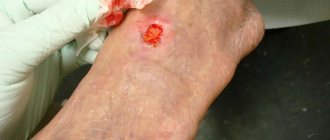
Advanced forms of venous insufficiency - trophic eczema and ulcers
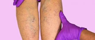
Spider veins and veins begin to appear through the skin, which with varicose veins become noticeable in the form of plexuses, similar to a bunch of grapes. The skin becomes drier and becomes covered with brown “islands” that grow as the disease progresses. Eventually, a trophic ulcer forms in their center.
The mechanism of the onset and development of the disease
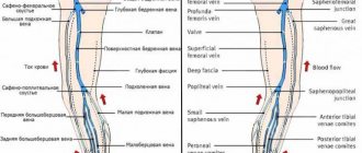
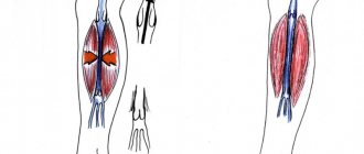
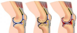
Under the influence of gravity, the blood in the vessels descends to the lower extremities, the body has to make efforts to lift it. Venous valves prevent blood from flowing downwards, which are actively helped in this by physical activity, muscle contraction and bending of the knees. The combination of these factors ensures normal blood flow.
Maintaining a constant resistance to gravity is possible due to physiological changes in the lumen of blood vessels when changing body position, the operation of the valve apparatus and the tone (elasticity) of the venous wall. If one of these component mechanisms is disrupted, pathological processes begin to affect the entire system as a whole. Loss of elasticity of the section of the vein below the valve, and its expansion leads to valvular incompetence, the inability to maintain blood flow for subsequent rise. Stagnation of fluid leads to increased pressure to move blood upward. But, over time, increased pressure increases the volume of the part of the vein that has lost its elasticity.
Venous reflux (reverse flow of blood from top to bottom) can join the pathological process. The liquid begins to stagnate and put pressure on the walls of the vessel. As a result, blood plasma leaks into the surrounding tissue, causing swelling. The situation develops similarly with initial valvular insufficiency.
Simultaneously with circulatory failure, the lymphatic system is also overloaded. Trophic disorders contribute to the formation of trophic ulcers. Trophic ulcers are long-term non-healing wounds (6 months or more) affecting the skin and tissues. They form on the lower leg, are surrounded by an area of inflammation, and have a high risk of infection.
CVI - stages of chronic venous insufficiency
The disease has several stages:
- first the venous wall changes;
- valves stop functioning;
- perverted blood flow occurs, leading to venous hypertension;
- varicose veins appear;
- skin pigmentation changes;
- Varicose eczema may appear;
- at this stage, thrombosis often develops, affecting the deep veins;
- complications include thrombophlebitis, pulmonary embolism.
The walls of the veins become more permeable, lymph enters the blood, trophism is disrupted and inflammation occurs (10-15% of cases). The last stage of CVI is a venous trophic ulcer (4% of cases)
Classification of the disease
Acute venous insufficiency has no classification by stages. Let's consider the degrees and stages of the chronic form of the disease. To fully describe the patient’s diagnosis, doctors use the CEAP classification, that is, assigning a kind of “code”, for example: C4a, S, Eс, Ad, Pr, 8, 10, 11, 17. It is deciphered based on the following classification characteristics.
C – pathology class:
- C0 – no visible symptoms;
- C1 – spider veins;
- C2 – dilated veins from 3 mm;
- C3 – swelling in the areas of the legs and ankles;
- C4a – dermatitis, pigmentation and other skin lesions;
- C4b – increased pigmentation, thickening of the skin;
- C5 – self-healing skin wounds;
- C6 – non-healing trophic ulcers.
The following index indicates whether the patient has complaints: A – without symptoms or complaints, S – there are complaints.
E – cause of disease:
- EC – congenital;
- Er – etiology unknown;
- Es – the reason is known.
A – localization:
- As – saphenous veins;
- Ar - vessels that connect the deep veins and subcutaneous veins;
- Ad – deep veins;
- Аn – there are no pathologies of the venous system.
P – type of pathology:
- Po – cessation of blood movement through the veins;
- Pr – valve insufficiency;
- Pr,o – both;
- Рn – no disturbances in the movement of blood through the veins were detected.
The number is the area of the venous system where the pathology is detected: from 1 to 18.
CVI - Diagnostics
Typically, patients turn to a phlebologist when obvious signs of CVI are visible to the naked eye, and the diagnosis is easy to make. Specialized clinics use ultrasound methods to diagnose CVI:
- the condition of blood vessels is studied;
- assesses how blood flows through them;
- Disturbances in the functioning of venous valves are considered.
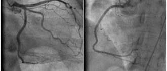
Ultrasound scanning gives a complete picture of the causes of CVI
With an ultrasound examination, the doctor receives a three-dimensional image of the vessel, reliable information about its functioning, and can make an objective diagnosis.
Classification
Clinical manifestations of venous insufficiency may vary. Therefore, for convenience, the clinic uses the international classification of chronic venous insufficiency according to the so-called CEAP system, which takes into account the causes, development, and manifestations of this pathology. Within the framework of such a system, a scale of decreased ability to work is also used, according to which the degrees of chronic venous insufficiency are distinguished:
- 0 – no symptoms;
- 1 – symptoms are present, but the patient is able to work;
- 2 – ability to work is possible with the use of supporting means;
- 3 – even with the use of supportive medications, the patient is unable to work.
CVI – complications of chronic venous insufficiency
Many patients have a common belief that CVI is not too dangerous, that it is just cosmetic. In fact, disturbances in the venous blood flow of the legs provoke the formation of blood clots. Therefore, CVI has two main complications: thrombosis and thrombophlebitis. In the first disease, the deep veins suffer:
- the affected area acquires a bluish tint,
- tissues swell
- movements are accompanied by pain.
With thrombophlebitis, blood clots form in the superficial veins. Wherein
- the skin turns red;
- Painful lumps form under the skin
- When walking, the patient experiences sharp pain in the legs.
A detached blood clot can clog a large vessel and cause death. Acute manifestations of these diseases are treated only in a surgical hospital. You can see the danger of CVI - photos, the results are on websites on the Internet.
What happens if venous insufficiency is not treated?
Venous insufficiency is a trigger for a number of disorders and diseases with more serious consequences:
- Skin changes (pigmentation)
- Eczema (skin inflammation)
- Ulcers (venous ulcers)
Compression jersey medi
Thanks to the breathable and elastic material, compression jersey provides high wearing comfort. Modern medical compression hosiery is visually indistinguishable from model hosiery, but provides high medical effectiveness when used.
The human body
How do veins work?
Vienna
Product Tips
Ideal compression product
Compression hosiery
CVI. How to treat the disease with creams?
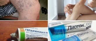
Treatment with all kinds of ointments is very common at the household level. Many patients stop treatment at this point. Most ointments only temporarily relieve swelling and pain. CVI – how to treat it? This question is asked by all patients. Such popular remedies as heparin ointment, venoruton, venitan, hepatrombin cope well with the external manifestations of CVI at an early stage, but cannot eliminate its cause. For effective treatment that will give long-term results, a visit to a phlebologist or vascular surgeon is mandatory.
Prevention
Venous insufficiency is easier to prevent than to treat. Simple but effective prevention methods are:
- restriction of being in a static position (this is standing and sitting with legs down);
- feasible physical activity;
- correction of body weight.
Patients at high risk for CVI should:
- wear elastic stockings;
- apply elastic bandaging of the lower extremities.
On our website you can find answers to the most common questions - for example, does venous insufficiency of the brain occur, how does the pathology progress in pregnant women, what complications can develop.
CVI - how to cure without consequences?
This can be done even in difficult cases; treatment of CVI in Moscow can be carried out at a high level. True, then surgery will be needed to restore blood flow in the veins. But in the early stages, conservative treatment of CVI without surgery is sufficient. In particular, elastic compression is used. For this purpose, special medical knitwear is used in the form of stockings, golf, tights (conservative treatment).
When treating CVI, it is very important not to waste time using various ointments that give a local and short-term effect, but to seek help from a qualified specialist.
Opportunities of the Innovative Vascular Center
- Thermal methods for treating varicose veins are endovenous laser coagulation (EVLC) and radiofrequency obliteration (RFO) of the veins of the lower extremities.
Endovenous laser coagulation is an effective treatment for varicose veins, the principle of which is based on the thermal effect of laser energy. This treatment was introduced in 2001 and is still the best method. With laser coagulation, the damaged vein is heated by a laser beam, which provides a strong effect of damaging the collagen of the venous wall, causing an inflammatory process in the vein and its overgrowth. Advanced varicose veins on the legs, which are treated with this method, regress completely and without a trace, and its main symptoms disappear: swelling, heaviness in the legs, hyperpigmentation of the skin. Varicose veins have been treated at the Innovative Vascular Center in Moscow using lasers since 2005, since the advent of this technology in Russia.
EVLT begins with the installation of a laser fiber into the lumen of a varicose vessel through a skin puncture, which is guided along the affected vein to the site of the incompetent valve. For the patient, this method is a safe, painless and reliable way to prevent further development of the disease and its complications. Complete elimination of varicose veins is observed in 98% of patients with proper use of the EVLT method. The capabilities of this method make it possible to both treat varicose veins on the legs in women and correct venous outflow in case of trophic ulcers.
Radiofrequency obliteration (RFO)
Treatment of varicose veins using the radiofrequency obliteration (RFO) method is a similar thermal method, but heating of the tissue of the venous wall occurs according to different physical principles due to radio wave energy. Radiofrequency obliteration allows you to remove varicose veins and eliminate its symptoms; such treatment in its immediate and long-term results does not differ from EVLT, but is more painstaking for a phlebologist.
Other thermal methods
When deciding how to treat varicose veins, phlebologists often used exotic methods. Varicose veins were treated with thermal effects using superheated steam and bipolar electrocoagulation. However, modern thermal methods are more effective and they allow the doctor to prevent further development of varicose veins, and the patient to be treated on an outpatient basis without disturbing his lifestyle. In the hands of a novice phlebologist, thermal ablation methods can cause unpleasant complications: decreased sensitivity, burns, seals. The effectiveness of this method in the hands of an experienced phlebologist is more than 98%, and the laser method and RFO make it possible to get rid of not only the initial form but also severely pronounced varicose veins on the legs without incisions. In the photographs from the “Treatment Results” section you can see the view before and after minimally invasive treatment.
Non-thermal methods for eliminating stem reflux
For many years, phlebologists have been thinking about how to cure varicose veins of the lower extremities without incisions and pain. The disappearance of the saphenous veins in the arms after frequent injections prompted the idea that certain substances can cause inflammation of the vein walls - thrombophlebitis and subsequent gluing of them with the disappearance of the lumen of the vein. After the advent of the Fegan method, when treatment began to be carried out based on the cause of varicose veins, the development of non-thermal scleroobliteration methods began. Since then, varicose veins on the legs, especially in women, are treated not only with a scalpel, but also with a syringe.
- Sclerotherapy
Sclerotherapy appeared in the practice of doctors at the end of the 19th century. In recent years, the method of treating varicose veins using injections of a special substance (sclerosant) has reached perfection. The main point of sclerotherapy is to inject a drug into a varicose vein, which causes inflammation and subsequent gluing of the varicose vein. Sclerotherapy does not involve eliminating the cause of venous insufficiency and is more suitable for certain forms of varicose veins or in the initial stages of the disease. Advanced varicose veins of the lower extremities are treated with more complex methods; damage to the trunk of the great or small saphenous vein does not allow one to count on a long-term effect of sclerotherapy, since a relapse is sure to occur due to reflux.
Sclerotherapy can be done if you are not allergic to tetradecyl sulfate or polidocanol. These substances are the main sclerosants. During sclerosing treatment, manifestations of thrombophlebitis may occur, especially if liquid forms of the drug are used. Sclerotherapy of perforating veins is highly effective in the treatment of venous trophic ulcers. It is possible to eliminate the manifestations of varicose veins of the lower extremities at any stage with the help of sclerotherapy, but the relapse rate is about 40% over the next 5 years.
The advantage of sclerotherapy is a good immediate effect and low cost of treatment. Injections of sclerosant lead to the gluing of veins and the cessation of the pathological process - reflux of blood through the saphenous veins. The drug is usually injected in the form of foam (Foam-Form) into varicose veins. A spasm of dilated subcutaneous vessels is formed, prolonged contact of the foam form of sclerosant with the vein wall and their subsequent inflammation and gluing. This process occurs unevenly and the degree of obliteration of the vein is not the same, so 40% of patients after sclerotherapy have relapses of varicose veins. After sclerotherapy, the affected area of the veins of the lower extremities closes and over time completely heals, and the blood flow in the opposite direction stops. To prevent the occurrence of skin necrosis due to the penetration of the foam form of sclerosant into the subcutaneous tissue, administration is carried out strictly under ultrasound control.
Foam sclerotherapy can be used as a stand-alone method or in combination with laser treatment to eliminate varicose veins. The number of sessions to eliminate varicose veins using sclerotherapy depends on the stage of varicose veins and the condition of the veins. The course of treatment usually consists of 2-3 procedures. The area of skin over the sclerotic vessel may take on a dark shade for 2-3 months (hyperpigmentation appears). It can ruin a woman's legs for several months, so this treatment is best done in the winter months. Drug treatment and ultrasound-guided vascular punctures can speed up the process of resorption of intravascular fluid accumulations (coaguls), the risk of which is about 10%. Coaguli form when there is insufficient compression, but will certainly go away over time. Many patients know that within a month after sclerotherapy, the signs of varicose veins of the lower extremities go away for many years, which is why sclerotherapy is still one of the most popular treatment methods.
- Using special glue (VenaSeal)
Since its inception, this method has aroused great interest among phlebologists. It involves gluing the trunk of the great saphenous vein with a special cyanoacrylate glue. In the lumen of the vessel, this glue polymerizes and fills the lumen of the dilated vessel. According to the developers, this method does not require any anesthesia, and a “plug” appears in the vessel, which reliably blocks blood flow. Taking this into account, half an hour is enough for the procedure to eliminate varicose veins on the legs. Venasil is the only technology for the treatment of varicose veins that does not require wearing compression stockings.
Most women can return to normal activities immediately. Symptoms of chronic venous insufficiency are relieved shortly after the procedure. The process of active promotion of this glue to the phlebological market should begin in the near future. However, there are certain disadvantages: The presence of a foreign body in the human body. The curdled glue remains in the vessel forever and can cause chronic allergies; sometimes there is inflammation of the vessel wall or rejection of the polymer with suppuration. Acute thrombophlebitis of the glued vessel may occur.
The use of glue in the trunk of the great saphenous vein does not eliminate the need to eliminate varicose tributaries, which is why doctors will have to remove signs of subcutaneous varicose veins with sclerotherapy or miniphlebectomy. The visible effect of using glue appears only when combined with other methods of eliminating varicose veins. The patient has to pay more. The unreasonably high cost of the gluing kit makes this procedure significantly more expensive than the modern laser or radio frequency method.
In our clinic, preference is given to thermal methods. We believe that it is better to give good local anesthesia than to treat varicose veins of the saphenous veins on the legs with an expensive and untested method. Moreover, the result is the same at best. If a relapse occurs, the patient will have to undergo a complex operation to remove the sealed vessel, since other methods will no longer be applicable.
- ClariVein mechano-chemical obliteration technology
The modern method of combined treatment of reflux along the subcutaneous venous trunks adds extra weight to conventional sclerotherapy. Mechano-chemical procedures mean a combination of mechanical damage to the inner surface of the venous wall and the introduction of a sclerosing drug. A catheter is inserted into the main saphenous vein through a puncture under ultrasound guidance. After installing the catheter in the desired location, the device is connected. The rotating sharp head of the catheter makes up to 3.5 thousand revolutions per minute, causing severe damage to the inner layer of the venous wall. At the same time, a sclerosing drug is injected through the catheter, which “mixes” in the lumen of the vessel and, using the rotating part of the catheter, acts on the vascular wall, causing its inflammation and gluing.
To date, the only advantage of this technology is the absence of the need for tumescent anesthesia. Mechano-chemical obliteration should, according to its inventors, trigger a stronger obliteration effect than foam sclerotherapy, although for some reason convincing data have not yet been presented. It is clear that such varicose veins can be treated with other minimally invasive methods, so its advantages are not obvious. The introduction of such therapy in Russia will begin in 2021, but statistical data on its advantages over EVLT and sclerotherapy do not yet exist. We have to wait for further studies from Europe or the USA in order to accurately determine the place of this technology.
- Miniphlebectomy
This is a modern microsurgical aesthetic method for removing varicose veins. It involves a delicate technique of puncturing and pulling out varicose veins using special tools. This operation is not for a novice phlebologist; you need to have the skills of delicate surgery. Miniphlebectomy is an operation without the use of a scalpel and is performed under local anesthesia. The punctures are carried out in the direction of the skin lines, so after 2 months they are practically invisible.
Miniphlebectomy has replaced the classical operation for varicose veins, which involves the use of 1-3 cm incisions, as it is aesthetically flawless, painless and very effective. By assuming how varicose veins manifest, the doctor can clearly plan micro-punctures and get by with minimal intervention. The patient can go home on his own feet immediately after the operation. Miniphlebectomy can be an independent effective method of treating varicose veins, or used in combination after laser coagulation of varicose veins. Removal of varicose veins is carried out using a special technique developed by Professor Varadi. This technique has been perfectly mastered by our phlebologists and allows for the removal of varicose veins on the legs - an effective treatment regardless of its cause.
CVI - treatment at home
Don't be afraid if you are diagnosed with CVI. Treatment with folk remedies will alleviate the situation. For example, venotonic drugs will help. They strengthen the walls of blood vessels, stimulate microcirculation, and quickly relieve the feeling of tired legs and swelling. Patients are also treated with blood thinning medications.
Do you need to relieve the pain of CVI? Treatment with traditional methods:
- Apple vinegar. Twice a day for 30 minutes. Apply gauze soaked in vinegar to your feet for 30 minutes. Keep your legs higher. Reviews about the product are contradictory, but the majority note a positive, weak effect.
- Bell pepper. Dilute a teaspoon of pepper powder in a glass of hot water and drink three times a day. The product stops leg pain and improves blood flow. But CVI does not go away that easily, treatment, the results are still not the same as when treated by a professional phlebologist.
Symptoms of the disease
Let's look at the general symptoms first. It should be remembered that in the initial stages of the disease they appear one at a time and are often “blurred”. The more the disease progresses, the more pronounced the symptoms, and they most often appear in groups. So:
- pain in the legs, feeling of heaviness, fullness;
- spider veins;
- swelling - first passing after rest, then permanent;
- night cramps in the legs;
- dryness, unhealthy shine of the skin, age spots or areas of discoloration;
- trophic ulcers at an advanced stage.
Consultation with a surgeon: when to contact, how is the appointment?
Symptoms of CVI by stage
| Stage | Description |
| 0 | The person is able to work, there are no symptoms, they do not appear after sports and other activities. |
| 1 | Slight pain, heaviness in the legs and swelling at the end of the day, which disappears in the morning, mild cramps. A person is not limited in physical activity and can work at the same pace. |
| 2 | Manifestations of venous insufficiency become pronounced. These are skin pigmentation, dermatological diseases, severe swelling, skin necrosis in some areas. It becomes difficult for a person to work physically or play sports. |
| 3 | Trophic ulcers appear and tissue metabolism is completely disrupted. A person loses his ability to work. |
Symptoms of AHF
Signs of the disease are more pronounced and appear much faster compared to CVI:
- Pain in the legs that increases with movement;
- just below the site of vein blockage there is large swelling;
- difficulties in any physical activity;
- pallor or bluishness of the skin;
- at the site of pathology, skin temperature decreases by 2-3℃;
- body temperature can rise to 40℃;
- increasing pain to unbearable;
- “referring” pain to the groin and pelvis area.
Important! To reduce the likelihood of a blood clot breaking off with subsequent thromboembolism, the patient needs to limit movement. Bed rest is recommended for up to 10 days; the affected leg should be higher than the body.
Prevention of chronic venous insufficiency
If you perform simple activities every day, the risk of developing CVI is reduced significantly. Even if CVI is discovered, treatment will not be so serious, especially in the early stages.
- You should lead a healthy lifestyle and play sports.
- Follow a diet.
- Wear comfortable shoes and use orthopedic insoles.
- Use elastic compression.
If the first signs of CVI still appear, you cannot postpone a visit to a phlebologist. He diagnoses the degree of CVI. Any delay in time is fraught with complications. Some of them are fatal. If the disease has nevertheless developed to a complex stage, you should not despair. Even complex cases can be cured using modern endovascular surgery, see the photo on our clinic’s website.
Treatment of venous insufficiency is available in the following branches:
Treatment of venous insufficiency in the Primorsky region
Address: St. Petersburg , Primorsky district, st. Repisheva, 13
Treatment of venous insufficiency in the Petrograd region
Address: St. Petersburg , Petrogradsky district, st. Lenina, 5
Treatment of venous insufficiency in Vsevolozhsk
Address: Vsevolozhsk , Oktyabrsky Prospekt, 96 A
Frequently asked questions from our patients on the Internet
My legs are constantly swollen, how can I restore blood circulation in my legs? Maria from Arkhangelsk asks:
Dear Maria! To restore blood circulation in the legs, you first need to find out the cause of the swelling. As a rule, from a modern point of view, you need to start with an ultrasound duplex scanning of veins and consultation with a phlebologist.
How to identify blood clots in the legs? Oksana from Vladimir is interested in:
Dear Oksana! If you have the slightest suspicion of thrombosis, you should urgently seek medical help. A good diagnosis begins and always includes vascular ultrasound. This technique allows you to accurately determine not only blood clots in the legs, but also the cause of their occurrence. However, the method has a pronounced operator dependence, and it is better to trust professionals in their field.
How to understand that there are blood clots in the veins? Maxim from Yekaterinburg is interested in:
Dear Maxim! Only a specialist can reliably understand that there are blood clots in the veins, or exclude this situation. Thrombosis can be suspected based on the following signs:
- Swelling of the limb.
- Pain and/or redness.
- Feeling of heaviness and fullness in the limb.
- Vein thrombosis has already been previously diagnosed, since blood clots in the veins often recur.
The best solution would be to consult a phlebologist with an ultrasound scan of the venous vessels. You can make an appointment at our center for a consultation at any time convenient for you by phone.
How to check veins for blood clots in the legs? Asks Valentina from Smolensk:
Dear Valentina! You can check the veins for the presence of blood clots in the legs using ultrasound duplex or triplex scanning of the veins of the lower extremities at an appointment with a phlebologist.
How to treat chronic venous insufficiency at home? Zinaida from Tambov asks:
Dear Zinaida! Chronic venous insufficiency should be treated at home only after a good consultation with a specialist who will diagnose and give recommendations. Otherwise, you have a good chance of getting complications from the disease. When you find yourself on websites that colorfully describe how and what to treat at home, remember that most often professional doctors have nothing to do with such recommendations.
How to deal with the symptoms of chronic venous insufficiency? Nellie from Ufa asks:
Dear Nelly! It is better to start fighting the symptoms of chronic venous insufficiency in the legs before these symptoms appear, that is, with prevention: regular physiological exercise, a balanced diet, the use of compression hosiery for excessive loads, air travel. If signs of CVI appear: swelling, heaviness, varicose veins, you should consult a good specialist. This is the only way you can count on success.
PS I do not advise you to treat the symptoms of CVI at home via the Internet.
How to relieve pain from chronic venous insufficiency at home quickly? Asks Alexandra from Tyumen:
Dear Alexandra! The appearance of pain with varicose veins is most often a symptom of a developed complication, varicothrombophlebitis or deep vein thrombosis. If pain occurs due to varicose veins, stopping it at home is an extremely dangerous undertaking. It is necessary to quickly seek medical help, possibly calling an ambulance. You can quickly relieve pain with any NSAID, for example, ketoprofen or meloxicam (Movalis). But this is purely a temporary measure.
Varicose veins have started, what should I do? Maria from Nizhny Novgorod asks:
Dear Maria! If you have developed varicose veins or there are pronounced signs of varicose veins, you need to contact a professional. The most competent diagnosis and subsequent treatment can be provided to you by specialized centers for the treatment of venous pathology. After the examination, the center specialist will explain to you in detail what and how to do.
What is a good prevention for varicose veins on the legs? Nadezhda from Orenburg is interested in:
Dear Nadezhda! Prevention of varicose veins on the legs includes the following aspects:
- The closest to physiological load on the lower extremities, the involvement of the calf muscles, reducing the time of static load.
- Complete, balanced nutrition.
- For long-term static conditions and air travel, wear compression hosiery of I-II compression class.
Establishing diagnosis
CVI is often equated with varicose veins, which is not correct. Varicose veins are one of the manifestations of pathology.
Diagnosis is based on medical history, examination and additional diagnostic methods. The specialist’s task is to determine the presence of chronic venous insufficiency, diagnosis according to the CEAP classification, and formulate treatment tactics.
An ultrasound of the veins is mandatory - only an ultrasound examination allows one to study the condition of the venous apparatus and make an accurate diagnosis.
The main ultrasound diagnostic methods include Dopplerography and angioscanning.
Doppler ultrasound
Used to study blood flow and diagnose reflux. Informative for the study of superficial veins with the presence of telangiectasia and reticular varicose veins (class C1). In patients with suspected damage to the deep veins of the extremities, with trophic ulcers, the method is often uninformative.
Ultrasound angioscanning
Visualization of deep and superficial veins, assessment of blood flow. According to the results of ultrasound scanning, unaffected veins, vessels with signs of reflux of non-post-thrombotic origin, and vessels changed as a result of previous thrombosis are determined. The method gives a complete picture of the condition of the superficial and deep veins of the lower extremities, and well visualizes the changed vessels.
During pregnancy, acute thrombosis, chronic arterial insufficiency, pathologies of the kidneys and liver and many other diseases, ultrasound angioscanning is an important tool for the differential diagnosis of CVI; it identifies pathologies with high accuracy, which allows you to prescribe the correct treatment.
X-ray contrast venography (venography)
Injection of a contrast agent into the vascular bed, followed by a series of x-rays. Currently, the technique is rarely performed, since ultrasound research methods have become much more informative and accessible.
People at risk
- Women are more susceptible to developing CVI than men - due to hormonal fluctuations, pregnancy, and childbirth.
- With varicose veins of the lower extremities, especially if there have been complications in the past.
- With diseases of the cardiovascular system - hypertension, atherosclerosis, heart rhythm disturbances.
- With metabolic disorders - diabetes, obesity, gout.
- Anyone whose ability to move freely or change body position is limited due to the nature of their profession, disability, diseases of muscles, joints, etc.
- Patients with a hereditary history of venous diseases.