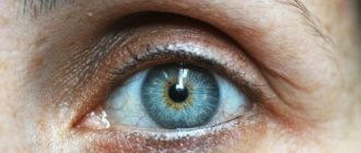
What is glaucoma
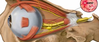
Glaucoma is a broad group of eye diseases characterized by constant or periodic increased intraocular pressure, atrophy of the optic nerve with the subsequent development of typical visual field defects and decreased visual acuity.
This disease occupies a leading place among the causes of irreparable blindness. Vision in glaucoma decreases gradually, and therefore the patient most often notices changes in visual functions already in the advanced stage of the disease. In this case, vision decreases, up to the onset of blindness, which is irreversible, as the optic nerve dies. It is no longer possible to restore sight to a patient who has gone blind for this reason!
Early diagnosis and treatment of this disease can compensate for the course of glaucoma, prevent damage to the optic nerve and associated vision loss. Therefore, it is very important to be regularly examined by competent specialists.
Primary open-angle glaucoma usually develops due to progressive impairment of the outflow of intraocular pressure. As a rule, the cause is age-related changes in the angle of the anterior chamber of the eye. IOP can also rise due to too active synthesis of aqueous humor, which is observed in people with myopia.
Angle-closure glaucoma occurs due to a sharp overlap of the anterior chamber angle by the root of the iris. What most often happens to people with farsightedness. Their eyeball is small, the anterior chamber is small, and the lens is large. These anatomical features contribute to the development of the disease.
The following factors can trigger an attack of angle-closure glaucoma:
- drinking large amounts of liquid at one time;
- long stay in a dark room;
- frequent work with a tilted head;
- instillation of mydriatics into the eye - drugs that dilate the pupil.
It should be noted that during an appointment with an ophthalmologist, a person often receives drops to help dilate the pupil and clearly see the fundus of the eye. In people with high intraocular pressure, these drugs can cause an attack of angle-closure glaucoma. That is why it is necessary to measure IOP before using mydriatics.
Complications
The most severe complication of secondary glaucoma is blindness, since people with this pathology have a high risk of optic nerve atrophy. The vascular and wound form of the disease is often complicated by vitreous hemorrhages and hyphema. Rubeosis of the iris and proliferation of newly formed vessels in the cornea are quite common. For uveal glaucoma, characteristic complications are inflammatory and infectious diseases - blepharitis, keratitis, conjunctivitis. With an eye contusion, subconjunctival bleeding often occurs due to a rapid increase in IOP. In the phacolytic form of glaucoma, in case of rupture of the lens capsule, plastic iridocyclitis may develop.
Causes of glaucoma development
Glaucoma can occur at any age and mainly affects people over 40 years of age. But this disease can also affect young people (juvenile glaucoma) and even newborns (congenital glaucoma), since most often it develops in those people whose parents also suffered from this disease. Glaucoma can be an occupational disease, develop due to age-related changes, injuries or concomitant diseases.
Risk factors include:
- age over 50 years;
- family history - the presence of glaucoma in close relatives;
- wounds, eye contusions;
- chronic ophthalmological diseases such as cataracts, high myopia, iridocyclitis, chorioretinitis;
- the presence of hypertension, hypotension, diabetes mellitus, obesity;
- sclerotic changes in blood vessels or deposition of atherosclerotic plaques in them;
- cervical osteochondrosis, leading to disruption of the innervation of the eyeballs.
How does secondary glaucoma differ from primary glaucoma?
Secondary glaucoma can also be open-angle or closed-angle, it has the same stages and signs as primary. However, a secondary increase in intraocular pressure has a number of characteristic differences from the primary forms of pathology:
- secondary glaucoma is always a consequence and complication of other diseases;
- most often the damage occurs asymmetrically, the disease affects one eye;
- vision loss occurs quite quickly;
- the processes are reversible if the underlying pathology can be treated.
Symptoms of glaucoma
Glaucoma is most often asymptomatic; deviations from the norm are often detected by chance, during an examination or visit to an ophthalmologist for some other reason.
Over time, a person begins to navigate much worse in space due to a narrowing field of vision. In the later stages of the disease, it may seem to him that he is looking at the world around him as if through a spyglass. Twilight vision is also greatly affected. The patient practically loses the ability to see something in the dark.
With angle-closure glaucoma, a person experiences severe pain in the eye, which soon spreads to the entire half of the head. The patient's body temperature rises, chills, nausea and even vomiting appear. When palpated, the eye turns out to be very hard, reminiscent of stone. All symptoms appear abruptly and develop rapidly.
Some nonspecific symptoms may indicate the presence of the disease:
- mild pain, feeling of heaviness in the eyes;
- rapid visual fatigue;
- deterioration of visual acuity at dusk;
- double vision;
- the appearance of rainbow circles when looking at the light;
- feeling of increased moisture in the eyes.
Diagnostics
At the initial appointment, the ophthalmologist will carefully examine the eyes, ask the patient about warning symptoms, inquire about the medical history, and find out the risk factors that contribute to the manifestation of the pathology. In addition to this, he will carry out the following diagnostic procedures:
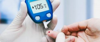
- Measure intraocular pressure using tonometry. All manipulations are simple and painless.
- Conducts testing for optic nerve damage using special instruments and equipment.
- Tests visual fields by examining features of the peripheral visual system.
- It will measure the thickness of the cornea (pachymetry), which will provide additional information in the diagnosis of glaucoma.
Dr. Trubilin’s clinic uses traditional and latest methods for diagnosing eye problems. An integrated approach is used for a comprehensive examination and making the most accurate diagnosis.
With us you can:
- undergo a visual acuity test;
- measure intraocular pressure;
- examine visual fields;
- do optical coherence tomography.
Additionally, biomicroscopy and gonioscopy of the affected area are performed. Based on the screening results, the doctor makes a diagnosis and prescribes individual treatment for glaucoma. When drawing up a scheme of restorative procedures, the patient’s age, medical history, as well as related details are taken into account.
Stages of glaucoma
According to the severity of the process, 4 stages of glaucoma are distinguished:
- I initial stage - the early stage is characterized by periodic surges in intraocular pressure, causing sharp dilation of the pupils and headaches.
- Stage II of advanced glaucoma - advanced glaucoma is manifested by a significant narrowing of the visual field on the nasal side or the formation of a large arcuate scotoma. Increased intraocular pressure provokes pain in the affected eye; sometimes the enlargement of the eyeball is even visible visually. Visual acuity decreases. At this stage, it is most often impossible to do without surgery.
- Stage III of advanced glaucoma - at this stage, glaucoma is accompanied by a concentric narrowing of the visual field and the complete loss of its large areas, that is, a significant increase in the blind spot. Sharply increased intraocular pressure leads to other pathologies: retinal detachment, clouding of the lens, and the formation of hemorrhages. Drug therapy and diet for the third stage of glaucoma serve only as an aid; the main method of treatment in this case is surgery.
- IV terminal stage of glaucoma - complete loss of central vision and complete loss of objective vision. With terminal glaucoma, there is a serious increase in intraocular pressure, it is accompanied by severe headaches and eye pain, enlargement and clouding of the eyeball - buphthalmos or bull's eye.
Depending on age, glaucoma is divided into congenital (in children under 3 years of age), infantile (in children from 3 to 10 years of age), juvenile (in persons aged from 11 to 35 years of age) and glaucoma adults (in persons over 35 years of age). All forms are acquired, except congenital.
Prognosis and prevention
With timely detection of secondary glaucoma and treatment of the disease in the early stages, the prognosis for the patient’s life and ability to work is favorable. The peculiarity of the disease is that timely treatment can restore partially lost visual functions.
The lack of specific methods for the prevention of secondary glaucoma places certain responsibilities on the person. Indeed, to prevent the disease, regular visits to the ophthalmologist are necessary to monitor the level of IOP. Regular measurement of intraocular pressure is recommended for patients in the postoperative period (within a year) after eye surgery, with traumatic eye injuries or with an unfavorable ophthalmological history (cataracts, ocular migraines, hemorrhages inside the eye).
Glaucoma treatment
Is glaucoma curable?
If there is a suspicion that a person has glaucoma, then it is possible to determine the stage of development of the disease during an examination by a specialist. The doctor will decide how to treat the patient based on the diagnostic data obtained. At the initial stage of development of the disease, it is possible to treat glaucoma without surgery, the so-called conservative method. Therapy is carried out using special eye drops that lower the pressure inside the eye, improve the outflow of fluid passing through the organ, or reduce its secretion.
A positive effect from such treatment is possible with the open-angle type of the disease, when there are no changes in the structure of the eyes. If a patient is diagnosed with angle-closure glaucoma, then medications are practically useless. And to eliminate problems, surgical treatment methods are already used. With their help, angle-closure glaucoma is curable both at the initial stage of the disease and at later stages.
Surgery for glaucoma
Surgery is indicated for open-angle glaucoma that responds poorly to prescribed medications. In case of an acute attack of angle-closure glaucoma, surgery is necessary if
if intraocular pressure cannot be normalized with drug treatment.
- Laser methods of treating glaucoma are considered the most modern and least traumatic operations:
1. Laser iridotomy is the removal of glaucoma without surgery. This type of treatment is highly effective, easily tolerated and safe. Removing glaucoma using a laser guarantees a complete cure for the disease. Laser iridotomy is performed without opening the eyeball and is a microsurgical intervention. The risk of complications is minimal.
2. Trabeculoplasty - the surface of the diaphragm is burned with a laser, which increases its tension and, as a result, permeability. As a result, more fluid swells from the anterior chamber, and the pressure decreases.
- Cryodestruction: the essence is similar to the previous method, but the effect is carried out not by a laser, but by cold, and the object is not the iris, but the sclera. It is exposed to cold, applying applications at several points at once. The operation is contraindicated for terminal stages of glaucoma, unsuccessful surgical interventions, or a history of pain. Cryodestruction is not as safe and more often causes complications. It is used if laser correction is contraindicated for a patient for some reason.
Prevention of glaucoma
Preventative measures will help reduce the damage that high blood pressure causes to the visual system, thereby preventing optic atrophy and blindness.
1. Do not overexert yourself - limit both physical and psycho-emotional stress. 2. Do not keep your head tilted - it is harmful for patients with glaucoma to engage in activities that require prolonged tilting of the head down. This applies to reading, drawing, drawing, knitting, embroidery and others. It is necessary to maintain a level head position when working at the computer or watching TV. 3. Set up the right lighting - it is dangerous for people with glaucoma to work in poor lighting. It is important to make it bright so as not to strain your eyes. 4. Give up bad habits, as smoking negatively affects the blood supply to the whole body. The transport of oxygen and nutrients to all elements of the eyeball is disrupted. 5. Choose loose clothing that does not interfere with blood circulation in the neck and head. 6. Avoid visual fatigue - it is important to take breaks while working at the computer, reading and watching movies. It is recommended to set aside 15-20 minutes every hour for rest. At this time, you need to really rest, and not change one strenuous activity to another. 7. Eat right - to prevent glaucoma, you need to include raw vegetables, fish, fruits in your diet, while reducing the amount of animal fats and sugar. 8. Consume moderate amounts of water and other drinks. You should not drink more than one glass of any liquid at a time. To be on the safe side, you can check your reaction to coffee or green tea: measure your blood pressure before and after. 9. Get proper rest, get a good night’s sleep, it is advisable not to stay up late, take a walk in the fresh air in the evening. You need to sleep on comfortable pillows. After waking up, it is recommended to do a warm-up without getting out of bed. 10. Do not refuse drug treatment. 11. Avoid sudden changes in lighting—changes in lighting intensity are a strong strain on the eyes, so before going to the cinema, for example, you need to use drops that prevent pupil dilation. 12. Constantly monitor your condition. Even with stable intraocular pressure, you should visit your doctor at least four times a year.
These measures will help avoid not only glaucoma, but also other diseases of the visual system. Prevention is recommended for everyone, since glaucoma can occur even in a completely healthy person. At Lensmaster you can undergo a free vision diagnostic by making an appointment through the website.
Methods for increasing the effectiveness of surgical treatment
In modern ophthalmic surgery, two new approaches to the treatment of glaucoma have been created, which have helped to increase the effectiveness of fistulizing operations.
In the first case, a small area of the episclera and sclera in the surgical area is excised. By reducing the number of cells in this area, the body’s fibroblastic reaction to the wound surface is also reduced.
The second approach is more radical and involves the use of antimetabolites during and after surgery. The most effective and promising in this case is the administration of cytostatics (fluorouracil and mitomycin). The experiment found that these substances suppress the activity of fibrocellular and collagen structures that can synthesize substances after antiglaumatosis operations.
Due to the use of cytostatics after surgical treatment of glaucoma, a rather pronounced avascular filtration cushion is formed.
As a result, the hypotensive effect becomes more pronounced, especially with previous unsuccessful operations, as well as in young patients.
Antimetabolites significantly reduce the risk of fibrous tissue developing in the filtering pathways created during surgery. Intraoperative administration of mitamicin was more effective than administration of 5-fluorouracil. However, the latter drug can also be used in the postoperative period, while mitamicin is not suitable for this. The high effectiveness of prescribing antimetabolites for non-penetrating sinusotomy has also been shown. These encouraging results will allow us to expand the indications for such surgical interventions and use them even in advanced glaucoma.
It should be noted that antimetabolites can negatively affect the cornea, causing toxic effects. In this regard, caution must be exercised when prescribing them.