Muscle weakness and muscle fatigue are among the most common symptoms for which neurological medical attention is sought. There are objective and subjective criteria to determine the presence of a pathological condition. When we talk about weakness, we usually mean a decrease in muscle strength, but patients often complain of general fatigue or difficulty moving in certain areas of the body.
Weakness can be present in individual muscles, affect many groups or the entire body. It occurs suddenly or increases gradually, occurs periodically or persists constantly. In paretic limbs, muscle tone changes, reflexes are quickened or weakened. Depending on the origin and localization of the pathological focus, the clinical picture is supplemented by other symptoms.
Classification
Muscle weakness is caused by a wide range of pathologies. It can be general (widespread) or local. The first implies a state of increased fatigue while maintaining muscle strength. In the second case, a violation of the pathways of nerve impulses from the central nervous system to certain muscles is noted. At the same time, for topical diagnosis it is important to understand the level of damage:
- Central motor neuron:
motor cortex, corticospinal and corticobulbar tracts. - Peripheral motor neuron:
anterior horns of the spinal cord, roots, nerves. - Neuromuscular synapse.
- Muscles.
If central or peripheral motor neurons are involved, paresis is noted with a decrease in muscle strength. In terms of severity, they can be mild, moderate, deep, or take on the character of paralysis (complete lack of movement). Traditionally, paresis is systematized taking into account the location of the lesion and clinical features. In neurology, the following classification is generally accepted:
- Central (spastic).
By eliminating the inhibitory effect of the cortex on the spinal cord, muscle tone and reflexes increase, pathological signs and synkinesis occur. - Peripheral (sluggish).
Damage to the peripheral motor neuron leads to rupture of the reflex arc, which is accompanied by hypotonia and hyporeflexia; upon examination, muscle atrophy and fascicular twitching are noticeable. - Mixed.
A combination of spastic paresis below the level of the lesion due to interruption of the corticospinal tract and peripheral, caused by a pathological process in the region of the anterior cords of the spinal cord.
Separately, psychogenic paresis is distinguished, which occurs without organic causes with complete preservation of the conductive tracts of the brain and spinal cord. Their development is associated with dysfunction of higher nervous activity. The involvement of individual limbs in the process is essential for diagnosis; therefore, in the clinic of nervous diseases, several types of paresis are distinguished:
- Monoparesis.
Weakness is observed in one limb. - Biparesis.
It is divided into hemiparesis, in which the arm and leg are affected on one side, and paraparesis, which involves symmetrical limbs - upper or lower. - Triparesis.
Muscle strength is reduced in three limbs (a combination of hemi- and paraparesis). - Tetraparesis.
Movement disorders affect both arms and legs.
Based on their prevalence, weakness of certain muscles (for example, fingers or facial muscles), entire groups (flexors, extensors) or sections (distal, proximal) are distinguished. It should be understood that the term “paresis” is used not only to describe the work of skeletal muscles, but is also applied to some internal organs (intestines, bladder).
Folk remedies
Photo: fitohome.ru
Paresis does not mean a complete loss of motor activity, so it is important to engage in therapeutic exercises and physical education for a long time, gradually increasing the load. The course of therapeutic exercises is selected by the attending physician and carried out in a medical institution. This is necessary so that each time the patient is under the supervision of a specially trained person who can assess the correctness of the exercise technique, as well as monitor the dynamics of treatment. In addition, it is important to exercise yourself and at home in order to improve and speed up the expected effect of treatment. A gradual increase in load should subsequently be complemented by movements with resistance. This will help in increasing muscle size and strength.
Therapeutic exercises should be combined with massage sessions, which improves tissue nutrition, promotes the formation of nerve impulses, and also prevents the development of muscle atrophy. You can sign up for a massage with a specialist, but given that this therapy is long-term, it is possible to teach massage techniques to relatives so that they can constantly practice at home.
For peripheral paresis, physiotherapeutic procedures have a good effect. They help improve blood circulation, stimulate metabolic processes and trophism, and help restore lost function. In this case, physiotherapy is used:
- diadynamic currents;
- electrophoresis;
- magnetotherapy.
Currently, acupuncture (acupuncture) is gaining momentum. This is one of the methods of alternative medicine, which still has a lot of critical views from various specialists, however, despite this, the demand for this procedure is progressively growing.
The information is for reference only and is not a guide to action. Do not self-medicate. At the first symptoms of the disease, consult a doctor.
SEARCH FOR TREATMENT AROUND THE WORLD WITH YELLMED
Why does muscle weakness occur?
Causes of weakness in the limbs
When considering the origin of skeletal muscle weakness, it is worth separating the concept of paresis associated with damage to nerve pathways and symptoms caused by defects in synaptic transmission and muscle damage. Often we are talking about blockade or increased destruction of acetylcholine, primary or secondary muscle pathology, reflex syndromes. Among the most well-known causes of weakness are the following:
- Myasthenia.
- Myasthenic syndromes:
Lambert-Eaton associated with slow closure of ion channels, familial infantile myasthenia. - Hereditary paroxysmal myoplegia:
hyper-, hypo- and normokalemic, Andersen-Tawil syndrome. - Infections:
botulism, tick-borne encephalitis. - Endocrine pathology:
hyper- and hypothyroidism, Conn's syndrome, Addison's disease. - Myopathies:
inflammatory (acute myositis, polymyositis, dermatomyositis), metabolic (including storage diseases), mitochondrial. - Muscular dystrophies:
progressive (Duchenne, Becker, Emery-Dreyfus), non-progressive (myotubular myopathy). - Diseases of the musculoskeletal system:
arthrosis, tendonitis, injuries. - Vascular pathology:
obliterating endarteritis, varicose veins of the lower extremities, Takayasu's disease. - Poisoning:
organophosphorus compounds, carbon monoxide, cyanides, aromatic hydrocarbons (toluene, benzene), plants (hemlock), nicotine, cocaine. - Taking medications:
D-penicillamine, antibiotics, antitumor drugs, statins, cortisone, colchicine, chloroquine.
Weakness in the muscles of the whole body is provoked by various systemic diseases and intoxications. It is noted in acute and chronic infections, autoimmune pathology, and malignant tumors. Low motor activity in older people, with immobilization, prolonged bed rest are common causes of widespread limb weakness.
Causes of facial muscle weakness
The facial muscles are innervated by the facial nerve (VII pair). Any pathology that disrupts the conduction of impulses along motor fibers - from the corticonuclear pathway to the peripheral part - can cause muscle weakness. It is also worth considering the direct damage to the muscles themselves. The list of probable pathologies includes the following conditions:
- Neuritis of the facial nerve (Bell's palsy).
- Facioscapulohumeral myodystrophy of Landouzy-Dejerine.
- Degenerative diseases:
progressive bulbar palsy, syringobulbia. - Tumors:
base of the skull (Garcin's syndrome), cerebellopontine angle, temporal bone and middle ear. - Hemorrhages and infarctions of the pons area.
- Congenital anomalies:
Chiari malformation, Klippel-Feil syndrome. - Infections:
herpetic ganglionitis of the geniculate ganglion (Ramsay-Hunt syndrome), polioencephalitis, Lyme disease, syphilis. - Meningitis:
bacterial, tuberculous, fungal. - Systemic pathology:
sarcoidosis, periarteritis nodosa, Behcet's disease. - Consequence of injuries, facial surgery, installation of a cochlear implant.
- The effect of chemotherapy drugs.
The facial nerve is often damaged by inflammatory processes in the ear and parotid salivary glands. Compression or rupture of its fibers is observed in TBI with a fracture of the base of the skull. The risk of facial nerve paresis increases in older people, with a long history of smoking, and the presence of concomitant pathologies (diabetes mellitus, arterial hypertension).
Causes of paresis
The upper or first neuron of the motor pathway begins in the motor cortex of the precentral gyrus, goes as part of the pyramidal tract through the internal capsule and trunk, ending in the anterior horns of the spinal cord. There, the impulse is transmitted to the second motor neuron, the axons of which make up the roots and peripheral nerves. Damage to these structures at any length is accompanied by paresis, which is typical for various pathologies of the nervous system:
- Strokes:
hemorrhagic, ischemic, subarachnoid hemorrhage. - Tumors and traumatic brain injuries.
- Demyelinating diseases:
leukodystrophies, multiple sclerosis, Devic's opticomyelitis. - Motor neuron diseases:
amyotrophic lateral sclerosis, spinocerebellar and bulbospinal atrophy. - Cerebral palsy.
- Neurodegenerative processes:
Wilson-Konovalov's disease, Shtrumpel's disease, Refsum's disease. - Myelopathy:
compression, spinal cord ischemia, transverse myelitis. - Damage to peripheral nerves:
polyneuropathies (metabolic, toxic, hereditary), tunnel syndromes, plexopathies. - Acute polyradiculoneuritis:
diphtheria, Guillain-Barré syndrome, ascending Landry's palsy. - Infections:
polio, meningococcal meningoencephalitis, rabies. - Vertebrogenic pathology:
intervertebral hernia, osteochondrosis, scoliosis. - Intoxication:
nerve poisons, salts of heavy metals (thallium, lead, arsenic).
Disturbances in spinal conduction are often caused by spinal cord injuries, which entail compression, ischemic damage, and swelling of the nervous tissue. There are also direct wounds - gunshots, bone fragments from fractures. Typically, the cervical spine is susceptible to such damage; the thoracic and lumbar spine are affected much less frequently. In case of concussion of brain tissue, the changes are transient in nature, in other cases they are more persistent.
Metastatic tumors (lung, breast or prostate cancer) are a common cause of compression of the spinal cord; lymphoma, multiple myeloma, and epidural hematomas are less common. If weakness appears in the arms or legs, tuberculous spondylitis, rheumatoid arthritis with subluxation of the atlantoaxial joint, and spinal vascular malformations are excluded.
Diagnostics
When conducting a clinical examination, true muscle weakness with a decrease in strength is distinguished from increased fatigue as a subjective sign. The anamnesis is used to find out information about the intensity and rate of development of the symptom, and the presence of additional signs. During a neurological examination, the doctor determines muscle strength (in points), range of active and passive movements, and reflexes. To determine the cause of muscle weakness, additional studies are prescribed:
- Blood analysis.
If an infection is suspected, a hemogram gives an idea of the leukocyte formula and ESR. Biochemical analysis allows us to identify electrolyte and hormonal disorders, the presence of antibodies to the pathogen. Toxicological examination shows the content of toxic substances in the blood. - Lumbar puncture.
Cerebrospinal fluid is taken for examination to exclude subarachnoid hemorrhages, infectious and inflammatory diseases of the brain and membranes. CSF pressure is assessed in case of volumetric processes (tumors, abscesses, hematomas). - X-ray.
X-ray of the spine is the first test prescribed for spinal injuries. Standard photographs do not show fractures of the odontoid process or the lower cervical segment, which requires special radiographs and the use of other imaging techniques. - Tomography.
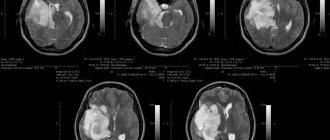
In case of damage to the central nervous system, MRI is considered the main method of neuroimaging. The method is highly informative for pathology of the posterior cranial fossa, inflammation of the meninges, and myelopathy. CT scan of the brain is more suitable for patients with basal skull fractures in the acute period of stroke. - Myelography.
This is an X-ray examination of the central canal of the spinal cord. The procedure is performed for herniated intervertebral discs, spinal injuries, and tumors. The technique shows any obstacles to normal liquor dynamics. - Electroneuromyography.
Damage to nerve trunks and fibers requires a functional study with analysis of impulse conduction to the muscles, which allows one to evaluate the speed of signal transmission, the location of damage, and the ability of muscles to contract.
If muscle weakness is accompanied by systemic disorders, the examination plan includes ultrasound of the kidneys and adrenal glands, thyroid and parathyroid glands. For vascular disorders, ultrasound and angiography are performed. The search for a malignant tumor may require radioisotope scintigraphy. Given the variety of causes, a neurologist has to carry out a thorough differential diagnosis with the involvement of related specialists.
Physiotherapy is used in the complex treatment of paresis.
Medicines
Photo: plastichno.com
In the treatment of paresis, neuroprotectors are used - drugs that help protect nerve fibers. For this purpose, B vitamins (B1, B6, B12) are prescribed, which are either used individually or in combination. An example of such a combination drug is Milgamma. This drug is able to restore metabolism inside cells, which helps slow down the process of destruction of myelin (the sheath of the nerve fiber), and also affects myelin regeneration. It is recommended to prescribe in 2 stages. At the first stage, an injection form of the drug is used, at the second, a transition to tablets is made.
Vinpocetine is used to improve cerebral circulation. Its effect is achieved by dilating cerebral vessels. In addition, the drug has antiplatelet and antihypoxic effects. Some experts consider this drug to be outdated and ineffective, but, nevertheless, it is still used to this day.
For neuroinfection, antibiotics are used if there is a bacterial etiology of the disease. The choice of antibacterial drug is made based on an analysis of the sensitivity of the microorganism that causes the infection to certain groups of antibiotics. Often, treatment begins before the test results are obtained, and broad-spectrum antibiotics are used. For example, cephalosporins may be prescribed.
It is important to understand that a phenomenon such as paresis can be a manifestation of various diseases, therefore the treatment is carried out by a qualified doctor who is able to select the necessary treatment taking into account each individual case.
Treatment of paresis
Help before diagnosis
Abruptly occurring severe local or general muscle weakness is a reason to seek medical help. In acute conditions associated with a direct threat to life, urgent measures are necessary. Suspected traumatic brain or spinal injuries require immobilization of the cervical spine with a head restraint or collar, and patients with probable spinal injuries are transported on a rigid stretcher.
In case of serious injuries with paresis of the respiratory muscles, hemorrhagic stroke, resuscitation measures are necessary - mechanical ventilation with intubation, chest compressions. At the prehospital stage, blood pressure is stabilized, cerebral edema is combated, vomiting and convulsions are stopped. Undifferentiated treatment of stroke involves the use of neuroprotectors.
Conservative therapy
A decrease in muscle strength requires a comprehensive solution aimed at eliminating the cause of motor dysfunction and restoring lost abilities. The basis of the conservative strategy is drug therapy, selected in accordance with clinical feasibility. Considering the causes and mechanisms of development of weakness, the following drugs may be prescribed:
- Neuroprotectors.
Used for strokes (ischemic, hemorrhagic), consequences of TBI, myelopathies. The main directions of neuroprotection are represented by antioxidant protection, inhibition of local inflammation (cytokine antagonists), improvement of trophism (nootropics, choline, carnitine preparations), blood circulation (nimodipine, vinpocetine). - Immunosuppressants.
Treatment of myasthenia gravis is carried out with immunosuppressants (azathioprine, cyclosporine), immunoglobulins. Interferons, monoclonal antibodies, and glucocorticoids are used to suppress autoimmune reactions in patients with multiple sclerosis. The latter are also used as replacement therapy for Addison's disease, cerebral edema, and radicular syndrome. - Antimicrobial.
Treatment of neuroinfections of bacterial origin requires the use of antibiotics. For tick-borne encephalitis, specific immunoglobulins are indicated; botulism is treated with the administration of antitoxic serum. In addition to etiotropic therapy for acute infections, detoxification agents are prescribed.
Correction of metabolic disorders is required for endocrine pathology, some myopathies, and polyneuropathies. B vitamins, NSAIDs, and cholinesterase inhibitors are actively used. The treatment regimen for vascular pathology includes vasoactive agents (pentoxifylline), venotonics, and antiplatelet agents. Poisoning is treated with active detoxification, including the use of extracorporeal methods.
At the rehabilitation stage, non-drug correction plays a special role. Massage and exercise therapy help restore muscle strength and range of motion. Therapeutic and activating regimens depend on the patient’s condition and the period that has passed since cerebral damage. In the complex treatment of paresis, kinesiotherapy, physiotherapy, and orthopedic correction are used.
Surgery
Surgery is often required to eliminate structural defects that cause paresis. For ischemic stroke, reperfusion techniques are recommended: selective thrombolysis, bypass surgery, endarterectomy. Hemorrhages are removed using puncture-aspiration, stereotactic, and microsurgical methods. In case of compression of the spinal roots, some neuropathies, and tunnel syndromes, decompression operations are necessary.
If conservative therapy for myasthenia gravis is ineffective, it is recommended to remove the thymus gland (thymectomy), which reduces autoimmune aggression. For post-traumatic and postoperative paresis of the facial nerve, reconstructive interventions (suturing, autoplasty) and plastic surgeries (lifting, muscle relocation) are performed to improve the aesthetic result.