Interesting Facts
| Options | Indications |
| Time from conception | 34 weeks |
| Period by month | 36 weeks |
| What month | 9 |
| Dimensions and weight of the fetus | 475 mm, 2600 g |
| Uterus dimensions | VDM - 32-34 cm |
| Pregnant weight | Gain from the beginning of pregnancy is 10-14 kg; over the last week 400-500 g |
Your baby is the size of
Papaya
475 mm Size
2600 g Weight
Congratulations! You are at the finish line. By the end of the 36th week of pregnancy, all organs and systems of the baby are fully formed. Now he is engaged in building up fat tissue. Your task is to accumulate strength, rest at the first opportunity and tune in to a positive mood.
Condition of mother and child in the postpartum period
The main medical measures that are carried out in relation to the newborn in the first hours are described in the table below.
| Name | Target | Execution technique |
| Suctioning the contents of the mouth and pharynx | Preventing aspiration - the entry of amniotic fluid into the respiratory tract | Using a sterile bulb or a special suction, in the first minutes of life. |
| Umbilical cord ligation | Preventing Infection | Applying sterile clamps in the first minutes of life Treatment with 5% alcohol solution of iodine or medical alcohol Newborns born to Rh-negative mothers are given a silk thread ligation instead of a brace (in case a blood transfusion is required). |
| Placement in an incubator | Creating favorable conditions for the development of a newborn | The child is placed in a transparent chamber, the necessary parameters are set - temperature, humidity If necessary, equipment is connected to provide breathing Monitoring the frequency and depth of the child’s breathing, heartbeat, body temperature using measuring instruments The length of stay depends on the condition of the baby and can range from 1-2 days to several weeks. |
| Treatment of the conjunctiva | Prevention of purulent inflammation of the conjunctiva | A 20% sodium sulfacyl solution is instilled onto the conjunctiva of the lower eyelid. |
| Measurement of anthropometric indicators | Monitoring the growth and development of the child | Measuring weight on a tray scale treated with hydrogen peroxide Measuring body length with paper tape Measuring head and chest circumference; in healthy children, the first value exceeds the second by 2-4 cm. |
| Identification | To recognize a child, record data | A bracelet made of sterile oilcloth with the mother's surname, date of birth and the above-mentioned data is placed on the wrist. |
In the first days, a premature baby may lack sucking and swallowing reflexes, so feeding is provided through a tube, pipette, or (less often) using droppers.
Birth at 36 weeks of pregnancy is considered early, so the baby is often placed in an incubator
Childbirth at 36 weeks of pregnancy, occurring without complications, is not a reason for longer hospitalization. If the child is practically healthy, his body weight is 2.4-2.5 kg, he can independently suck a woman’s breast or bottle, his breathing, heartbeat, and thermoregulation have stabilized, then discharge from the maternity hospital is possible 7-8 days after birth.
Less healthy children are transferred to a specialized department of the hospital (along with their mother) or discharged after 1-4 months. Immediately after giving birth, all women feel very tired, drowsy and thirsty. If the labor process is normal, the condition may return to normal already on the second day.
Even in the absence of tears in the perineum and rectum, pain may be felt due to swelling, tissue overstretching and pushing. This goes away in 2-3 days. If a pregnant woman had an episiotomy, the stitches will heal in about 7 days.
There is bloody discharge from the vagina. They should not be abundant, otherwise you need to inform your doctor about it. Periodic, palpable contractions may occur in the uterus. They intensify during breastfeeding. Before discharge from the hospital, the size of the uterus should be restored almost to its original size.
Childbirth at 36 weeks is still considered premature, but the birth of a baby at this stage of pregnancy is accompanied by a lower likelihood of complications than in previous months. The lungs can already open on their own, and vital organs are also functioning. The best prognosis is observed in those children who weigh 2.5 kg and above.
Article design: Vladimir the Great
Feelings of the expectant mother
Due to your heavier belly, enlarged breasts and stiffness in your movements, you may feel tired and overwhelmed at 36 weeks. But breathing, most likely, has become easier, and the heartburn has subsided, because at this stage the stomach drops into the pelvis, and the uterus no longer puts pressure on the diaphragm and stomach.
Precursors of labor in first-time mothers
About 2 weeks before the expected birth, instead of progesterone, estrogens begin to play a dominant role in the hormonal background, the concentration of which determines the rate of dilatation of the cervix at the onset of labor. A pregnant woman may feel the following changes in sensations and well-being:
- weight loss and disappearance of swelling - this is how the body gets rid of excess fluid before childbirth;
- changes in stool - thinning to the point of diarrhea and frequent urges - another way to eliminate fluid;
- discharge of the mucus plug from the cervical canal - this is a jelly-like discharge, light or yellowish, sometimes mixed with blood;
- training contractions are chaotic contractions of the uterus that last a few seconds; the woman notes that she is pulling in her lower abdomen, but the pain goes away on its own.
Multiparous women experience the same symptoms, the only difference is that they may appear 1-2 weeks earlier and be more pronounced also because an experienced mother can already distinguish them from other conditions.
Analysis of perinatal outcomes in preterm birth
In modern obstetrics, miscarriage remains one of the pressing problems. In developed countries of the world, the frequency of premature births ranges from 5% to 9%, in various regions of the Russian Federation - from 6% to 15%, in Moscow - about 6% of observations [1]. In the structure of early neonatal mortality, 60–70% are premature babies, 50% of them have varying degrees of severity of neurological disorders [2, 3].
The problem of premature birth has always been of great social importance. Contrary to the efforts of scientific and practical obstetrics, the incidence of premature births does not tend to decrease [3, 4]. The birth of a premature baby, especially with extremely low body weight (ELBW) and very low body weight (VLBW), is a psychological burden for family members and those around them, and a social burden for society. The problem is associated with the high cost of caring for such children and the high incidence of disability.
Antenatal therapy with corticosteroids, tocolytics and antibiotics has reduced perinatal morbidity and mortality, but despite all efforts, the incidence of preterm birth remains significant, and the management of preterm birth requires monitoring and retrospective analysis in order to determine uniform principles of delivery in the gestational age from 22 to 37 weeks .
With preterm birth, stillbirth occurs approximately 10 times more often than with timely birth. According to G. T. Sukhikh et al. (2011), due to the development of perinatal services, the mortality rate of children in this group is decreasing and noticeably, for example, with ELBW (up to 1000 g) from 90% to 20%, with VLBW from 50% to 5% of cases.
Choosing a method of delivery for preterm birth is sometimes a difficult task. According to the literature, only about 25% of pregnant women with a gestation period of 28–37 weeks are delivered through the vaginal birth canal. This group, as a rule, consists of premature births without obstetric complications or extragenital pathology. In 75% of cases of premature birth, delivery occurs by cesarean section [2, 5, 6].
Preterm birth is a birth that occurs between 22 and 37 weeks of gestation, with fetal weight ranging from 500 to 2500 g.
In connection with the transition from January 2012 to the standards of the World Health Organization (WHO), we are faced with the problem of delivery from 22 weeks of pregnancy and, accordingly, the birth of children with EBBT and VLBW.
The purpose of our work was to study the perinatal outcomes of preterm birth depending on the method of delivery.
Materials and research methods
A retrospective analysis of the histories of premature births was carried out on the basis of branch No. 1 of the Center for Pregnancy and Rehabilitation of the Moscow Department of Health, “Maternity Hospital No. 10”, at gestational ages from 22 to 37 weeks. We analyzed 841 histories of preterm births for the period 2012–2014. Of these, 73 (0.56%) were preterm births resulting in stillbirth, and therefore these birth histories were excluded from our analysis.
Thus, our study included 768 (5.9%) preterm births that resulted in a live birth.
To carry out the analysis, these cases of preterm birth were divided into four groups, according to the classification of preterm birth based on the gestational age of the newborns:
- Group 1 - from 22 to 28 weeks (27 weeks 6 days) - newborns with ELBW, up to 1000 g, extremely unfavorable prognosis, high rates of perinatal mortality and morbidity - 68 (8.9%) births, 70 newborns (2 twins );
- Group 2 - 28–30 weeks 6 days - VLBW, up to 1500 g, where the outcome of birth for the fetus is more favorable - 45 (5.8%) births, 48 newborns (3 twins);
- Group 3 - 31–33 weeks 6 days - moderate prematurity - 151 (19.7%) births, 156 newborns (5 twins);
- Group 4 – 34–36 weeks 6 days – 504 (65.6%) births, 509 newborns (5 twins).
Research results
The age of the patients we examined ranged from 19 to 41 years, the average age was 27 ± 1.7 years, no significant differences in age were noted in all groups.
We carefully studied the medical history of the examined patients; in our study, the dependence of the influence of harmful production factors on the course of pregnancy and premature birth was not determined. However, it should be noted that in the 1st and 2nd groups, students of secondary and higher professional institutions predominated - 19 (27.9%) and 23 (26.6%) patients, and in the 3rd and 4th groups ( 10.3% and 9.6%) respectively. The professions of white-collar workers were equally common - healthcare, education and office work - 12.7%, 13.4%, 15.1% and 15.6% of cases, respectively.
A clinical and statistical analysis of the state of somatic and reproductive health of the examined patients showed that, in general, the groups were comparable in terms of the main analyzed parameters.
In the somatic status of the patients, chronic arterial hypertension should be noted, which was significantly more common in groups 1 and 2 (21.3% and 20.4%, respectively), compared with groups 3 and 4 - 5. 8% and 4.6%, respectively (p < 0.05). Chronic pyelonephritis was significantly more common in groups 1 and 2 (21.3% and 19.4%) of observations, compared with groups 3 and 4 (10.6% and 9.1%) of observations (p < 0.05). The nature of menstrual function in all patients did not have any significant disturbances. It is noteworthy that among gynecological diseases, carriage of a viral infection occurred with high frequency in groups 1 and 2 (25.3% and 18.9%) of cases, in comparison with groups 3 and 4 ( 15.3% and 13.1%) respectively. Chronic endometritis in groups 1 and 2 occurred significantly more often (23.1% and 19.3% of cases), compared with groups 3 and 4 (11.6% and 9.8% of cases) respectively (p < 0.05), which also indicates unfavorable factors for planning pregnancy and is a high-risk factor for pregnancy.
Studying the parity in the study groups, it was established that there was a predominance of multiparous patients in the 1st group - 36 (52.6%), in the 2nd group 219 (43.2%), in the 3rd group - 78 (50%), in group 4 - 218 (43.2%), respectively. An intergenerational interval of less than 1 year occurred in group 1 - in 32 (47.4%), in group 2 - in 38 (25.4%), in group 3 - in 29 (19.5%) , in the 4th group in 23 (4.5%) patients. Thus, the data obtained give us the right to consider parity and intergenerational interval as risk factors for miscarriage.
From the obstetric history it was revealed that all patients had a high percentage of uterine curettages (two or more): in the 2nd group - 50.4%, in the 3rd group - 42.1%, in the 4th group - 37 .2% and in group 1 in 30.7% of patients. Patients in groups 1 and 2 had a history of perinatal losses due to premature birth significantly more often (Table 2).
The majority of pregnant women were registered at the antenatal clinic: in the 1st group - 58 (84.9%) patients, in the 2nd group - 35 (78.3%) patients and in the 3rd and 4th groups - 139 (89.3%) and 416 (82.5%) patients, respectively. Of these, the following were registered before 12 weeks of pregnancy: in the 1st group - 54 (92.3%) patients, in the 2nd group - 27 (75.8%) patients and in the 3rd and 4th groups - 116 (85.3%) and 330 (79.4%), respectively. Attendance at the antenatal clinic was regular in all groups; the average number of visits was not assessed due to the difference in gestational age in the groups. This fact indicates a high interest in pregnancy.
In total, 120 (15.6%) patients from all four groups were not registered for pregnancy at the antenatal clinic, of which 74 (61.7%) were citizens of other countries.
The course of this pregnancy was complicated in the first trimester by acute respiratory viral infection (ARVI) with a rise in body temperature above 37.5 °C significantly more often in group 1 compared to other groups: in group 1 - in 36 (52.9% ) patients (p < 0.05), in the 2nd - in 7 (16%), in the 3rd - in 8 (5.2%), in the 4th - in 8 (1.7%), respectively. The threat of miscarriage was significantly more common in groups 3 and 4 in 79 (52.3%) and 113 (22.2%) patients, respectively, compared to groups 1 and 2 in 14 (21% ) and 14 (30.4%) patients, respectively (p < 0.05), that is, starting from the first trimester of pregnancy, the patients we examined had factors leading to complications during pregnancy. In the second trimester of pregnancy, the most common pathology was also the threat of miscarriage: in the 2nd group - in 23 (50.23%), in the 3rd group - in 23 (15.2%) patients, in the 1st and in groups 4 in 7.9% and 8.4%, respectively (p < 0.05).
The results of our studies are confirmed by literature data, where the threat of miscarriage in the first and second trimesters is a common complication of pregnancy ending in premature birth, ranging from 38.9% to 43.8% [1, 7, 8].
Isthmic-cervical insufficiency (ICI) was significantly more common in group 3 - in 57 (36.8%) patients (p < 0.05), compared with groups 1, 2 and 4 (9 .3%, 8.5% and 9.4%). In the 1st group, surgical correction of ICI was performed in 16 patients (23.5%), in the 2nd group - 5 (11.1%) patients underwent surgical correction and 7 (15.6%) - with an obstetric pessary, in the 3rd group y - in 22 (14.1%) patients, surgical correction of ICI, correction with an obstetric pessary in 15 (9.6%) patients, in the 4th - 7 (1.4%) cases of surgical correction of ICI and in 13 (2, 5%) an obstetric pessary was used.
One of the most serious complications of pregnancy is preeclampsia. Preeclampsia was significantly more often noted in group 4 - in 186 (37.1%) patients, compared with group 2 (2-0.9%), 3 (5-3.3%) and 1 (2–2.9%) groups (p < 0.005), which seems to be more related to gestational age.
According to the literature, multiple pregnancy is one of the risk factors for premature birth. It has been proven that with multiple pregnancy, the threat of miscarriage develops in every 2nd patient already from the first trimester of gestation, which subsequently often leads to premature birth, the level of which varies, according to different authors, from 36.6% to 50% of cases [9–11]. In our study, we drew attention to the fact that multiple pregnancies occurred in all groups: in the 1st group - in 2 (2.9%), in the 2nd - in 3 (6.6%), in the 3rd - 1st - in 5 (3.3%), in 4th - 5 (1.0%) patients.
Placental insufficiency prevailed in the 2nd group and amounted to 43.4%, while in the 1st group it was 14.7%, in the 3rd group - 13.2% and in the 4th group - 3. 4% respectively (p < 0.005). Summarizing the analysis of the course of this pregnancy, we came to the conclusion that the risk factors for premature birth are ARVI with a rise in body temperature in the first trimester, the threat of miscarriage, especially in the first trimester, multiple pregnancy and placental insufficiency.
Further research was aimed at studying the course of labor and, accordingly, analyzing the outcome of birth for newborns in the corresponding groups, depending on the method of delivery.
Delivery through the natural birth canal was carried out in the 1st group in 50 (74.5%), in the 2nd group in 24 (52.4%), in the 3rd group in 26 (17.1%) patients, in group 4 in 273 (54.2%), respectively. In group 3, vaginal delivery was significantly inferior to abdominal delivery.
Analysis of the duration of labor through the natural birth canal showed that in the 1st, 2nd and 4th groups, rapid labor took place up to 4 hours in 27.3%, 24.1% and 19.6% of multiparous patients, respectively. In connection with this, the average duration of labor in these groups was significantly shorter and amounted to 6 hours 42 ± 12 minutes than in the 3rd group, in which the average duration of labor was 9 hours 12 ± 4 minutes; rapid labor was not observed.
Considering that patients with threatened preterm labor often have a complication in the form of premature rupture of amniotic fluid, we determined the duration of the anhydrous interval. Thus, in group 1, the duration of the anhydrous interval ranged from 35 minutes to 107 hours 30 minutes, the average duration of the anhydrous interval was 21 hours 20 ± 35 minutes. In groups 2 and 3, the anhydrous interval ranged from 17 hours 17 minutes and 12 hours 27 minutes, the average anhydrous interval was 14 hours 43 ± 29 minutes, 12 hours 33 ± 17 minutes, respectively. In group 4, the anhydrous interval ranged from 10 minutes to 28 hours 20 minutes, with an average of 7 hours 31 ± 29 minutes.
In all groups, when the anhydrous interval lasted 12 hours or more, antibacterial therapy was carried out, before which material was collected from the cervical canal for bacteriological examination. According to the results of the bacteriological study, opportunistic microflora were most often sown: Escherichia coli - 18.4%, Staphylococcus aureus - 12.6%, Enterobacter spp. — 5.0%, Enterococcus spp. - 4.6%. We drew parallels between the duration of the anhydrous interval, the results of bacteriological culture and the development of chorioamnionitis. In the 1st group, chorioamnionitis was diagnosed in 30.6%, in the 2nd group in 18.9%, in the 3rd group in 14.3%, in the 4th group in 3.4% of patients. The highest percentage of complications in the form of chorioamnionitis was observed in group 1 in 30.6% of patients. This group had the largest percentage (28.8%) of patients with positive results of bacteriological examination. The lowest percentage of complications in the form of chorioamnionitis occurred in group 4 (5.4%) of cases; in this group, a positive bacteriological culture result was observed in 3.1% of patients.
Thus, the development of chorioamnionitis directly depends on the microflora of the birth canal and the duration of the anhydrous interval.
The guiding principle for preterm birth is its careful management. Childbirth through the birth canal is carried out in such cases against the background of epidural anesthesia, antispasmodics, without perineal protection. The method of choice for pain relief for preterm labor was epidural anesthesia in all study groups: in group 1 - in 35 (52.1%), in group 2 - in 36 (79.2%), in group 3 - in 133 ( 87.8%), in the 4th - in 285 (56.5%) patients. Perineal dissection was performed in group 3 in 108 (72%), in group 4 in 228 (45.3%) patients; in group 1 and group 2 episiotomy was not used.
Delivery by cesarean section was significantly more common in group 3 - in 125 (82.9%) patients (p < 0.05), in group 4 - in 231 (45.8%) patients, in group 2 - in the 1st and 1st groups there were 21 (47.6%) and 18 (25.5%) patients, respectively. Indications for the surgical method of delivery were: a set of relative indications (placental insufficiency, uterine scar, preeclampsia, history of infertility, in vitro fertilization, chorioamnionitis), as well as premature abruption of the normally located placenta (PONRP), acute fetal hypoxia, fetal breech presentation.
Significant differences were observed in relation to such an indication as “preeclampsia”: with increasing gestational age, the frequency of this pregnancy complication significantly increases from 10.3% (in 16 patients) in group 3, against 28.3% (in 142 patients) in group 4 (p < 0.05). We have already noted that in group 4, the most common complication of pregnancy was preeclampsia.
Regional methods of anesthesia predominated in the structure of anesthesia services in all groups. Endotracheal anesthesia was used as anesthetic support in the 1st and 2nd groups in 7 (39.5%) and 8 (40.2%) cases, respectively. In groups 3 and 4, spinal anesthesia was predominantly used - in 76 (60.7%) and 174 (75.4%), respectively. Spinal epidural anesthesia, in cases of need for prolonged pain relief, was used significantly more often in group 4 - in 34 (14.8%) patients (p < 0.05) compared to other groups.
Whenever possible, we prevented respiratory distress syndrome (RDS) with dexamethasone in short and long courses at a rate of 24 mg of the drug per course. In the 1st group, prophylaxis of SDR was carried out in 43 (63.2%) cases, in the 2nd group in 41 (90.2%), in the 3rd group in 76 (50.1%) and in the 4th group in 149 (29.6%) cases. In terms of the number of cases, prevention of fetal SDD was carried out more often in group 4. The lack of prevention of fetal SDD is explained by the time from admission to delivery, from 40 minutes to 6-8 hours, when it was necessary to deliver in an emergency or a gestation period of more than 34-36 weeks. Repeated courses of prevention of respiratory distress syndrome were not carried out. According to the literature, prevention of fetal SDD is recommended during gestation from 27 to 34 weeks of pregnancy [1, 7].
After delivery, we assessed the condition of the premature baby using the Apgar score. An increased incidence of depression of vital signs in the newborn child correlates with a low birth score. Assessing the child’s condition 5 minutes after birth has important prognostic significance; if it remains low, the prognosis is poor. Adaptation of the cardiovascular system to extrauterine life occurs simultaneously with adaptation of the lungs. In group 1, the average Apgar score was 3.3 ± 1.7 points in the first minute, and 4.2 ± 1.4 points in the fifth minute. In group 2, the average Apgar score was 5.3 ± 1.9 points in the first minute, and 6.2 ± 1.6 points in the fifth minute. In groups 3 and 4 at the 1st minute 6.7 ± 1.5 points; 7.2 ± 1.7 points, at the 5th minute 7.6 ± 1.4 points; 8.1 ± 1.2 points, respectively. When comparing the frequency of cesarean section and perinatal outcomes, it was noted that the assessment of the condition of newborns on the Apgar scale is higher with abdominal delivery. According to the results of the analysis, in group 1 there were no significant differences in the assessment of the condition of newborns depending on the method of delivery. In groups 2 and 3, delivery by cesarean section increased the newborns' Apgar score (6.3 ± 1.3 points; 6.9 ± 1.4 points; 6.7 ± 1.5 points; 7. 2 ± 1.7 points, respectively) compared with vaginal delivery (5.1 ± 1.1 points; 5.3 ± 1.2 points; 6.1 ± 1.2 points; 6.5 ± 1. 4 points accordingly).
In group 4, the Apgar score was higher in newborns delivered vaginally (7.1 ± 1.1 points; 8.2 ± 1.3 points) compared to patients delivered by cesarean section (6 .3 ± 1.3 points; 6.9 ± 1.4 points). Our results correspond to the literature data [1].
In group 1, one of the methods of delivery cannot be considered decisive in assessing the condition of the newborn, since the indications for delivery by cesarean section were urgent conditions (PONRP, placenta previa, bleeding) and the condition of the newborn could most likely be affected by the volume of blood loss before delivery and the development of against this background, acute fetal hypoxia rather than the method of delivery itself.
Newborns of the 1st group had an average birth weight of 715 ± 106 g, in the 2nd group - 1190 ± 211 g, in the 3rd group - 1692 ± 171 g, in the 4th group - 2320 ± 182 g.
An analysis of resuscitation measures provided to newborns showed that their volume depends on the gestational age, on the adaptation of the pulmonary system and the need of the newborn for artificial pulmonary ventilation (ALV), and on the timely prevention of SDR. In group 1, prophylaxis of SDR does not have a significant impact on the volume of resuscitation care. In groups 2, 3, 4, the need for mechanical ventilation significantly (p < 0.05) decreases with increasing gestational age and correlates with the prevention of SDR with dexamethosone (Table 3).
The need for Kurosurf administration directly correlates with the gestational age and the prevention of fetal SDD. Kurosurf was used: in the 1st group - in 48 (68.2%), in the 2nd group - in 14 (29.7%), in the 3rd group - in 33 (21.4%), in 4 In the group, only 6 (1.2%) newborns needed this drug (p < 0.05).
Based on the data obtained, we can conclude that the prevention of fetal SDD significantly reduced the need for the administration of Curosurf to newborns of groups 2 and 3. Newborns of the 4th group require the administration of Kurosurf, as well as other resuscitation measures, extremely rarely due to their gestational age.
The most serious complication of childbirth for a premature baby is birth trauma, which occurs 7 times more often in premature births than in timely births [12]. The following features of premature newborns predispose to birth trauma: a relatively large head, mainly due to the cranium, soft bones of the skull, and exposed sutures and fontanelles, including lateral fontanelles [13]. The vessels of the brain have a subependymal germinal layer located above the head and body of the caudate nucleus; it becomes thinner after the 30th week of gestation and disappears almost completely by the 36th week. This area is the source of most cases of intraventricular hemorrhage in premature infants [14].
Analysis of perinatal outcomes in the study groups once again indicates a direct dependence on gestational age and method of delivery. According to the data obtained, in the period of 22–27 weeks of gestation (group 1), there was a high percentage of perinatal mortality in 31 (44.5%) cases. A total of 17 (40.4%) newborns from group 1 were transferred to the second stage of nursing. In the period of 28–30 weeks (group 2), perinatal outcomes are optimistic, and a direct dependence on the method of delivery by cesarean section was noted; there were no cases of perinatal losses in this group. In groups 3 and 4, there was one case each of perinatal loss caused by the following obstetric complications (1 case - decompensation of placental insufficiency, 1 case - PONRP, bleeding). All other newborns of the 3rd group were transferred to the second stage of nursing in the premature wards. In group 4, 302 (59.4%) newborns were discharged home. The transfer of newborns to the second stage of nursing was carried out mainly in the 1st group on the 7th day, in the 2nd, 3rd, 4th groups on the 3rd–4th day.
When transferring to the second stage of nursing in group 1, the leading diagnoses were: central nervous system (CNS) depression syndrome, prematurity and severe and moderate SDD (100%), intrauterine infection in 15 (21.4%) newborns, malnutrition I –II degree in 8 (11.4%). In the 2nd and 3rd groups, the leading diagnosis during transfer was CNS depression syndrome, prematurity and moderate SDD - in 23 (47.92%) and 59 (37.8%) newborns, respectively, also in the 3rd group the diagnosis of grade II malnutrition was significantly more common - in 78 (50%) (p < 0.05); in the 4th group, prematurity, intrauterine pneumonia, central nervous system depression syndrome, and grade II malnutrition appeared with the same frequency.
The structure of the pathological diagnosis in group 1 was dominated by: severe asphyxia - 57.7%, pulmonary atelectasis - 23.4%, pulmonary distelectasis - 23.4%, equally often multiple petechial hemorrhages, congenital generalized infection - productive portal hepatitis, small focal productive pneumonia, intraventricular hemorrhages of II–IV degrees - 22.7%. There were no perinatal losses in groups 2 and 4; in group 3 there was 1 (0.6%) case of perinatal loss due to grade II intraventricular hemorrhage in combination with congenital heart disease (hypoplasia of the left heart).
A histological examination of the placenta showed that in groups 1 and 2, purulent chorioamnionitis was more common - in 16 (34.8%), widespread exudative parietal chorioamnionitis - in 12 (27.6%) studies. In groups 3 and 4, chronic placental insufficiency was observed in 54 (72.3%) and 101 (86.5%) studies, respectively.
conclusions
- The risk of preterm birth is determined in accordance with the risk strategy in obstetrics and perinatology. Our analysis identified the leading medical and biological risk factors for preterm birth:
- somatic pathology - chronic inflammatory diseases of the urinary system, chronic arterial hypertension;
- chronic inflammatory diseases of the genitals;
- history of early reproductive losses,
- intergenerational interval is less than 1 year.
- Modern obstetric tactics in the event of a threatened miscarriage, aimed at prolonging pregnancy under control of the functional state of the fetus, make it possible to carry out a full course of prevention of SDR, thereby improving perinatal outcomes of premature birth. However, we noticed that in group 1, prophylaxis of SDR with dexamethoson did not have the expected effect on the perinatal outcome; most likely, this is due to extreme prematurity of the fetuses.
- The method of choosing delivery for premature birth in pregnant women at high perinatal risk should be surgical delivery, since it reliably reduces the perinatal mortality rate. However, in the gestation period from 22 to 28 weeks, cesarean section, as our results showed, does not increase the viability of the newborn. In the gestation period from 29 weeks to 34 weeks, a cesarean section should be considered a more gentle method of delivery, based on perinatal risk. After 34 weeks, the method of operative delivery does not affect the perinatal outcome. In the structure of indications for operative delivery, there were those indications that are equally common in the general population and were not determined by gestational age. The method of choice for delivery after 34 weeks of gestation is vaginal delivery.
- In order to solve the identified problems, it is recommended:
- pre-conception preparation, which necessarily includes a full examination;
- pregnancy planning, increasing the intergenerational gap;
- careful collection of anamnesis, identification of risk groups for miscarriage;
- detection of ICI in the early stages of gestation, an individual approach to the management of pregnancy in patients with a compromised cervix;
- an individual approach to drawing up a delivery plan based on perinatal risk;
- timely microbiological analysis of the parietal microflora of the birth canal with an antibiogram, prescription of antibacterial therapy in accordance with the sensitivity of microorganisms to the antibiotic.
Literature
- Kozlov P.V., Makarov O.V., Volodin N.N. Premature pregnancy. complicated by premature rupture of membranes. M.: Man-PRINT. 2012. P. 132.
- Radzinsky V. E., Kostin I. N. Premature birth // Obstetrics and gynecology. 2009. No. 4. pp. 16–19.
- Mackeen AD, Seibel-Seamon J., Grimes-Dennis J. et al. Tocolytics for preterm premature rupture of membranes // Cochrane Database Syst Rev. 2011. V. 5. No. (10). CD007062.
- Jay D. Iams, Roberto Romero, Jennifer F. Culhane, Robert L. Goldenberg. Primary, secondary, and tertiary interventions to reduce the morbidity and mortality of preterm birth // The Lancet. PretermBirth. January 5, 2008.
- Lukaev A. A., Pastarnak A. Yu., Bolibok N. V., Orazmuradov A. A. Delivery of women with premature birth // Electronic scientific journal “Modern problems of science and education”. 2014. No. 2.
- Fatkullin F.I. The choice of method of operative delivery for premature birth // Kazan Medical Journal. 2008. T. 89. No. 5. P. 610–613.
- Kulakov V.I., Murashko L.E. Premature birth. M.: Medicine, 2002. P. 176.
- Robert L. Goldenberg, Jennifer F. Culhane, Jay D. Iams, Roberto Romero. Epidemiology and causes of preterm birth // The Lancet. Preterm Birth. January 5, 2008.
- Koroteev A.L., Mikhailov A.V., Konstantinova N.N. Modern ideas about the tactics of managing multiple pregnancies in the early stages // Journal of Obstetrics and Women’s Diseases. 2001. No. 2. P. 27–30.
- Makarov E. E., Gudimova V. V., Glinyanaya S. V. Outcomes of multiple pregnancy for the fetus and newborn // Ros. Bulletin of obstetrician-gynecologist. 2001. No. 1. P. 46–49.
- Sidelnikova V. M. Current problems of miscarriage (a series of clinical lectures). M., 2001. P. 170.
- Makarov O.V., Bakhareva I.V., Kuznetsov P.A., Romanovskaya V.V. Modern approaches to predicting preterm birth // Ros. Bulletin of Obstetrics and Gynecology. 2007. No. 7. pp. 10–15.
- Timoshenko V.N. Premature newborns: a textbook. Rostov-on-Don: Phoenix, 2007. 192 p.
- Sidelnikova V. M., Antonov A. G. Premature birth. Premature baby. M.: Geotar-Medicine. 2006. pp. 192–206.
S. B. Kerchelaeva1, Doctor of Medical Sciences, Professor O. V. Kuznetsova, Candidate of Medical Sciences A. V. Tyagunova, L. V. Popova, Candidate of Medical Sciences M. V. Burdenko, Candidate of Medical Sciences G. Yu Aristov
GBOU VPO RNIMU im. N. I. Pirogova Ministry of Health of the Russian Federation, Moscow
1 Contact information
What happens to the fetus
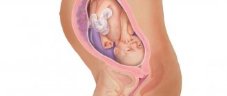
At 36 weeks of pregnancy, the baby's weight averages 2.6 kg, and it becomes increasingly difficult for him to move freely. Therefore, the mother feels all the movements and even the way the child hiccups quite clearly. Usually by this time the fetus is turned head down, arms and legs are pressed to the body. He will remain in this position until he is born.
There is less lanugo and cheese-like lubricant on the baby’s body. The baby swallows them along with the amniotic fluid, which is how meconium is formed - the baby's first stool.
Possible consequences
Childbirth at 36 weeks of pregnancy can lead to the following abnormalities in the baby:
- poor temperature balance;
- hypovitaminosis (lack of vitamins);
- anemia (develops in almost everyone, and in full-term children - in 20% of cases);
- problems with feeding;
- rickets associated with immaturity of the musculoskeletal system and lack of calcium and phosphorus;
- respiratory disorders (in 1-2% of newborns);
- encephalopathy;
- hypoglycemia (reduced blood glucose concentration);
- increased risk of mortality and morbidity.
In the delayed period, the development of cerebral palsy is possible, the prevalence of which in babies born at 36 weeks is about 6%. According to studies conducted abroad, such children may subsequently experience difficulties in learning, their motor and speech skills are reduced, and visual impairment and emotional instability are more common.
Tests and ultrasound
The thirty-sixth week of pregnancy is the optimal time for the following studies:
- biochemical blood test - to assess the general health of the mother, hemoglobin levels, hormones, etc.;
- test for HIV, sexually transmitted infections - to determine labor tactics;
- gynecological smear - will reveal the presence of pathogenic microflora;
- A general urine test will indicate possible problems in the functioning of the kidneys and urinary system.
You will also need to undergo a CTG: the fetal heartbeat can be used to diagnose hypoxia and other disorders in a timely manner.
Ultrasound at the 36th week of pregnancy is performed according to indications, if it has not been prescribed previously or for additional diagnostics with accompanying symptoms.
Nutrition
Nutrition of a pregnant woman at the 36th week of gestation
As we said earlier, nutrition from the 36th week needs to be reviewed. A woman should reduce the amount of meat she eats and increase the proportion of vegetables. It is advisable to consume them raw. This is necessary to increase the amount of fiber, which allows you to maintain normal digestion.
Additionally, from this period it is worth limiting the consumption of exotic fruits. It is recommended to organize nutrition in the last weeks before childbirth according to certain rules:
- Eat simple, easily digestible food;
- Last hearty dinner 2-3 hours before bedtime;
- Stop eating until you feel full;
- Do not include harmful foods in your diet: processed foods, smoked foods, pickles, fast food;
- Drink about 1 liter of fluid per day;
- Minimize the amount of salt consumed;
- Do not drink strong tea or coffee unless otherwise prescribed by your doctor.
Vitamins
In most cases, taking vitamins from the 36th week has already been discontinued. If the gynecologist did not do this at the previous appointment, it is worth asking him about the need to receive additional beneficial substances in the form of dietary supplements directly.
The removal of fortified complexes is carried out in order to prevent excessive weight gain of the fetus, so that the woman can avoid difficulties during childbirth. By this week, the fetus has completed its development, and the incoming nutrients from food will be enough to maintain the normal weight of the unborn child.
What to discuss with your doctor
- If tests reveal that you have low hemoglobin levels, talk to your doctor about what medications you can take. This condition cannot be corrected with diet, and your lack of iron means the same deficiency in your child.
- Breech presentation, that is, with the buttocks toward the cervix, is the reason for cesarean section in many cases. But in general, the baby has another 1-2 weeks to roll over. Find out from your gynecologist what exercises can help with this and when you should come for a follow-up ultrasound to check how the fetus is lying.
- Is intimate relations possible at 36 weeks of pregnancy? Check with your doctor to see if you have any contraindications. Usually, experts in the third trimester advise “not to get carried away,” and also to use a condom during sex, as the risk of infection is high, especially if the mucus plug begins to come off.
Good to know
Why does premature birth occur?
Which baby is considered premature?
How to test for amniotic fluid leakage at home
What is “oligohydramnios” during pregnancy?
Malpresentation of the fetus
How does childbirth occur with a breech presentation of the fetus?
All texts for pages about mother and baby were kindly provided by RAMA Publishing - these are chapters from the book by Svetlana Klaas “Your Favorite Little Man from Conception to Birth”, reviewer Irina Nikolaevna Kononova, Candidate of Medical Sciences, Associate Professor of the Department of Obstetrics and Gynecology of the Ural State Medical Academy (Ekaterinburg).
Possible complications
Many pregnant women complain of the following unpleasant symptoms at 36 weeks.
Pubic bone hurts
This happens under the influence of the hormone relaxin, which prepares muscles and ligaments for childbirth, making them softer and more elastic.
The belly becomes hard at 36 weeks of pregnancy
This feeling is associated with contractions - true ones, when labor begins, and training ones. You can tell them apart by measuring the time between contractions. During childbirth, they occur at regular intervals, and the pain increases. Training sessions are usually chaotic and will soon pass on their own. Uterine tone at 36 weeks may also be associated with this symptom. It is characterized by severe and prolonged pain.
The child moves less
This feeling may be subjective and due to the fact that the baby has become more crowded in the womb. However, if you really have doubts, it is better to tell your gynecologist about it. Using CTG or ultrasound, he will be able to quickly assess the condition of the fetus.
Legs become very swollen
This is sometimes inevitable in the third trimester of pregnancy. Don't limit yourself to fluids. Contrary to popular belief, this will only increase swelling if it appears. But in this case it is necessary to reduce salt consumption. If, against the background of swelling, nausea occurs, blood pressure or temperature increases, immediately consult a gynecologist: these are symptoms of gestosis in pregnant women.
Helpful advice for a future dad
You are already accustomed to the fact that in this section we talk about mom - her well-being, emotions, useful and not so useful actions.
However, today this page is intended for future dads - please give it to your spouse to read or send him the link. So... Dear future dad! The baby has not only occupied your partner’s belly and is making his presence known more and more - he has “zombified” her brain and feelings! If conversations are only about the Baby, if feelings are only towards the Baby, it’s time to sort it out.
To do this, you need to form several undeniable attitudes:
- I am and will be the most important person in the life of my half, no matter how strong feelings for the Kid she may be overwhelmed at the moment;
- I allow my soul mate - the mother of my Baby - to talk about him and express her love for him, because I understand that this is how she shares these feelings with me and expects support from me. This helps me find common topics for conversation with her, and also be sure that a loving mother will give birth to a healthy and strong heir (heiress);
- I will not hesitate to ask you to express your love for me as much as for the Baby, but I will not abuse such a request;
- I will become more and more imbued with the idea that a new meaning has appeared in my life - a child who will be similar to me in many ways. And it depends on me what his life will be - joyful, happy or full of anxiety;
- only I can ensure the full safety of the mother and the Baby, and for the time being, their material well-being;
- I am the main representative among men for my Baby and soon I will be able to give him what his mother cannot. I will teach the Kid (be it a son or a daughter) to be strong and not be afraid to overcome obstacles, play football and fish, hold a hammer and drive a car;
- I will raise my son to be a real man, and for the time being I will talk about this through the wall of my mother’s belly;
- I’ll tell my daughter right now that she’s beautiful and that I’m looking forward to seeing her in a dress and bows.
Regardless of what your significant other says and feels in connection with the Baby, your Baby expects that he and you will develop his own unique friendly (and not competitive) relationship, into which the mother - for the sake of her own well-being - sometimes need not be initiated .
Preparing the breasts for lactation
The recommendation to “harden your breasts” by rubbing your nipples with a towel after a shower is hopelessly outdated. This method will not only not help in any way, but will also harm the delicate skin of the areola: irritation will appear, and during feeding, cracks will appear.
But what you should definitely pay attention to is the shape of the nipple. If it is flat or retracted, you will need to be patient and persistent because your newborn will have a hard time holding the breast properly in his mouth at first.
We advise you to buy a nipple shaper in advance - a shell-shaped device that will help it rise. Learn comfortable feeding positions and ways to help your baby latch on correctly.
Care
If before this time you prepared your breasts for feeding with the help of special massages, now you should no longer do this, since massage of the mammary glands can provoke premature release of the hormone oxytocin, which can lead to premature labor.
But you shouldn’t give up walking; movement and fresh air are necessary not only for you, but also for your baby.
Benefit or harm? Scientists explain how epidural anesthesia affects the course of labor
Checklist for 36 weeks of pregnancy
- Don't miss any opportunity to relax. It is better to lie down, raising your legs or sitting on your left side. This way you will restore strength, improve blood circulation and cope with swelling in your legs.
- Install a contraction counter on your phone. Easy-to-use apps will help you differentiate between practice contractions and real ones.
- Sing, read and just talk to your child. This has a positive effect on both his well-being and your emotional state. There is an opinion that the baby, while in the womb, remembers the sounds of the mother’s lullaby, and will calm down and fall asleep easier after birth.
Do you have any questions? We will be happy to answer them during a consultation at any time convenient for you. Leave a request on the website.
What should you do when the birth process begins?
Prenatal contractions, accompanied by nagging pain in the lower abdomen, occur in pregnant women approximately 5-7 hours before delivery. If you put your hand on the abdominal wall, you can feel the muscles tense.
It is necessary to prepare documents (birth certificate, exchange card, passport, compulsory health insurance policy, sick leave, SNILS, contract for paid childbirth), a bag with things and call an ambulance.
The duration of contractions varies from person to person; some women may experience rapid labor. If they become regular or the plug or water breaks, or there is a desire to push, then you need to immediately call an ambulance. If the contractions were only training contractions and labor activity of the uterus is not recorded during the examination, the pregnant woman can be sent back home.