The causes of chest deformation are divided into two groups - congenital and acquired. Deformity can be a consequence (complication) of a number of spinal diseases.
Among the main causes of deformation are the following:
- kyphosis,
- scoliosis,
- chronic obstructive pulmonary diseases,
- marfan syndrome,
- anomalies of osteogenesis,
- achondroplasia,
- Turner syndrome,
- Down Syndrome,
- emphysema,
- rickets,
- keeled deformity,
- funnel chest,
- cleft sternum,
- Poland syndrome,
- Wife Syndrome,
- congenital rib anomalies,
- asthma,
- incomplete fusion of the fetal sternum,
- congenital absence of the pectoral muscle,
- ankylosing spondylitis,
- inflammatory arthritis,
- osteomalacia
As we can see, in most cases, the deformity is caused by genetic reasons leading to abnormal development of the upper spine; it develops against the background of existing anomalies and syndromes.
Genetic causes, in turn, lead to the asymmetric development of bone and cartilage tissue and cause the development of asymmetry of the ribs and sternum, as a result - convexity or concavity of the chest.
Scoliosis, tuberculous bone infection, rickets, as well as all kinds of injuries and damage, including those associated with a person’s professional activity, are among the causes that cause acquired deformation of the chest.
Types of chest deformities
In children, two types of congenital pathology most often occur: funnel-shaped (also called “shoemaker’s breast”) and keeled:
- The funnel chest is characterized by a sunken shape of the front part, in which it is pressed inward and takes the form of a depression. In this case, the sternum visually appears expanded. Science does not know the exact reasons for the appearance of this defect.
- Keeled deformity appears already at birth and is expressed in the form of a convex shape of the bone corset of the chest. The cause of pectus carinatum is believed to be excessive growth of the ribs or osteochondral cartilage. Usually the disorder is accompanied by abnormal development of the spine, curvatures (scoliosis, kyphosis), and various types of heart defects. The keeled deformity can also be asymmetrical, with the shape of the sternum being sunken only on one side and protruding on the other.
Most experts agree that congenital chest deformity originates during intrauterine development. A child's rib cartilage grows too quickly, causing increased pressure, resulting in a sunken rib cage. The disease makes itself felt soon after the birth of the baby or in his first years of life. Associated syndromes may include rickets, abnormal development of the spine, heart disease, asymmetric development of bones and cartilage of the sternum. Over time, if left untreated, the deformity can rapidly progress, causing physiological discomfort and disturbances in the functioning of internal organs.
One of the most serious diseases is Poland syndrome. This is a rare congenital syndrome, which is characterized by a complete or partial absence of the muscular frame on one side and an accompanying malformation of the hand. Its cause is considered to be impaired blood circulation in the vertebral arteries during fetal development. In this case, the heart muscle and main arteries are not protected by the bone skeleton of the chest and are at risk of mechanical damage.
Chest deformity - treatment in Moscow!!!Treatment of chest deformity by famous thoracic surgeons Dr. Rudakova S.S. and kmn Koroleva P.A. :
Make an appointment for consultation and surgery - 8 (495) 517-66-26 | Dmn Rudakov Sergey Sergeevich, thoracic surgeon |
Current state of the problem of treatment of keeled and rare deformities of the chest
(according to literature data)
Pileated chest deformity
Definition and pathogenesis
Peeled chest deformity (PCD) is characterized by symmetrical or asymmetrical curvature of the anterior sternum and the ribs articulating with it. CDHA causes only a cosmetic defect in children; A functional examination does not reveal any deviations from age norms. However, in adolescents and adults, CDHA can cause functional disorders (decreased vital capacity of the lungs, increased minute volume of respiration, decreased oxygen consumption, etc.) due to decreased mobility of the ribs and the presence of the entire sternocostal complex in a state of “constant inspiration.”
KDGK classification
There are several classifications of CDGK, the most complete and practically significant of which were proposed by G.A. Bairov and A.A. Fokin. They distinguish three types of CDHA: manubriocostal, corporocostal and costal. The manubriocostal type is characterized by forward bending of the manubrium of the sternum and 2-3 costal cartilages articulating with it. The body of the sternum and the xiphoid process are often displaced posteriorly. With the corporocostal type, the sternum is either obliquely directed downward and forward in a straight line with maximum protrusion in the lower third, or arched forward in the middle and lower third. The cartilaginous sections of the ribs are often curved inward. The costal type is caused by the forward bending of the costal cartilages. Curvatures of the sternum are not pronounced and are often rotational in nature.
Treatment methods for CDHA
The vast majority of surgeons are of the opinion that the treatment of CDGK is only surgical. The methods proposed by several authors for correcting CDG with the help of “pressure” corsets are not widely used. The first thoracoplasty for CDG was performed by Lester in 1953. In our country, the first operation was performed by G.A. Bairov in 1967. Below we provide a description of the most well-known methods of surgical treatment of CDHA.
Thoracoplasty according to Ravitch M.
A transverse submammary skin incision is made. The rectus abdominis muscles are cut off from the sternum and costal arches. The pectoralis major and minor muscles are cut off from their insertions on the chest. All costal cartilages are resected subperichondrially on both sides, starting from 2 including the costal arches. Catgut sutures are applied to the perichondrium remaining in the places where the cartilage is removed, shrinking it into an “accordion”. Thus, the intercostal spaces and perichondrium are shortened, and the sternum is lowered to its normal position. If there is severe curvature of the sternum, a wedge-shaped sternotomy is performed. Parts of the sternum at the site of sternotomy are fixed with two U-shaped bone nylon sutures. The pectoralis major muscles are sutured to each other along the midline over the sternum in order to fix it in a corrected position. The rectus abdominis muscles are sutured to them from below. The wound is sutured in layers.
Thoracoplasty according to N.I. Kondrashin.
A vertical skin incision is made along the anterior surface of the chest from the manubrium of the sternum in its center and 4 cm below the xiphoid process. The xiphoid process of the sternum is cut off and, together with the rectus abdominis muscles, is retracted downwards. A transverse sternotomy is performed subperiosteally between the 1st and 2nd ribs. In the longitudinal direction, subperiosteally between the upper and lower sternotomy, the keeled deformed body of the sternum is resected, leaving its lateral plates at the junction of the II-VII ribs with the sternum. The II-VII ribs are dissected subperichondrially on both sides. After this, the crossed ribs are transposed along with the remaining plates of the body of the sternum to the center and sewn together with thick Mylar threads using interrupted sutures. Sutures are placed on the sternum corresponding to the II-VII ribs. In this case, the ribs at the places of their dissection are not sutured. The second ribs are fixed to the remaining part of the manubrium of the sternum with two interrupted thick lavsan sutures (Fig. No. 1).
a B C
Rice. No. 1. Scheme of thoracoplasty surgery according to N.I. Kondrashin for keeled chest deformity.
a) skin incision; b) resection of the body of the sternum, dissection of ribs II-VII on both sides; c) transposition of the crossed ribs along with the remaining plates of the body of the sternum to the center and suturing them together.
Thoracoplasty according to O.V. Dolnitsky and L.N. Dirdovskaya.
A wavy skin incision is made with an additional vertical one along the midline. The sternum and deformed costal cartilages are exposed throughout. The pectoralis major muscles, rectus abdominis muscles, and sometimes the external oblique muscles are cut off from their insertions. The costal cartilages from 2-3 to 7 ribs on the right and left are excised subperichondrially. At the level of the 3rd-4th intercostal space, a transverse sternotomy of the sternum is performed. Then the distal end of the sternum is resected within 1-1.5 cm, a triangular groove is formed at its end, into which the xiphoid process is sutured with Mylar sutures. Tension of the rectus abdominis muscles causes the sternum to descend posteriorly. The pectoralis major muscles are connected in the midline to each other, and the rectus and external oblique abdominal muscles are sutured to them below. Thus, a kind of muscular frame appears, exerting pressure on the sternum in front (Fig. No. 2).
a B C
Rice. No. 2. Scheme of thoracoplasty surgery according to O.V. Dolnitsky and L.N. Dirdovskaya at the KDGK.
a) skin incision; b) resection of costal cartilages, transverse sternotomy, shortening of the distal end of the sternum; c) fixation of the sternum, suturing the pectoralis major, rectus and external oblique abdominal muscles.
Thoracoplasty (metallosternochondroplasty) according to V.A. Timoschenko.
The operation is performed for corporocostal and costal forms of CDHA. A transverse submammary skin incision is made from the left nipple line to the right. The xiphoid process is cut off from the sternum. The parietal pleura is peeled off on both sides using a blunt method. The pectoral muscles are separated, exposing only the deformed costal cartilages. The latter are resected subperichondrially. The costal arches are cut off from the sternum. A transverse wedge sternotomy is performed to correct its deformity. To securely fix the sternocostal complex in a corrected position, a titanium plate is installed in front of the sternum, which is bent individually to the shape of the patient’s chest and fixed to the bony part of the ribs. The costal arches are fixed to the sternum with nylon sutures. The plate is removed after 6 months.
Application of an external “pressure” corset according to Haje SA.
The technique is based on the plastic properties of the sternocostal complex in children and consists of gradual compression of the chest in the sagittal direction with an external corset. The author describes the results of treatment of 21 patients under the age of 13 years, who were wearing a corset of an original design and, gradually reducing the length of the lateral rods, corrected the keeled deformity. The corset can be worn for up to 2 years.
Rice. No. 3. Method of correction of CDHA according to Haje SA.
The vast majority of methods for surgical correction of CDG are very traumatic, since they involve manipulation of the osteochondral and muscle tissues of the chest. Many authors do not use any additional fixation devices to stabilize the sternocostal complex. The feasibility of using the external corset proposed by Haje raises some doubts, given the need to constantly wear a device that compresses the chest for up to two years.
Rare congenital chest deformities
Rare congenital deformities of the chest include costomuscular defect (Poland syndrome), sternum defects (split and bifurcation of the sternum), Currarino-Silverman syndrome. There are isolated reports in the literature about these defects, and M. Ravith believes that they constitute approximately 2% of all DHA.
Costomuscular defect - Poland syndrome
Poland syndrome (PS) is a complex of defects including absence of the pectoralis major and minor muscles, syndactyly, brachydactyly, atelia (absence of the nipple of the mammary gland) and/or amastia (absence of the mammary gland itself), deformation or absence of several ribs, absence of hair in the armpit depression and reduction in the thickness of the subcutaneous fat layer. The individual components of this syndrome were first described by Lallemand LM (1826) and Frorier R (1839). However, it is named after the English medical student Alfred Poland, who in 1841 published a partial description of this deformity. A complete description of the syndrome in the literature was first published by Thompson J in 1895.
SP occurs with a frequency of 1:30,000 - 1:32,000 newborns and is right-sided in 80% of cases. With the left-sided variant, various manifestations of the reverse arrangement of internal organs are sometimes observed, from dextracardia to the full form of situs viscerum inversus. Chest deformity in SP varies from mild hypoplasia to aplasia of costal cartilages or entire ribs on the affected side.
Treatment of SP is a technically complex surgical intervention. It has three goals: to eliminate the rib defect and restore the bone frame, to eliminate the retraction of the hemithorax, to create the correct anatomical relationships of the soft tissues with the modeling of the nipple and mammary gland.
In 1961, Sulaama M. et al. cited 5 observations of the joint venture. In 4 patients, plasty was performed with rib grafts taken subperiosteally from the surrounding region with muscle grafting (Fig. 4). In one patient with a total absence of two ribs, after subperiosteal mobilization, the rib above the defect was crossed at the proximal end, and the rib lying under the defect at the distal end. The free edges of the ribs are secured as a bridge over the defect (Fig. 5).
Rice. 4. Scheme of the Sulaama 1 operation for Poland syndrome.
1. - rib defect, 2. - perichondrium remaining after cartilage resection, 3. - displaced autografts.
Rice. 5. Scheme of the Sulaama 2 operation for Poland syndrome.
A. — view before surgery; b. — movement of the ribs (direction shown by arrows); c — view after surgery.
Ravitch M. used Teflon-coated split rib grafts for reconstruction. Urschel HC used latissimus dorsi flaps in addition to rib grafts. Haller JA wrote that complex deformity of the chest wall requires intervention in early childhood to replace missing ribs and eliminate the paradoxical movement of the anterior chest with the formation of a pulmonary hernia. To stabilize the chest wall, autogenous rib grafts and silicone prosthetic tissue were used to replace the missing endothoracic fascia. While restoring physiological functionality, this method does not eliminate the cosmetic defect at all due to the absence of the pectoralis major and sometimes minor muscles. However, the author himself notes that the use of silicone does not give good results in childhood and leads to a number of complications. In the author's opinion, the only technique that allows one to immediately eliminate rib defects and cosmetic imperfections is the use of rib autografts with one-stage plastic surgery of part of the latissimus dorsi muscle. Three patients aged 10, 14, and 16 years old who were operated on using this technique achieved good results.
G.A. Bairov gave a description of 16 patients with SP, operated on between the ages of 4 and 11 years. Among his patients there were 8 girls and 8 boys. Technique of operation according to G.A. Bairov is as follows: an oblique incision through the place of maximum retraction along the intercostal spaces with an upward turn at the sternum exposes the sternocostal complex. The ends of the affected ribs are isolated. Bone grafts are cut out from the above and below the ribs, which, together with the periosteum, are moved to the site of the defect and sutured to the ends of the damaged ribs on one side and to the sternum on the other (Fig. 6).
Rice. 6. Scheme of surgery for Poland syndrome according to G.A. Bairov
In 2 patients, in order to correct the deformity, traction threads were passed through the ribs, which were fixed on a Marshev splint with a slight traction. In one patient with a large rib defect, a tibial allograft was used. The results of the operations were considered good.
Congenital cleft sternum
Congenital sternal cleft (CSC) is a rare pathology. With this anomaly, there are usually no heart defects, the sternum is partially or completely split, and the pericardium, as well as the skin covering the sternum, is intact. A partial defect of the sternum is usually localized in its upper part and in the region of the manubrium, in contrast to thoracic and thoracoabdominal ectopia of the heart, in which the lower part of the sternum is predominantly split. In most patients with incomplete splitting of the sternum, its lower third and xiphoid process are preserved. In addition to a pronounced cosmetic defect, a split sternum does not perform a protective function. The anterior surface of the heart and the great vessels turn out to lie directly under the skin, which undoubtedly poses a certain danger to the child’s life, given the high level of childhood injuries.
The literature describes 5 types of VRG (Fig. 7): 1. Splitting of the manubrium of the sternum only. 2. Splitting of the manubrium and the upper third of the body of the sternum (U-shape). 3. Subtotal splitting of the sternum (V-shape). 4. Total splitting of the sternum. 5. Splitting of the lower third of the sternum and xiphoid process.
Figure 7. Types of sternal clefts according to G.A. Bairov.
There are two classical methods for closing a midline defect. The Longino L method consists of suturing along the midline of the rudiments of the sternum after their partial marginal excision (Fig. 8). This operation is used in young children, when the chest is pliable, sufficiently plastic, and the halves of the split sternum can be joined together without difficulty.
Rice. 8. Scheme of the Longino operation for ARH.
Sabiston thoracoplasty differs from the previous one in that, in addition to partial marginal excision and suturing of parts of the sternum, a parasternal oblique subperichondrial chondrotomy is performed to increase (expand) the size of the chest and prevent compression of the internal organs (Fig. 9).
Rice. 9. Scheme of the Sabiston operation for ARH.
Along with this, there are a number of modifications. Knox L., with a U-shaped cleft, suggested resecting the horizontal (lower) part of the sternum, and then placing it on top of the newly formed two-part sternum to increase strength (Fig. 10). One patient was operated on using this technique with a good result.
1 2
Rice. 10. Scheme of the Knox L. operation for ARH. 1. — lines of dissection of the horizontal branch of the sternum, 2. — autografts on the sternum.
G.A. Bairov published the results of surgical treatment of 11 patients aged 1 month and above. up to 7 years. Indications for surgery according to G.A. Bairov are: paradoxical pendulum-like movements of the mediastinal organs, leading to circulatory disorders and venous stagnation, respiratory disorders, lack of bone protection for the heart, lungs and great vessels, increase in the defect with age, cosmetic defect. In children under 1 month of life, G.A. Bairov performs only suturing of the rudiments of the sternum according to Longino. At a later age, a Sabiston-type operation was performed, supplemented by cutting off and cross-fixation of mm. sternoclaidomastoideus to the sternum (Fig. 11). Valla JS proposed, in addition to the first plastic method, to separate the periosteum from the connected parts of the sternum and fix it on the opposite side.
Rice. 11. Scheme of the operation according to G.A. Bairov under the VRG.
The main disadvantage of performing all the operations described above, especially in older children, is to varying degrees a decrease in the volume of the chest, especially with significant diastasis between the parts of the split sternum. Simple mathematical calculations show that when diastasis of 5 cm is eliminated, the volume of the chest in a 5-year-old child decreases by approximately 400 ml3. Although the undoubtedly high compensatory capabilities of the child’s body help patients cope with increased intrathoracic pressure, this nevertheless complicates the course of the postoperative period. Formed from split parts, the sternum is thinner and less durable than normal. This circumstance definitely affects the functional outcome of treatment.
Currarino-Silverman syndrome
Currarino-Silverman syndrome (CSS) is an early synostosis between the constituent parts of the sternum. As a result, the sternum develops relatively smaller than normal. And as a result, deformation of the entire bone frame of the chest occurs, often of the combined type (a combination of keeled deformation of the sternum with retraction of the costal arches). The costal arches are longer than normal and approach the lower part of the sternum at a very acute angle. The xiphoid process is displaced posteriorly, towards the mediastinum. Sometimes this syndrome is combined with congenital heart defects.
The first description of surgical correction of this type of DHA was given by Ravitch M. in 1952. He considered SCS as a variant of keeled chest deformity and proposed an original method for its correction. Since then, there have been isolated reports of this pathology in the literature. From 1966 to 1999, only three literature sources were found in the Medline database that described this syndrome and methods for its surgical correction. Chidambaram B. described 5 patients with SCS and congenital heart defects. All patients had severe DHA. Unfortunately, in this article the main emphasis is on the treatment of heart defects, without mentioning at all the correction of DHA. In the next article, Mehta AV also provides data on the treatment of heart defects in patients with SCS. And in only one article, Shamberger RC, Welch KJ described 5 patients operated on for this syndrome. The authors used resection of costal cartilages from 2 to 7, transverse wedge resection of the sternum, followed by suturing of the sternotomy incision with strong silk. No additional sternal fixation was performed. Long-term results are not described.
Acquired deformities
Acquired chest deformities (ACD) are curvatures of the sternum and ribs that arise as a result of external influences on the child’s body (inflammatory diseases, injuries, surgeries, etc.). Most often, this type of DHA is mentioned as a complication after thoracoplasty for various types of congenital deformities and cardiac operations. It is noteworthy that all authors do not single out this type of DHA as a separate problem and the approach to their treatment is quite standard.
Deformations after suffering purulent diseases of the chest cavity and chest wall
These deformities are a complication of advanced inflammatory processes caused by pathogenic microorganisms. Yu.F. Isakov et al described 15 patients with chronic empyema. All these patients had significant deformations of the chest, consisting of a sharp decrease in its circumference on the affected side, narrowing of the intercostal spaces, and scoliosis of the thoracic spine. 4 of them also had rib deformities due to osteomyelitis. According to the authors, these 15 patients made up 2.8% of the total number of children with bacterial forms of lung destruction. This kind of DHA is now extremely rare, which is explained by the effectiveness of modern antibacterial drugs.
Post-traumatic deformities
Chest injuries in children are quite common. According to N.G. Damier chest injuries account for 3.4% of all injuries in children. Due to the mobility and softness of the bone frame, they can be easily corrected in the acute period of injury. Rib fractures in children rarely cause DHA. More often, post-traumatic deformities occur after fractures of the manubrium and body of the sternum. Such DHAs rarely lead to pronounced cosmetic defects and most often do not require surgical correction. However, Norotte G describes a 14-year-old patient with a transverse fracture of the body of the sternum, who, two months after the injury, developed a chest wall deformity of the VDHA type. For which he underwent corrective surgery.
Relapses after thoracoplasty for various types of congenital DHA
The occurrence of relapses is directly related to the degree of deformity, the presence of concomitant pathology, the age of the patients, the severity of the surgical intervention and the method of thoracoplasty. Considering the fairly common occurrence of congenital DHA, especially in combination with various hereditary syndromes (Marfan or Ellers-Danlos), the number of complications after their surgical treatment also remains at a high level. Willital (1977) after performing 235 operations using the Paltia plate, when analyzing the results after 4 years, noted 25% of satisfactory and poor results. Oelsnitz (1976), examining 120 patients operated on using the classical Rehbein technique in the long-term period, noted good results in only 83 (69.2%). Satisfactory and poor results (30.8%) were due to varying degrees of recurrence of deformity. Sbokos (1975) indicate relapses of DHA after Rehbein thoracoplasty in 15% of cases. It has been noted that if Kirschner wires are used to fix the sternum, relapses occur in 32% of cases (Dolnitsky, 1978). If we summarize the literature data on the results of thoracoplasty using metal fixators, we can note that satisfactory and poor results were obtained in 23.8% of cases. Krause and Papiow (1977) have the greatest experience in performing thoracoplasty with sternum fixation with bone grafts. The authors performed 87 operations on children aged 2 to 15 years. Long-term results were monitored in 33 patients, and only 41% achieved a good result. A satisfactory result was noted in 48%. And in 4 patients (3.5%) there was a pronounced relapse of the deformity. After thoracoplasty without the use of special fixatives, relapses also occur. Ravitch M. (1977) noted 20% of unsatisfactory results after his operations, Pena (1981) and Ottolenghi (1982) - 15% and 20%, respectively. Robicsek (1974) slightly modified Ravitch M.'s operation, but nevertheless obtained 31% of varying degrees of relapse. Haller (1978) performed 254 operations using the Ravitch M method. Recurrences of deformity occurred in 32%. And only 10 children underwent relapse correction. The summary data presented in the literature show that after thoracoplasty without the use of fixatives, relapse occurs in 19.9% of patients.
<<< Back <<
(c) A.V. Vinogradov 2004
Chest deformity - treatment in Moscow!!!Treatment of chest deformity by famous thoracic surgeons Dr. Rudakova S.S. and kmn Koroleva P.A. :
Make an appointment for consultation and surgery - 8 (495) 517-66-26 | Dmn Rudakov Sergey Sergeevich, thoracic surgeon |
Symptoms
The first symptom of chest deformation is external changes. The cosmetic defect may be barely noticeable in newborn babies: it can be detected when inhaling, and the ribs recede. As the child grows older, the disease becomes more pronounced and reaches its peak by 3 years.
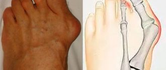
A sunken chest in a child accounts for more than 90% of cases of the disease. With this pathology, the costal cartilages are not fully developed, the sternum is increased in width, the chest cavity is reduced, and curvature of the spine occurs. As a result of these processes, a child may lag behind his peers in physical development, often suffer from respiratory infections, suffer from autonomic disorders, and respiratory failure. Children react poorly to physical activity, get tired quickly and are not able to play sports on a par with their peers. If there is a disturbance in the upper or middle region, motor function also suffers.
Causes
Most chest deformities in children are a genetic pathology. In other words, the genes already have a program that triggers abnormal growth and development of chest cartilage. Quite often, parents tend to blame themselves for the appearance of any deformities in their baby. But, for the most part, chest deformation is a genetic congenital defect, which, fortunately, can be corrected. If the deformity is congenital, then in such situations the shape of the anterior part of the chest is changed. Such disorders are accompanied by underdevelopment of the ribs or their absence, underdevelopment of the muscles and sternum. The causes of acquired deformities include various diseases (rickets, scoliosis, chronic lung diseases, bone tuberculosis), injuries, burns that occur in the chest area. When the formation of bone structures is disrupted, the most severe forms of deformation can occur.
Diagnosis of pathology
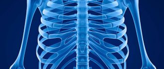
Diagnosis of diseases associated with the bone corset of the sternum is usually not difficult and can be carried out by studying external manifestations. At the initial stage, the specialist prescribes an x-ray examination in two projections, which gives a complete picture of the curvature and the severity of the disease. Next, a computed tomography may be prescribed, the results of which will reveal not only the degree of deformation of the bones and cartilage, but also the nature of the displacement of the mediastinum, the heart muscle, and how compressed the lung is. Magnetic resonance imaging gives a complete picture of the condition of bone, cartilage and soft tissues.
Functional examinations of the heart and respiratory system are prescribed as additional methods: electrocardiogram, echocardiogram, spirographic examination, examination of respiratory function and determination of lung volume. The complex of these methods makes it possible to identify functional disorders and track the dynamics of recovery after operations.
Clinic
With keeled deformity, the shape of the chest protrudes forward, depending on the degree of the disease. You can observe symmetrical and asymmetrical protrusion of the sternum and costal arches. The symptom complex includes poor development of the pectoral muscles. What becomes noticeable in adolescence. Sternum defects also include ectopia cordis and cleft sternum, which are serious manifestations of the disease.
How to correct a chest deformity
In the treatment of the spine and joints, a distinction is made between conservative and surgical treatment methods. The tactics are chosen based on the severity of the pathology and the presence of concomitant diseases of the respiratory and cardiovascular systems. Treatment of chest deformity at home includes performing a set of exercises that strengthen the muscle corset, physical therapy complexes, breathing exercises, swimming, wearing a corset, and a massage course. Conservative methods cannot completely correct the situation, but they can significantly slow down the process and maintain the normal function of internal organs. Treatment should be carried out under the constant supervision of an orthopedic surgeon. Drug therapy is aimed at eliminating those problems that have arisen in the internal organs as a result of their deformation under the pressure of the bone skeleton.
In traumatological practice, three stages of deformation are distinguished. With age, the child’s pathology will progress, so conservative treatment can give results only at the first stage. At the second and third stages, conservative methods do not show effectiveness, so doctors recommend surgical reconstruction of the chest. In this way, further deterioration can be avoided and normal conditions for the functioning of the heart and lungs can be created. As a rule, surgery is performed when the child reaches 7 years of age. In the treatment of chest deformity in adolescents, the method of placing two magnetic plates has been actively used over the last few years. One is implanted behind the sternum, and the second is placed on a special corset. The outer plate attracts the inner plate due to the magnetic field, and a gradual change in the position of the bones occurs.
2. How does keeled breast shape affect health?
Keeled breasts can be diagnosed at birth, but initially it does not affect the functioning of the heart and respiratory system. The following defect-related conditions may develop with age:
- dyspnea;
- fast fatiguability;
- tachycardia during physical activity;
- decreased vital capacity of the lungs;
- drop in oxygen consumption coefficient.
If the patient is not engaged in heavy physical work, he is usually only concerned about the external manifestation of the disease, since in a calm state the listed disorders are not expressed.
Visit our Pulmonology page