Hydrocephalus, also called hydrocephalus in children, is a pathological condition in which excess cerebrospinal fluid (CSF) forms in the child’s head. Hydrocephalus itself is not always an independent disease, and can exist as a syndrome in other pathological conditions of the brain . In severe cases, hydrocephalus can have serious consequences and require surgery.
Get an MRI for children in St. Petersburg
Types of hydrocephalus
There are several variants of the disease depending on the cause of occurrence and localization. By origin, there is a congenital form, which develops as a result of problems suffered by the baby in the prenatal state, and an acquired form, provoked after birth by external factors (injuries, infections).
According to the type of location, they are distinguished:
- external hydrocephalus, when in a child cerebrospinal fluid accumulates mainly in the space under the membranes of the brain;
- internal form, in which cerebrospinal fluid flows into the ventricles of the brain, causing them to expand;
- mixed or general hydrocephalus, which is characterized by a combination of external and internal forms of the disease.
There are also open (communicating) and closed (obstructive) types of pathology: in the first case, the communication between the ventricles and the subarachnoid space is not broken, in the second it is absent. According to the speed of flow, acute, subacute and chronic dropsy of the brain are distinguished.
What causes hydrocephalus
Cerebrospinal fluid is produced in our body constantly and circulates throughout the brain and spinal cord, supplying necessary substances to the cells and washing away the waste products of brain cells. In addition, this liquid protects the brain from damage, softening all effects on its cells.
Cerebrospinal fluid flows through special areas in the human brain called ventricles, which communicate with each other through special openings. Eventually the fluid is absorbed (absorbed) into the subarachnoid (subarachnoid) space that surrounds the brain. This space is permeated with capillaries, so excess fluid enters the bloodstream.
In the case of a normal process, fluid production and absorption should be the same. In this case, the total amount of liquor will remain unchanged or almost unchanged. If the output of cerebrospinal fluid into the bloodstream decreases, it begins to accumulate in the ventricles - hydrocephalus occurs.
Hydrocephalus can have a number of causes, such as:
- Disorders during intrauterine development (Chiari malformation, neural tube defects, Dandy-Walker syndrome);
- Arachnoid cyst;
- Various infections, such as meningitis or encephalitis;
- Intraventricular hemorrhages;
- Head injuries;
- Tumor in the posterior cranial fossa.
Hydrocephalus can be either congenital or acquired, that is, occurring after the birth of the child.
In addition, dropsy is classified according to its effect on intracranial pressure:
- Hypertensive (associated with increased pressure in the cranial cavity);
- Normotensive (not affecting blood pressure);
- Hypotensive (leading to a decrease in blood pressure).
Symptoms of hydrocephalus of the brain in a child
Classic signs of hydrocephalus in children over 1–3 years of age are symptoms of increased pressure on the brain, which include:
- intense headache;
- nausea and vomiting;
- spastic paresis, when muscles involuntarily tense and limb movements become sluggish;
- constant fatigue;
- urinary incontinence;
- gait disturbances;
- convulsions;
- strabismus;
- nystagmus;
- developmental delay.
Hydrocephalus, which occurs as a consequence of a brain injury or infection, can cause in an older child a deterioration in memory and attention, decreased visual acuity, irritability, dizziness and head pain that does not go away after taking painkillers.
The following symptoms help to suspect hydrocephalus in infants:
- bulging and pulsation of the fontanel;
- divergence of the sutures of the skull;
- deformation of the head with a predominance of the cerebral region over the facial region;
- rapid increase in head circumference with a significant deviation from the norm;
- increased excitability, moodiness;
- poor appetite;
- frequent volumetric regurgitation, similar to vomiting;
- exotropia;
- Graefe syndrome;
- a distinct pattern of saphenous veins on the head.
In some cases, an additional sign may be a slow heartbeat, tremor (trembling) of the chin and limbs, which persists for a long time.
Hydrocephalus - what symptoms should alert you?
Since the disease in most cases develops in the first months of life, doctors carefully monitor the growth rate of the child's head. Excessively rapid enlargement of the head, occurring ahead of the age norm, is an undoubted sign of hydrocephalus ; this in itself is a formidable symptom that requires immediate medical intervention.
At a later age, the bones of the skull lose their plasticity and the disease develops without external visual manifestations. However, the increase in intracranial pressure does not go away, which is manifested by a variety of neurological disorders, such as decreased vision, noise or ringing in the ears, and impaired coordination of movements. Sometimes the disease proceeds covertly, then no obvious symptoms appear, but the child’s mental development still lags behind the age norm, and learning problems appear. There is also an aggressive course, in which case the intracranial pressure rises so much that the eyes seem to protrude from their sockets.
Reasons for the development of hydrocephalus in children
Factors that can lead to the internal form of the disease are:
- anomalies and malformations of the fetus during the period of intrauterine formation, associated with infections, injuries, and the influence of bad habits of the mother;
- various birth injuries;
- prematurity and premature birth with low body weight.
The causes of acquired hydrocephalus in a child can be infectious diseases of the brain and its structures: meningitis, malignant and benign neoplasms, encephalitis. In some cases, the starting mechanism is traumatic brain injuries and bruises resulting from falls, blows, accidents and other injuries.
Diagnosis of hydrocephalus
- The main method of examination is an x-ray, the results of which will confirm the dehiscence of the sutures of the skull.
- It is also recommended to do a craniogram - an X-ray examination of the skull in different projections without contrast. Such a diagnosis will not only identify hydrocephalus, but also determine its type in order to prescribe effective therapy as soon as possible.
- Fundoscopy may be required to identify optic disc congestion.
- For differential diagnosis, angiography of cerebral vessels is prescribed.
Get diagnosed with hydrocephalus of the brain at Clinic No. 1
- X-ray
- Craniogram
- Ophthalmoscopy (as prescribed by a doctor)
- Angiography
For one-time payment for services - 20% discount
Call
Diagnosis of dropsy of the brain
In infancy, a visual examination is sometimes sufficient to determine the nature of the pathology: the doctor, by examining the baby’s physiological parameters and listening to the parents’ complaints, can make a preliminary diagnosis at the initial appointment.
To clarify the disease in children with an open fontanel, including to identify pathology during intrauterine development, ultrasound is used to assess the degree of enlargement of the ventricles. Additional methods are:
- fluoroscopy;
- computed and magnetic resonance imaging;
- lumbar puncture, necessary for a detailed analysis of the cerebrospinal fluid.
A child with suspected hydrocephalus must be examined by an ophthalmologist or pediatrician; if necessary, concomitant diseases may require consultation with a cardiologist, surgeon, oncologist and other specialists.
Subgaleal reservoir.
The essence of the method is to install a silicone catheter into the lateral ventricle and remove it into a reservoir formed under the aponeurosis. Liquor from the ventricular system rushes into the subaponeurotic reservoir, from where it can be periodically evacuated painlessly.
The reservoir is punctured with a thin intradermal needle 1 to 3 times a week, removing accumulated cerebrospinal fluid and introducing solutions of antiseptics and antibiotics. If possible, I transfer the patient to a day hospital for psychological rehabilitation of the mother. The reservoir acts as a receiver, port and shock absorber and allows for control of ventriculomegaly.
The advantage of this technique compared to external drainage is that it allows sanitization of the liquor system with minimal risk of infection and greatly facilitates patient care. Compared to external drainage, we obtained more encouraging results.
Treatment of hydrocephalus in children
Depending on the type and nature of the pathology, treatment for hydrocele can be conservative or surgical. Conservative tactics are used only in cases with a non-progressive open form of the disease, which is caused by external factors. It involves medication to relieve symptoms and eliminate the cause.
In all other situations, it is rational to use surgical treatment methods, which are aimed primarily at eliminating the obstacle that impedes the outflow of cerebrospinal fluid (tumor, abscess, intracranial hematoma, developmental anomalies). In cases where it is not possible to eliminate the cause of the pathology, a special operation is performed - bypass surgery. The technique involves introducing a system of tubes and valves into the brain, which will drain excess cerebrospinal fluid to other parts of the body.
In situations that threaten the child’s life, when immediate assistance is required, external ventricular drainage is performed - an operation during which excess cerebrospinal fluid is quickly pumped out of the ventricles of the brain using the drainage system.
With timely diagnosis and adequate treatment, a child after hydrocephalus grows and develops in accordance with the norms; the disease does not affect the mental or mental state in any way.
Treatment
Currently, occlusive (obstructive) hydrocephalus is treated surgically. Surgical intervention consists of draining excess cerebrospinal fluid outside the cerebrospinal fluid system: into the abdominal (abdominal) cavity or into the atrium. Sometimes CSF may be drained into the pleural cavity. In these cavities, cerebrospinal fluid is absorbed and excreted along with waste products of the body.
To drain the CSF, the surgeon implants a drainage system (shunt). The system material is silicone and polypropylene. Both of these materials are well tolerated by the body. All elements of the system are implanted under the skin; there are no external areas.
Disease prevention
The list of measures that can prevent the development of pathology includes:
- pregnancy planning;
- prevention of birth injuries;
- giving up bad habits before conceiving a baby and during pregnancy;
- vaccine prevention of infectious diseases of the brain (meningitis, encephalitis);
- early perinatal diagnosis;
- regular monitoring of children at risk by a pediatrician and neurologist;
- timely completion of preventive examinations up to a year;
- providing the child with safe living conditions to prevent household traumatic brain injuries.
Parents need to closely monitor the development and condition of young children, promptly contacting doctors in case of infectious diseases, falls and injuries affecting the head and neck area.
Specialists at the SM-Doctor clinic will conduct a detailed diagnosis if hydrocephalus is suspected, plan treatment and monitor the child throughout therapy and rehabilitation.
Excessive drainage
Excessive drainage of cerebrospinal fluid occurs when the valve is incorrectly selected according to the pressure parameter. If the valve opening pressure is too low, it can cause excessive drainage, causing the cerebral ventricle to compress and deforming the brain tissue. The patient experiences headaches that are most severe when standing.
In addition, nausea, vomiting, drowsiness and nervous system disorders, in particular double vision, appear. School-age children experience a decline in mental abilities.
Signs of the disease
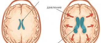
The symptoms of the disease are dictated mainly by reduced perfusion of brain tissue, overstretching of groups of nerve fibers (conducting pathways) due to increased ICP.
- In acute pathogenesis, weak microcirculation (hypoperfusion) leads mainly only to functional disorders of intracranial metabolism. This is
a change in energy metabolism, a reduction in the level of creatine phosphate and ATP, an increase in the concentration of lactic acid and inorganic salts of phosphoric acids. The acute clinical picture is reversible. - The long-term existence of hypoperfusion causes irreversible transformations at the structural level. These are
defects in the vascular endothelium and disruption of the BBB, axonal damage (destruction of axons, up to their complete disappearance). Dropsy of prolonged duration ultimately causes brain atrophy. - The morphology of signs in hydrocephalus in combination with high intracranial pressure is characterized, first of all, by atrophy of the substance of the brain and periventricular edema. There is also damage to the vascular mesenchyme, disruption of brain homeostasis, axonal lesions, and in rare cases, neuronal death. These signs are combined with the clinical picture of the primary pathology that provoked hydrocephalic syndrome.
The symptomatic complex characteristic of hydrocephalus in early childhood includes such distinctive features as:
- increased head size;
- frequent regurgitation;
- restless behavior of the child;
- bulging of a large fontanel;
- divergence of cranial sutures;
- the severity of the venous pattern on the scalp;
- delayed psychomotor development, less often physical;
- throwing the head back;
- “setting sun” syndrome (Graefe);
- congestive optic disc;
- paraplegia of the lower extremities (in severe, advanced conditions).
In adults and older children, the clinical picture depends on the rate of progression of hydrocephalus. In the acute form of the disease, combined with high ICP, the following are observed:
- a bursting and pressing headache that spreads to the orbits of the eyes (one of the features is the peak of pain in the morning after a night's sleep, and then, during the day, the severity of the pain syndrome decreases);
- nausea, which usually accompanies morning headaches (vomiting often occurs in the morning, after which a person notices an improvement in the condition);
- visual disturbances usually include blurred vision, blurred vision, double vision and a burning sensation in the eyes;
- fatigue, drowsiness, lethargy;
- convulsive phenomena (like an epileptic seizure);
- when the brain stem is compressed due to dislocation of brain structures - oculomotor disorders, forced head position syndrome, clouding of consciousness (up to a coma), respiratory failure.
Hydrocele of the brain in the chronic stage manifests itself:
- signs of dementia (dementia), emotional instability;
- apraxia of walking, more often it manifests itself with a shaky and uncertain gait, disproportionately large steps (being in a lying position, patients often do not experience difficulties in imitating walking and twisting a “bicycle” with their legs);
- decreased muscle strength, sometimes patients complain of pain in the neck;
- severe imbalance (in the final stages), which is expressed by a person’s inability to move and sit independently;
- partial or complete loss of sensitivity (not always!):
- urinary and/or fecal incontinence (with a massive lesion).
The pathology is dangerous due to its life-threatening complications! In no case should you ignore an urgent visit to the doctor if one or more symptoms from the lists provided are identified. Timely visit to the hospital for the purpose of diagnosis and receiving adequate medical care increases the chances of a favorable prognosis, including complete recovery.
On average, out of 10 patients who do not receive treatment at the right time, 6-7 people die soon (this also applies to children). Those who did not undergo therapy but survived are doomed to disability with neurological disorders, mental and physical disabilities with a tendency to progress.
How is the diagnosis made?
Diagnosis of hydrocephalus is based on the clinical picture, examination of the fundus, as well as additional research methods, such as neurosonography (NSG), ultrasound examination of the brain (in infants under 2 years), computed tomography (CT) or magnetic resonance imaging (MRI) of the brain brain The primary diagnosis can be made by a neonatologist, pediatrician, neurologist or neurosurgeon.
The most common operation is ventriculoperitoneal shunt (VPS).
Neurosonography is an effective method for diagnosing the state of the brain and ventricular system in children under 1.5-2 years of age, until the large fontanelle and other “ultrasound windows” - areas of the skull where the bones are very thin (for example, the temporal bone) and transmit ultrasound. It allows you to detect expansion of the ventricular system, intracranial space-occupying formations (tumors, hematomas, cysts), and some malformations of the brain. However, it should be remembered that NSG is not an entirely accurate method. The image of the brain is obtained with significantly lower resolution (less clear) than with CT and MRI.
If any brain pathology is detected, a CT or MRI is necessary. Without them, it is impossible to make an accurate diagnosis, identify the cause of hydrocephalus, and even more so carry out treatment. This equipment is expensive and is still not installed in all hospitals. In this case, parents should insist on having a CT or MRI scan performed at other centers or perform them themselves on a commercial basis. It should be borne in mind that a clinic that undertakes the treatment of children with hydrocephalus is required to have this equipment. Otherwise, we can recommend that parents choose another, more equipped hospital, at least in another city.
More information about shunt systems for the treatment of hydrocephalus
The result of the operation largely depends on the quality of the shunt system. In recent years, in many hospitals, neurosurgeons have been using shunt systems produced by the largest American company Medtronic. The company's engineers, together with leading US neurosurgeons, have developed a range of valves of various models and sizes (including valves for newborns).
One of the latest achievements in the production of liquor drainage devices is the Delta valve. This is a unique valve, has no analogues from other manufacturers. It was created to avoid such a common complication as excessive CSF drainage. If all other valves pass as much fluid as they are designed for pressure, then the Delta valve allows as much fluid as is needed to remain in the ventricle to maintain intracranial pressure within physiological limits. When implanting the Delta valve, the patient is maintained at normal pressure, regardless of the rate of cerebrospinal fluid production and, most importantly, regardless of the patient’s body position (lying/standing).
The technological features of the materials from which the system valves are made prevent deformation and sticking during pumping; the dome of the valve reservoir is designed to withstand repeated punctures with a thin needle (the holes self-tighten). Catheters are made of high-quality latex-free silicone, so they do not stick together or form loops, which significantly reduces the risk of system blockage.
The valves are equipped with connectors for connection to catheters; their design facilitates connection and reduces the possibility of disconnection and disconnection of the system.
A radiopaque mark is applied along the length of the catheters, this allows you to see the shunt on an x-ray. The same substance is applied to the valve with a dot code indicating the pressure of the valve. There are no metal parts in the shunt system. This is very important when conducting CT and NMR studies, because metal will produce artifacts, and a magnet in NMR may shift the location of the system (if it had metal parts).
All systems are sterile and supplied in double sterile packaging. To reduce the risk of infection, Medtronic has developed a unique BioGlide hydrogel. It is applied to the inner and outer surfaces of catheters, as well as the outer surface of the valve, and does not peel off. Before implantation, the neurosurgeon can treat the system parts with antibiotics, and the hydrogel will hold them for 3 days for postoperative antibiotic therapy inside the patient’s body. This way, the risk of infection is minimized.
Myths about increased intracranial pressure
The diagnosis of “increased intracranial pressure”, “intracranial hypertension (ICH)” or “hypertensive-hydrocephalic syndrome”, as already mentioned, is often made and in some cases - without reason. How does increased intracranial pressure (ICP) manifest? As already noted, in children under 2 years of age such manifestations are, first of all, accelerated growth of head circumference, bulging and enlarged fontanelle, possible eye movement disorders, and delayed psychomotor development. Most often, all these disorders manifest themselves in a complex. In children over 2 years of age, these are headaches with nausea and vomiting, more often in the morning, changes in the fundus of the eye (detected during examination by an ophthalmologist). Of course, the clinical picture may be different, but without the above symptoms, the diagnosis of “increased intracranial pressure” is doubtful.
This is how pediatrician Evgeniy Komarovsky describes the diagnosis of “increased intracranial pressure.” “Since the main symptom of hydrocephalus is a rapid increase in head size, measuring head circumference is included in the standards of any preventive examination, starting, of course, from the moment of birth. It is very important to emphasize here that it is not the specific size expressed in centimeters that matters, but rather the dynamics of this indicator. That is, stating the fact that the boy Petya at 3 months has a head circumference of as much as 45 cm is not at all a reason to become depressed and urgently save this boy. But the fact that the head circumference has grown by 7 cm over the past month is already alarming and dangerous, and requires both serious attitude and active control. Let me emphasize once again - not immediate treatment, but control. And if the trend continues, then measures should be taken.”
Symptoms such as sleep and behavior disorders, hyperactivity, attention deficit, bad habits, poor academic performance, hypertonicity in the legs, “marble” skin pattern, including on the head, nosebleeds, chin trembling, tiptoe walking, are not in themselves indicate increased intracranial pressure. And yet, some neurologists make a diagnosis of ICH based on these complaints. Neurosonography, having become a huge boon for pediatrics and neurology, has made a significant contribution to the excessive and false diagnosis of “hypertensive-hydrocephalic syndrome.” NSG makes it possible to quickly obtain an image of the brain substance and measure the size of the ventricles. However, to clarify the diagnosis, as we have already said, CT and MRI are mandatory.