Features and mechanism of disease development
Hydrocephalus or dropsy of the brain is a disease that is accompanied by the accumulation of excess cerebrospinal fluid inside the ventricles, in the meningeal spaces of the brain and leads to their expansion, and also causes an increase in intracranial pressure.
The ventricles of the brain produce cerebrospinal fluid. It serves as a kind of pillow, protects the brain and spinal cord, delivers nutrients, and is responsible for removing waste. Normally, the fluid moves before being absorbed into the bloodstream. With hydrocephalus, its movement is blocked and absorption is limited. In the absence of adequate treatment, complications develop, including death.
Types of disease
There are several types of hydrocephalus of the brain. Pathology can be congenital or acquired. The first progresses under the influence of unfavorable factors during intrauterine development. The second is formed under the influence of various pathologies and external factors.
Depending on the location of the disease, it can be:
- External - with accumulation of fluid under the meninges;
- Internal – with a violation of the outflow of CSF from the ventricles;
- Common – combines the characteristics of both previous forms.
According to the mechanism of development, hydrocephalus can be open, with preservation of the connection between the ventricular system and the subarachnoid space, or closed, with a disruption of this connection.
How is the diagnosis made?
If a patient complains of a constant headache and nausea, experiences fainting and changes in emotional background, he should seek help from a neurologist. First, the specialist examines the patient: checks motor reflexes, joint and muscle reactions. Impaired outflow of venous blood, swelling of the face, and complaints of increased fatigue suggest a diagnosis of “external hydrocephalus.”
To clarify the diagnosis, additional examination is carried out:
- Ultrasound of the head and neck for a preliminary assessment of the condition of the vessels: basilar artery, carotid, vertebral.
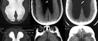
- CT scan. It helps to establish the degree of damage to brain tissue, to assess how widened the subarachnoid fissures are due to excess cerebrospinal fluid, and whether there are neoplasms in the cavity of the cranium that impede the outflow of cerebrospinal fluid.
- Magnetic resonance imaging. A study that allows you to see changes in the soft tissues of the head with maximum accuracy. It helps to accurately diagnose, classify hydrocephalus, determine how quickly the pathology develops, and select the optimal treatment.
- X-ray of the head with the introduction of a contrast agent. Helps to identify disturbances in the outflow of venous blood, damage to the vascular bed, and the formation of hematomas, which caused a deterioration in the absorption of cerebrospinal fluid.
- Ophthalmological examination. Allows you to determine the presence of congestion, swelling of the optic nerve, atrophy of the tissues of the ocular apparatus, indicating a pathological increase in intracranial pressure.
- Lumbar puncture . Most often it is carried out if there is a suspicion that meningitis or encephalitis provoked the development of dropsy. A puncture is performed to determine the level of cerebrospinal fluid pressure in the spinal cord.
If a patient is diagnosed with chronic external hydrocephalus, a thorough examination should be performed approximately every six months. The frequency of visits to the doctor is determined individually, depending on the course of the disease, the type of pathology and the characteristics of the patient’s body.
Forms of pathology
The form of hydrocephalus determines the nature of the disease and its severity. The following criteria are most often used for classification:
- The intensity of the manifestation of the pathology - when severe, a lot of fluid accumulates, which disrupts the activity of the nervous system. In this case, the disease is accompanied by severe neurological symptoms. A moderate degree is characterized by the accumulation of a small volume of cerebrospinal fluid and the absence of symptoms;
- The degree of impact on brain structures - in a compensated form, the liquid has no effect on the brain; in a decompensated form, it worsens its functioning;
- The nature of the course - hydrocephalus of the brain in the chronic form is accompanied by a gradual increase in symptoms, in the acute form it leads to a sharp deterioration in the person’s condition.
Modern diagnostic measures allow determining the type of disease, its form and developmental features.
Symptoms of hydrocephalus
The main symptom of hydrocephalus is an enlarged skull. It appears in children under two years of age, since their bones have not yet fully fused, becoming deformed under the influence of high intracranial pressure.
Many symptoms are visible to the naked eye. A sharp discrepancy between the facial and cerebral skulls, bulging eyes, bulging fontanels, divergence of cranial sutures, protruding and pulsating veins on the head should alert parents.
Children's mood worsens, they become whiny, eat poorly, burp often, and cannot sleep at night.
In a child with hydrocephalus, normal development is disrupted. He does not show curiosity, reacts poorly to toys, begins to walk and pronounce sounds late, and over time his vision deteriorates, even to the point of blindness.
At an older age, when the bones of the skull grow together, symptoms of increased intracranial pressure appear, since the cerebrospinal fluid, due to the blockage of the cerebrospinal fluid pathways, accumulates in large quantities inside and does not find a way out. Therefore, headaches, seizures and vomiting come to the fore. These symptoms occur as a result of compression of brain structures.
Stages of development
External hydrocephalus has 3 stages of development, each of them has its own characteristics:
- Mild - the size of the dropsy is small, the body itself tries to cope with disturbances in the flow of CSF. A person usually feels a headache that is not too intense, he periodically feels dizzy, feels slightly worse, and sometimes gets dark in his eyes;
- Moderate – manifestations become more noticeable, the intensity of headaches increases, especially during physical activity. Possible disturbances in visual function, rapid onset of fatigue, pressure changes, depression and severe irritability;
- Severe - at this stage, hydrocephalus of the brain can cause seizures, apathetic state, fainting, apathy, and also lead to the development of conditions in which a person is no longer capable of self-care.
At the initial stages of development of the pathology, neurological symptoms may be absent. They appear due to organic processes that occur in the cerebral substance, as well as against the background of circulatory disorders and atrophy of some areas.
Hydrocephalus in children
Signs of hydrocephalus are determined by the person’s age and stage of the disease. In children, including newborns, the development of pathology may be indicated by capricious behavior and poor appetite, marbled skin and slow reactions, eyelid retraction and gaze almost always directed downward, as well as swelling of the fontanelle and excessively enlarged head size.
It is the size and shape of a child’s head that often indicates pathological processes. By the age of six months, babies with hydrocephalus have a head circumference greater than the chest, the skull becomes oblong, pear-shaped, and this is noticeable to the naked eye.
With hydrocephalus in children, the normal functioning of the brain is blocked, which leads to a lag in mental, physical, and mental development. The risk of pathology increases in newborns born prematurely with low body weight. The cause may also be intracranial birth injuries or infectious diseases suffered by the mother during pregnancy.
A child with this disease begins to roll over, hold his head up, and sit up later than his peers. In severe forms of the pathology, these skills are not formed, muscle strength in the limbs decreases up to paralysis, and muscle tone increases. After fusion of the fontanelles, a severe headache begins, often in the morning, frequent vomiting, and nosebleeds.
Symptoms of the disease in adults
Hydrocephalus in adults manifests itself:
- Headaches for which neither sleep nor painkillers can help;
- Nausea, feeling of approaching vomiting;
- Feeling of constant fatigue;
- Sudden changes in mood, tendency to irritability;
- Changes in behavior;
- Fogging of consciousness, difficulties with memory, the thinking process;
- Balance imbalance;
- Inability to control urination;
- Seizures and convulsions;
- Sleep disorders;
- Problems with appetite;
- Blurred vision, a feeling of double vision, movement of the eyeballs downwards.
Headache with hydrocephalus intensifies when changing a horizontal position to a vertical one. Fluctuations in blood pressure are possible. With normal pressure hydrocephalus, signs of dementia, urinary incontinence, and gait disturbances appear in combination with pronounced dilatation of the cerebral ventricles, but without an increase in CSF pressure.
Diagnostics
If you have signs of hydrocephalus, you should consult a neurologist. To assess the patient’s health status, a set of diagnostic procedures is prescribed:
- X-ray of the skull;
- computed tomography or MRI;
- echoencephalography;
- Ultrasound;
- ophthalmoscopy.
Hardware techniques help to identify possible anomalies in the structure of the cerebrospinal fluid system, the presence of trauma, cysts, hematomas, tumors. A lumbar puncture may be performed to identify the cause of the disease. This is an invasive procedure in which cerebrospinal fluid is taken for examination. Such an analysis will determine the infectious nature of the disease and identify the degree of increase in intracranial pressure.
Causes of pathology
The main reasons for the development of hydrocephalus in adulthood are:
- Pathologies of the vascular system;
- Infectious diseases;
- Neurosurgical operations;
- Head injuries;
- Tumor processes;
- Previous stroke;
- Anomalies of the development of the central nervous system.
Hydrocephalus can be a complication of meningoencephalitis, meningitis, rupture of an aneurysm or intraventricular hemorrhage, the formation of a cyst or tumor in the brain.
The cause of hydrocephalus in adults may be regular intoxication, for example, frequent consumption of alcohol. Alcohol has a damaging effect on neurons, causing tissue areas to die. An increased risk is created by chronic diseases such as multiple sclerosis, diabetes, metabolic disorders, and encephalopathy. Often the disease develops against the background of age-related changes in the body associated with the aging of brain tissue and blood vessels.
What is hydrocephalus?
The trophism of nervous tissue is ensured by the continuous circulation of cerebrospinal fluid. Cerebrospinal fluid moves along a certain path within the cerebral structures, participating in metabolism. Liquor protects tissue from mechanical stress, provides stable concentrations of electrolytes, acid-base balance, and acts as an immunobiological barrier.
Hydrocephalus is a pathological condition characterized by disturbances in the production, outflow and/or circulation of cerebrospinal fluid, its excessive accumulation, often with an increase in the level of intracranial pressure. The disease is characterized by neurological symptoms. Over time, hydrocephalus leads to the development of secondary changes. Synonym: dropsy of the brain. Uncontrolled course of the disease is fraught with death.
Manifestations of dropsy:
- attacks of cephalalgia (headache);
- fluctuations in blood pressure;
- nausea;
- vomit;
- urinary incontinence;
- unsteadiness of gait;
- mental disorders (dementia, etc.);
- in young children - a progressive increase in head size, a symptom of the setting sun, etc.
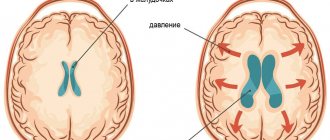
Schematic representation of the ventricles of the brain normally and with internal hydrocephalus.
In the early stages of the development of pathology, changes occur in the brain, but there are no neurological symptoms. Manifestations arise against the background of organic processes in the cerebral substance, circulatory disorders, and atrophy of individual areas.
Hydrocephalus is a sign of the disease, because is a complication of the latter. The idiopathic form of the pathology practically does not occur. Hydrocephalus and its causes are identified using neuroimaging methods. MRI is considered the most informative.
The pathology is most often diagnosed in children in the first 3 months of life. The disease also occurs in adults, especially after 60 years. Reasons for the development of hydrocephalus:
- traumatic brain injuries;
- tumors;
- vascular pathologies;
- infectious diseases;
- previous neurosurgical operations;
- strokes and hematomas;
- developmental abnormalities of the central nervous system.
Treatment tactics for cerebral hydrocephalus depend on the type of disease. If the course is favorable, drug therapy is prescribed. If the drugs are ineffective and there is a high risk of complications, surgical intervention is performed urgently.
Effectiveness of MRI in hydrocephalus
Magnetic resonance imaging is used to diagnose hydrocephalus. This is the most informative imaging method, allowing an accurate diagnosis to be made, regardless of the stage of the pathology.
The essence of MRI is to obtain layer-by-layer images of the area used by scanning. The operating principle of tomography is associated with the influence of a magnetic field and recording radio frequencies emanating from the human body.
Unlike X-rays, MRI does not have a negative effect on the body. This is a non-invasive and absolutely safe method that allows you to obtain high-quality images, including three-dimensional ones. They clearly show brain tissue of different densities and the boundaries between them, tumors and blood vessels. Thanks to the study in several projections at a given depth, it becomes possible to assess the condition of any part of the brain.
How is the procedure performed?
MRI of the brain is performed with or without contrast enhancement. In the first case, the patient is injected intravenously with special drugs that create a clear image of the affected areas and improve the quality of visualization. For example, with this approach, not only the tumor itself is clearly visible, but also its exact boundaries.
No special preparation is required for the procedure, but before it begins, you will have to remove all metal jewelry. The tomography machine itself looks like a large cylinder, inside of which there is a movable platform. The patient is placed on it lying on his back. To protect against the noise produced by the device, earplugs are used.
The table slides inside the tomograph, and the doctor performs the procedure from an adjacent office, controlling the device using a computer. Throughout the examination, voice communication with the doctor is maintained, so the patient can report unpleasant or uncomfortable sensations at any time.
The tomograph sequentially takes a series of images, which ultimately form the finished image. In order for the images to be of high quality, the patient must remain completely still. On average, the procedure takes about 30-45 minutes.
Evaluation of results
This technique makes it possible to identify not only hydrocephalus of the brain, but also concomitant diseases, and accurately determine the type of pathology. The picture shows both direct and indirect signs of the disease. Direct ones include enlargement of the ventricles of the brain and subarachnoid space.
Indirect signs are:
- The value of the interventricular index is 0.5 or more;
- Downward displacement of the hypothalamus;
- Swelling with intense dropsy;
- Partial protrusion of the upper part of the lateral ventricles.
Based on the results of the study, a diagnosis is made and treatment is prescribed. MRI may show replacement hydrocephalus. This means that a certain amount of fluid is present in the subarachnoid space, but its presence is not a sign of dropsy, but a manifestation of a compensatory mechanism associated with age-related or organic changes in tissue.
Consequences of hydrocephalus
The complications of hydrocephalus are very serious, so treatment should not be delayed. The neuropsychic development of a child, his intelligence, motor skills, vision and even life depend on a correct diagnosis. In many cases, hydrocephalus is the result of a more serious problem, such as a brain tumor.
To prevent the negative consequences of the disease, parents are advised to regularly examine their child with a pediatrician and neurologist. Routine examinations will help identify pathology at an early stage. The Into-Sana clinic employs the best specialists who care about the health of each patient. Here you will receive qualified assistance at the European level.
Methods and principles of treatment
The main task is to eliminate the accumulated excess cerebrospinal fluid in order to restore its normal flow.
In some cases, drug therapy is performed. Improvement from conservative treatment of hydrocephalus usually occurs when the disease is a complication of an infectious or inflammatory disease or traumatic exposure. When medications do not provide the desired effect, surgery is performed to install special shunts. This element ensures the drainage of fluid to the abdominal cavity, where it is absorbed by the body. As a result, pressure decreases and the size of edema in the brain decreases.
Another surgical treatment option is to create an opening in the 3rd cerebral ventricle to allow cerebrospinal fluid to flow between brain regions. Both methods allow you to relieve symptoms and block the development of dangerous complications. If the disease is caused by the appearance of a tumor, it is removed.
The earlier hydrocephalus is detected, the better the prognosis for treatment.
If the operation is timely and successful, the patient can return to normal life, and a full recovery is possible. Advanced stages of the disease not only worsen the quality of life, but also lead to dangerous consequences, including death. That is why accurate diagnosis, primarily MRI, plays an important role. Rate this article: (1 rated 5 out of 5)
Treatment of hydrocephalus
For this disease, medications are ineffective. Hydrocephalus is treated surgically. During the operation, the doctor installs a shunt system that allows cerebrospinal fluid to flow normally from the cavities of the brain into the abdominal cavity, and less often into the heart chamber or pleural cavity. The shunt system is equipped with a valve that prevents liquid backflow.
In some patients, ventriculostomy may be used. This is an endoscopic operation during which the surgeon makes a small hole in the wall of the 3rd ventricle of the brain. The normal outflow of cerebrospinal fluid then occurs through it.
Folk remedies for hydrocephalus have limited use. You can use tinctures of mint, lemon balm, cornflower, astragalus, calamus, and buckthorn berries.