This article will talk about scaphocephaly, a rather rare developmental pathology that occurs in one child out of 2000. The largest percentage of children born with this disorder are boys.
Scaphocephaly - (scaphocephaly, from the Greek “skull in the shape of a boat”) is the process of premature fusion of one or more sutures of the skull. The pathology is one of the subtypes of craniostenosis, a disease characterized by pathological formation of the skull.
The sutures must fuse as the child grows; at birth, the bone elements are connected by fibrous tissue, and as the brain grows larger during growth, the skull expands proportionately. Gradually the tissue heals, forming a seam.
With scaphocephaly, overgrowth occurs earlier than expected and the skull is deformed, causing the child’s head to take on a strange shape, abnormally narrow and long.
About the disease
Among the deformations of the skull there are congenital and acquired.
Congenital ones appear during pregnancy, and the child is born with a certain malformation. Acquired ones occur after childbirth, most often as a result of injury or certain intervention, such as surgery. Craniostenosis in children can be either congenital or acquired after birth. This defect is a premature fusion or absence of sutures in the bones of the skull, which normally should remain plastic for the natural growth and development of the child's brain. As a result, intracranial pressure increases and the shape of the skull changes. If we are talking about a developmental defect, craniostenosis in newborns may be accompanied by other defects that affect the brain and other organs of the body. In the case of an acquired disease, suture fusion occurs due to injury or surgery.
Craniostenosis occurs in one in 1000 newborns.
3242
send an SMS with the donation amount to this number
Craniostenosis, craniosynostosis and cranial deformities
Craniostenosis, craniosynostosis and cranial deformations are the result of premature or uneven ossification of cranial sutures (Greek kranion - skull; synostos - fusion; stenos - narrow, close). The incidence of craniostenosis is 1 in 1000 newborns. The immediate cause of the development of craniostenosis is seen in: a) metabolic disorders, which entails accelerated osteosynthesis of the skull bones (Greek osteon - bone; synthesis - connection, combination, composition); b) disorder of the blood supply to the bones and membranes of the brain; c) reducing the force acting through the dura mater on the cranial vault and promoting its stretching.
There are primary, or idiopathic (Greek idio - special; pathos - suffering, disease), and secondary craniostenosis. Primary craniostenosis is hereditary or associated with a hereditary predisposition. This, for example, is the symptom complex of Tersil (1942), which includes: 1) tower skull; 2) exophthalmos; 3) nystagmus; 4) mental retardation; 5) epileptic seizures; 6) intracranial hypertension and 7) optic atrophy with blindness. Primary craniostenosis is included in the clinical picture of many hereditary mental retardation syndromes with different types of inheritance. Secondary craniostenosis develops for various external reasons, such as inflammatory processes, vitamin D-deficiency rickets, phosphorus deficiency in the blood, overdose of thyroid hormone (in the treatment of hypothyroidism), X-ray exposure in the first half of pregnancy.
Normally, in newborns, all the bones of the skull are not fused, the anterior and posterior fontanelles are open. The posterior fontanel closes by the end of the 2nd month, the anterior one - during the 2nd year of life after childbirth. By the end of the 6th month, the bones of the calvarium are connected to each other by a dense fibrous membrane (Latin fiber - thread). By the end of the 1st year, the child’s head size is 90%, and by 6 years it reaches 95% of the adult’s head size. The closure of the sutures by connecting the jagged edges of the bones begins by the end of the 2nd year and is completely completed by the age of 12–14 years.
Premature overgrowth of the fontanelles and cranial sutures leads to a narrowing of the skull and a decrease in the volume of the cavity of the cranium. This interferes with normal brain development and creates conditions for liquorodynamic disturbances. With an increase in intracranial pressure, an associated hypertensive headache occurs (Greek hyper + Lat. tension - tension). The development of congestion in the fundus of the eye, concentric narrowing of the visual fields, and subsequently secondary atrophy of the optic nerves with a progressive decline in vision up to its loss are also possible. Typically, exophthalmos occurs on both sides, disorders of the oculomotor nerve, thinning of the skull bones, and the appearance of digital impressions in the bones of the calvarium, visible on x-rays. There is a high risk of developing mental retardation.
In case of secondary craniostenosis in the early stages of its formation, conservative treatment of the underlying disease can be effective. In case of primary craniostenosis, as well as secondary craniostenosis in the case of already developed significant intracranial hypertension, shunting and decompression therapy are indicated: the formation of craniectomy passages up to 1 cm wide along the line of suture ossification. Timely surgical treatment for craniostenosis ensures further normal development of the brain.
Premature or uneven overgrowth of the fontanelles and cranial sutures also leads to deformation of the skull. The shape of the skull is assessed taking into account the cranial index (CI) - the ratio of the transverse and longitudinal dimensions of the skull. Normally, or mesocephaly (Greek mesos - average), the CI is 76–80.0 in men, 77–81.9 in women. With premature overgrowth of the sagittal (Latin sagittal - arrow) suture dividing the skull into the right and left halves, dolichocephaly (Greek dolichos - long) occurs with a CI of less than 75. A variant of dolichocephaly is scaphocephaly (Greek skaphe - boat) or cymbalocephaly (scaphoid head), in which the elongation of the head is complemented by a protruding forehead and nape. There may also be a saddle-shaped skull - it is elongated and depressed in the parietal region.
With premature overgrowth of the coronal sutures (coronal or coronal synostosis), brachycephaly occurs (Greek brachys - short) - an increase in the transverse size of the head with a CI of more than 81. In this case, the face of children is flattened, and exophthalmos often occurs. With premature overgrowth of the coronal suture on one side, plagiocephaly (Greek plagos - oblique), or cross-headedness is formed: the skull is asymmetrical, the frontal bone on the side of the synostosis is flattened, on the same side there may be exophthalmos, as well as an increase in the middle and posterior cranial fossae.
If both the sagittal and coronal sutures heal prematurely, the growth of the skull continues only in height. As a result, a high conical skull is formed, somewhat flattened in the anteroposterior direction - acrocrania (Greek akron - limb), or tower skull. A variant of acrocrania is a pointed skull, or oxycephaly (Greek oxis - sharp) - a high skull, tapering upward and with a sloping forehead.
Premature fusion of the frontal suture is characterized by a narrow frontal and broad occipital bone. In this case, the frontal bones grow together at an angle, and a “ridge” is formed at the site of the frontal suture. If the posterior parts of the skull enlarge compensatoryly and its base deepens, trigonocrania occurs (Greek trigonon - triangle).
Brachycephaly, scaphocephaly and trigonocephaly are usually not accompanied by neurological pathology. If there is no brain damage, then there is no psychiatric pathology.
Return to Contents
Why is craniostenosis dangerous?
The absence of movable sutures in the skull severely limits the brain. Therefore, the sooner help comes for craniostenosis, the higher the likelihood of a complete cure for the child in the future. Compensation mechanisms in infants under 2 years of age are very high, but rehabilitation after this age turns out to be more complex and lengthy. If not treated promptly, the disease can cause the following conditions:
If you do not receive treatment in a timely manner, the disease can cause the following conditions:
- delayed physical, mental, intellectual development;
- disruption of normal brain functions;
- skeletal underdevelopment;
- compression and atrophy of the optic nerve up to complete loss of vision;
- headache;
- ophthalmological diseases;
- death.
Establishing diagnosis
A specialist can identify signs of scaphocephaly during the first examination of the child. With the development of pathology, the skull has a non-standard, elongated shape with obvious deformation of the facial skeleton.
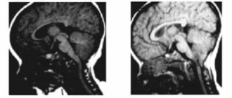
To confirm the initial diagnosis, research methods are prescribed:
- radiography;
- magnetic resonance imaging;
- computed tomography.
Studies make it possible to determine the degree of violation and establish sutures affected by pathological changes.
What is craniostenosis in children?
First of all, it depends on whether the child has other developmental defects. Sometimes pathology accompanies various syndromes - then it is called syndromic. For example, it can occur simultaneously with fusion of fingers or toes, cleft palate or lip, or cerebral hernia.
If fusion of the sutures in the bones of the skull occurs without other developmental defects, it is considered non-syndromic, that is, independent.
Doctors classify the disease into types based on which cranial sutures are fused:
- metopic,
- lambdoid,
- coronary,
- sagittal.
Synostosis - or suture fusion - can involve one to several sutures. There is a concept of pansynostosis - complete overgrowth of all seams. This type of pathology can be considered the most severe; it is less common than others.
There is a solution - surgery
With a disorder such as scaphocephaly, surgical intervention most often arises.
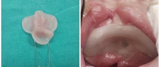
The essence of the operation is to separate the sutures that have grown together and restore the normal shape of the skull. During the postoperative period, it is necessary to wear a special helmet. The scars from the operation are almost invisible and are completely hidden after the hair grows back.
In addition, after the operation, the child needs to undergo dental treatment and physiotherapeutic procedures.
In most cases, one surgical intervention is enough to completely eliminate the defect and ensure normal growth and formation of the skull.
Depending on the patient’s condition, follow-up examinations with a neurosurgeon are prescribed 3-6 months after the operation. It is also possible to involve other specialists if the neurosurgeon sees a need for this.
Photos of children diagnosed with scaphocephaly before and after surgery:
An alternative to surgery is endoscopy. This method is used as a gentle method to reduce the degree of damage to the skull. But endoscopy can only be performed when the child has not reached the age of six months.
How is craniostenosis treated in children?
There is only one way to eliminate the pathology - surgery. It is performed to restore the shape of the skull bones. After the bones take their natural shape, they are held together with mini-screws or mini-plates and surgical wires, which are removed after a year. They are removed after a year. During the operation, doctors use modern, technologically advanced biodegradable material, which does not need to be removed surgically - it gradually dissolves on its own, and in its place, the patient’s own bone tissue grows. This greatly simplifies the patient's recovery.
The best results can be expected if the surgery is performed between 3–4 months and 2 years of age. Severe deformations of the skull can sometimes be noticed only after the bones have completed their formation, that is, at the age of 5–6 years. In such cases, the pathology manifests itself as severe headaches, blurred vision and increased intraocular pressure, increased fatigue and irritability.
If the disease occurs as a response to craniotomy or after injury, the deformed section of the bone is removed and an implant made of modern material - polymer, metal or ceramic - is installed in its place.
Supposed reasons
Medicine does not give a clear answer to the question of why such a disorder as craniostenosis and, in particular, scaphocephaly occurs.
This disease has been little studied by science and we can only talk about the hypothetical factors of its occurrence. Possible reasons include:
- intrauterine, hormonal and hereditary disorders;
- defects in growth factor receptor genes;
- mechanical factor of compression of the fetal head by the walls of the uterus.
The most common theory is about genetic defects.
Forecast and recovery
In cases where scaphocephaly is detected in a timely manner and proper treatment is carried out, we can talk about a favorable prognosis.
Defects in the structure of the skull are eliminated surgically or alternatively, which allows the child to develop normally.
Without treatment, patients experience various disorders, both intellectual and physiological. For example, vision may be significantly reduced or blindness may occur.
For this reason, if a child has visual deviations from the norm in the development of the shape of the skull, its occipital, frontal or facial zones, you should immediately seek help from specialists.