An artery in the pulmonary circulation that carries deoxygenated blood from the heart to the lungs
| Pulmonary artery | |
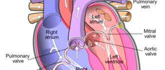
Anterior (frontal) view of an open heart. White arrows indicate normal blood flow. (The pulmonary artery is indicated at the top right.) | |
| Details | |
| Predecessor | truncus arteriosus |
| System | Cardiovascular, Respiratory |
| Source | Right ventricle |
| Identifiers | |
| Latin | pulmonary artery |
| MeSH | D011651 |
| TA98 | A12.2.01.101 A12.2.01.201 |
| TA2 | 4077, 4091 |
| F.M.A. | 66326 |
| Anatomical terminology [edit in Wikidata] | |
And the pulmonary artery
is an artery in the pulmonary circulation that carries deoxygenated blood from the right side of the heart to the lungs.
The largest pulmonary artery is the main pulmonary artery
or
pulmonary trunk
from the heart, and the smallest are the arterioles, which lead to capillaries that surround the pulmonary alveoli.
Structure
The pulmonary arteries are blood vessels that carry blood from the right side of the heart to the capillaries of the lungs. The transported blood, unlike other arteries, does not contain oxygen (“deoxygenated”). The main pulmonary arteries emerge from the right side of the heart, and they divide into smaller arteries that gradually divide and become smaller until they become arterioles and eventually capillaries.
Main pulmonary arteries
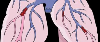
Volumetric rendering of a high resolution computed tomography scan of the chest.
The anterior chest wall, airways, and pulmonary vessels anterior to the root of the lung were digitally removed to visualize different levels of pulmonary circulation. In order of blood flow, the pulmonary arteries begin as the pulmonary trunk
or
the main pulmonary artery
. The main pulmonary artery begins at the base of the right ventricle. It is short and wide - approximately 5 centimeters (2.0 in) long and 3 centimeters (1.2 in) in diameter.
The main pulmonary artery divides into true
and
the left main pulmonary artery
.[1] The left main pulmonary artery is shorter and somewhat smaller than the right, passing horizontally in front of the descending aorta and left bronchus to the root of the left lung. The superior left main pulmonary artery is connected to the concavity of the proximal descending aorta by means of the ligamentum arteriosus.[2]
The opening of the pulmonary artery (or pulmonary trunk) is round in shape and located at the apex of the conus arteriosus, close to the interventricular septum.
It is located above and to the left of the atrioventricular orifice, and is guarded by the pulmonary semilunar valves.
Pulmonary arterial tree
It then divides into two lobar arteries, one for each lobe of the left lung. The right main pulmonary artery has a longer and more horizontal course as it crosses the mediastinum. It passes under the aortic arch, behind the ascending aorta and in front of the descending aorta. It passes posterior to the superior vena cava and in front of the right bronchus. Upon reaching the hilum of the right lung, the right main pulmonary artery divides into two branches:
- truncus anterior - supplies blood to the right upper lobe
- interlobar artery - the lower and greater branch, supplies blood to the middle and lower lobes of the lung
At the far end, the pulmonary arteries (labeled below) become capillaries to the pulmonary alveoli.
The right and left main pulmonary arteries arise from branches that roughly correspond to the lobes of the lungs and in such cases may be called lobar arteries
.
The lobar arteries branch into segmental arteries
(approximately 1 per lobular segment), which in turn branch into
subsegmental pulmonary arteries
.[3] They eventually form
the intralobular arteries
.[4]
Contraindications to CT angiography of the pulmonary arteries
If the patient is pregnant or suspects that she may be pregnant, the doctor must be notified about this, because CT angiography can harm the fetus. CT angiography is also not recommended for women who are currently breastfeeding.
In addition, there is a risk of allergies if there is an individual intolerance to the components of the contrast agent. Patients who experience an allergic reaction to iodine should inform their doctor. Patients suffering from renal failure or other kidney diseases should also tell their radiologist in advance. In addition, the doctor should not be left in the dark if the patient is taking medications and herbal supplements. Contraindications to CT are people with elevated creatinine levels, diabetes mellitus (type II), thyrotoxicosis, and blood clotting problems.
Development
The pulmonary arteries originate from the truncus arteriosus and the sixth pharyngeal arch. Truncal arteriosis is a structure that forms during the development of the heart as a successor to the conus arteriosus.[5]
By the third week of development, the endocardial tubes developed swelling in the area closest to the heart. The swelling is known as Bulbus cordis and the superior portion of this swelling extends into the truncus arteriosus.[6] The structure is ultimately mesodermal in origin.[5] During cardiac development, the heart tissue undergoes folding and the truncus arteriosus is exposed to the left and right ventricles. As the septum develops between the two ventricles of the heart, two bulges form on either side of the truncus arteriosus. They gradually increase in size until the trunk divides into the aorta and pulmonary arteries. [7]
During early development, the ductus arteriosus connects the pulmonary trunk and aortic arch, allowing blood to bypass the lungs.[8]
Preparation for CT angiography of the pulmonary arteries
— Three hours before the procedure, you must stop eating or taking pills; only light drinking is allowed.
— The doctor must be provided with the necessary medical documentation and statements with the results of ultrasound, previous CT or MRI studies. This is necessary to ensure that CT angiography is performed with high quality.
— The patient should take care of preliminary blood donation in order to conduct a biochemical analysis that reveals the level of urea and creatinine. To do this, you can provide analysis results no more than two weeks old or take a rapid analysis.
— To get the most accurate result, the patient should remove metal objects and things from the chest area. We are talking about jewelry, clothes with metal fittings or thread, and mobile phones.
Function
The pulmonary artery carries deoxygenated blood from the right ventricle to the lungs.[9] Here the blood passes through the capillaries next to the alveoli and becomes oxygenated as part of the breathing process.[10]
Unlike the pulmonary arteries
, then the bronchial arteries supply nutrition to the lungs themselves.[11]
Pressure
B pulmonary artery pressure
(
PA pressure
) is a measure of the blood pressure found in the main pulmonary artery. This is measured by inserting a catheter into the main pulmonary artery.[12]:190–191 The mean pressure is usually 9–18 mm Hg. Art.[13] and the wedge pressure measured in the left atrium can be 6-12 mmHg. Wedge pressure may be increased in left-sided heart failure,[12]:190–191 mitral valve stenosis, and other conditions such as sickle cell disease.[14]
Procedure for CT angiography of the pulmonary arteries
Before performing a CT angiography, a contrast agent with an iodine-containing dye is injected into the patient's elbow area. During the administration of the substance, the patient may experience a feeling of warmth spreading throughout the body, a salty or metallic taste in the mouth, and mild nausea. Such sensations are usually short-lived.
CT scan of the pulmonary arteries is performed on an outpatient basis. The patient and the doctor are in different rooms. While the patient is in a supine position on the CT scanner table, the radiologist operates the machine and instructs the patient from the next room. The doctor and patient see each other through the window located between the rooms. They talk through a speakerphone located in the tomograph itself. If necessary, the patient has the opportunity to call a doctor. The scanner is shaped like a ring and is open on both sides, so the patient has the opportunity to monitor what is happening. Due to the fact that image acquisition is a very precise process, to ensure maximum quality, the radiologist asks the patient from time to time to remain still and hold his breath for a few seconds. The duration of the entire procedure is about an hour.
After a CT scan, you should drink as much water as possible to prevent dehydration and speed up the removal of the contrast agent.
Clinical significance
The pulmonary artery is relevant in a number of clinical conditions. Pulmonary hypertension is used to describe an increase in pulmonary artery pressure and can be defined as a mean pulmonary artery pressure greater than 25 mmHg.[12]:720 As measured on a CT scan, a diameter greater than 29 mm is often used as a cutoff to indicate pulmonary hypertension.[15] This may occur as a result of a heart problem such as heart failure, a lung or airway disease such as COPD or scleroderma, or a thromboembolic disease such as a pulmonary embolism or the emboli seen in sickle cell disease.[12]:720 –721
A pulmonary embolism is an embolus that becomes lodged in the pulmonary circulation. This may occur due to deep vein thrombosis, especially after a period of immobility. Pulmonary embolism is a common cause of death in patients with cancer and stroke.[12]:720–721 A large pulmonary embolus that lodges in the bifurcation of the pulmonary trunk with extensions into the left and right main pulmonary arteries is called a saddle embolus
.[16]
Diagnosis of pulmonary hypertension
Pulmonary hypertension is difficult to diagnose at an early stage, so it is rarely detected during a routine physical examination. Even the symptoms of more advanced pulmonary hypertension are often similar to other heart and lung diseases.
The doctor performs a physical examination and asks questions about your medical and family history. After that, medical tests are prescribed that will help make a diagnosis of pulmonary hypertension and determine its cause. Tests for pulmonary hypertension may include:
- blood tests;
- chest x-ray;
- electrocardiogram (ECG);
- an echocardiogram (sometimes while exercising on a stationary bike or treadmill);
- right heart catheterization;
- computed tomography (CT);
- magnetic resonance imaging (MRI);
- pulmonary function test (measures how much air the lungs can hold and the flow of air into and out of the lungs);
- polysomnogram (during sleep);
- histological examination of biopsy material from an open lung;
- genetic tests (if the test result is positive, the doctor may recommend that other family members be tested as well).
Additional images
- Show image main pulmonary artery
ventrally to the aortic root and trachea, and
the right pulmonary artery
passes dorsally to the ascending aorta, while
the left pulmonary artery
passes ventrally to the descending aorta. - Pulmonary circuit
- Cross section of the chest showing the relationship of the pulmonary artery.
- Pulmonary artery
- Pulmonary artery. Deep dissection. Front view.
- computed tomography scan of a normal lung with different levels of pulmonary arteries.
- Bronchial anatomy
Recommendations
- "Pulmonary vasculature". University of Virginia School of Medicine
. 2013. Retrieved 2017-06-24. - D. Chaitlin, Melvin; K. Ursell, Philip (2011). "Cardiac Anatomy". In Chatterjee, Kanu (ed.). Cardiology: an illustrated textbook
. JP Medical Ltd. page 6. ISBN 9789350252758. - "Anatomy of the pulmonary artery." University of Virginia School of Medicine
. 2013. Retrieved 2017-06-24. - Takahashi, M., Fukuoka, J., Nitta, N., Takazakura, R., Nagatani, I., Murakami, Y., Otani, H., Murata, K. (2008). "Imaging of pulmonary emphysema: an illustrated review." Int J Chron Obstruct Pulmon Dis
.
3
(2): 193–204. Doi:10.2147/COPD.S2639. PMC 2629965. PMID 18686729. - ^ a b
Larsen 2009, p. 157. - Larsen 2009C. 159-160.
- Larsen 2009C. 176-179.
- Braunwald, Eugene. Heart Disease: Textbook of Cardiovascular Medicine
(Fourth ed.). item 791. - "22.4 Gas exchange - anatomy and physiology." opentextbc.ca
. Received 2019-05-22. - "Exchange of oxygen and carbon dioxide - diseases of the lungs and respiratory tract." Custom version of the MSD manual
. Received 2019-05-22. - Braunwald, Eugene. Heart Disease: Textbook of Cardiovascular Medicine
(4th ed.). item 790. - ^ a b c d e
edited by Nicky R. College, Brian R. Walker, Stuart H. Ralston;
illustrated by Robert Britton (2010). Davidson's Principles and Practice of Medicine
(21st ed.). Edinburgh: Churchill Livingston/Elsevier. ISBN 978-0-7020-3084-0 .CS1 maint: multiple names: list of authors (link) CS1 maint: additional text: list of authors (link) - Edwards Lifesciences LLC> Normal Hemodynamic Parameters - Adult Archived 2010-11-10 on the Wayback Machine 2009
- Pashankar F.D., Carbonella J., Bazzi-Asaad A., Friedman A. (April 2008). "Prevalence and risk factors for elevated pulmonary artery pressure in children with sickle cell disease." Pediatrics
.
121
(4):777–82. Doi:10.1542/peds.2007-0730. PMID 18381543. S2CID 26693444. - Frank Gaillard; and others. "Pulmonary hypertension." Radiopedia
. Received 2017-05-27. - Jeremy Jones; and others. "Saddle pulmonary embolism." Radiopedia
. Retrieved 2017-10-08.
- Schoenwolf, Gary C.; and others. (2009). Larsen's human embryology
(4th ed., thoroughly revised and additional ed.). Philadelphia: Churchill Livingston/Elsevier. C. Development of the genitourinary system. ISBN 9780443068119.


![Table 2. Hemodynamic definition of PH (adapted from [2] with modifications)](https://ms-pi.ru/wp-content/uploads/tablica-2-gemodinamicheskoe-opredelenie-lg-adaptirovano-iz-2-s-izmeneniyami-330x140.jpg)