Polycystic kidney disease with an autosomal recessive pattern of inheritance (PKD-AR) belongs to the group of congenital hepatorenal fibrocystic diseases. The kidneys and liver are always primarily affected, but as the disease progresses, other organs may also become involved in the pathological process. The disease is characterized by bilateral kidney enlargement due to the presence of small multiple cysts and liver damage.
What is polycystic disease
The term polycystic kidney disease or polycystic disease refers to a serious defect in the development of organs in which the correct formation of the renal tubules that collect urine and direct it to the pelvis is disrupted.
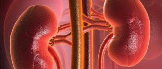
Small cysts appear in the area of the tubules - vesicles filled with liquid. Kidney damage in polycystic disease always occurs on both sides; the size of the cysts can be from a match head to a large cherry and even more. They contain a light or brown jelly-like mass inside. The growth of cysts with age leads to the fact that the number of functioning cells (nephrons) is reduced, and this leads to the development of renal failure. Often, with polycystic kidney disease, the spleen, pancreas, seminal vesicles, liver, lungs or other organs are also affected.
Classification
Depending on the time of onset of the first manifestations and the degree of liver damage, the following types of disease are distinguished:
1. Perinatal type - the most common:
- accompanied by oligohydramnios and pulmonary hypoplasia;
- 75% die within 24 hours of birth;
- minimal degree of liver fibrosis;
2. Neonatal type - minimal degree of periportal liver fibrosis;
3. Infantile type - moderate periportal fibrosis;
4. Juvenile type - pronounced periportal fibrosis in combination with portal hypertension, splenomegaly and portosystemic varicose veins.
Causes of polycystic kidney disease in adults
“Polycystic disease is a genetic disease caused by the degeneration of the kidney parenchyma with the formation of multiple cysts of various sizes,” says urologist-andrologist Alexander Danilov.
– This occurs as a result of a disruption in the formation of the functional units of the kidneys - nephrons - in the prenatal period. Some nephrons in polycystic disease do not connect to the collecting system of the kidney. This causes liquid to accumulate in them. They turn into cavities with fluid - cysts. There are from 20 to 50 such cysts. Externally, the affected kidney resembles a bunch of grapes. Cysts can be of different sizes and shapes, and the affected organ can weigh up to 2 kilograms. However, in adults with polycystic disease, the kidney retains a sufficient amount of fully functioning kidney tissue. Therefore, clinical manifestations of the disease may appear late or be completely absent.
Pathological anatomy
Macroscopic structure
The fetus has a characteristic appearance (Potter's face): narrow eyelid slits, a characteristic groove under the eyelid line, micrognathia, a flattened nose, soft large ears of an abnormal shape. The kidneys are increased in size, while maintaining their bean-shaped shape, but cannot perform their function. Poorly functioning kidneys do not produce enough fetal urine, leading to oligohydramnios and pulmonary hypoplasia. The hydrostatic pressure of the amniotic fluid ensures the normal development of the respiratory system.
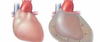
Figure 1 | Fetus, 23 weeks. Fetal death after premature birth was due to pulmonary hypoplasia, caused by oligohydramnios due to PBP. Enlargement of both kidneys with displacement of the abdominal organs in combination with lung hypoplasia.
Figure 2 | Longitudinal section of a kidney with PCB.
Microscopy
Kidneys Radially oriented dilated collecting ducts form renal cysts with a diameter of 1–2 mm, between which normal glomeruli and tubules are visible.
The size of cysts can vary depending on age. In the early stages, the disease begins with microcysts, which then grow and turn into macrocysts. Cystic kidney disease is accompanied by slight interstitial fibrosis of the renal parenchyma.
Figure 3 | Microscopic specimen of a kidney from a patient with PBC-AR. Twenty-fold magnification, hematoxylin-eosin staining. ✱ — radial renal cysts; ▼—renal capsule. Arrows indicate normal glomeruli between dilated collecting ducts.
Liver Histological changes in the liver include the following malformations of the ductal plate: biliary hyperplasia, biliary ectasia and periportal fibrosis.
Disturbances in the morphogenesis of the development of the bile ducts lead to their dilatation. With subsequent progression of the disease, the dilated ducts turn into macrocysts connected by normal ducts, which allows them to be verified quite well using MRCP.
Figure 4 | Pathological changes in the liver. a) The structure of the normally branched portovenous and ethmoidal systems of the biliary tract (left) is disrupted due to a defect in the development of the ductal plate and errors in the terminal differentiation of cholangiocytes (right); b) Coronal T2-weighted image of the abdominal cavity: - the arrow indicates a cystic fusiform dilatation of the bile ducts; — arrow like an arrowhead — nephromegaly with small cysts; ✱ — splenomegaly. c) Preparation of a liver fragment, hematoxylin-eosin staining, 40x magnification:
✱ — extensive fibrosis of the portal area;
arrow - dilated tortuous bile ducts; arrowhead type arrow —hypoplasia of tributaries of the portal vein.
Symptoms of polycystic kidney disease in adults
In many ways, the symptoms depend on the age at which the first signs of pathology appear.
If they occur in the newborn period, the pathology has an unfavorable course and quickly leads to death due to the development of uremia (poisoning with metabolic products that should be excreted by the kidneys). If it is adult polycystic disease, it typically develops slowly and progressively. The disease occurs in several stages - compensated, subcompensated and then transition to decompensation.
In the compensation stage, there are initially no symptoms. Over time, you may experience:
- feeling of pressure in the lumbar region;
- abdominal pain of varying nature;
- urinary disorders due to stretching of kidney tissue;
- frequent fatigue;
- headache;
- blood in urine.
Basic kidney functions are not impaired at this stage.
At the stage of subcompensation, the first signs of kidney failure appear:
- nausea and dry mouth, thirst;
- attacks of headache or migraine;
- very persistent increase in blood pressure;
- polyuria (excessive urine output) with low density;
- blood in the urine, casts, leukocytes (if pyelonephritis is associated);
- fever, toxicosis, chills, if the cysts suppurate;
- attacks of renal colic if urolithiasis is associated.
In the stage of decompensation, constant uremia (urinary bleeding) is formed. Without hemodialysis, the patient feels extremely unwell and serious complications arise.
Compensation stage
In the compensatory stage, manifestations of polycystic disease are most often characterized by general symptoms: fatigue, general weakness, dysuric phenomena. The patient may also complain of pain in the lumbar region (aching, pressing, prolonged, not relieved by taking antispasmodic drugs). Pain may radiate to the lateral abdominal wall. The occurrence of painful sensations is associated with stretching of the renal capsule with an increase in the volume of the kidney affected by cysts of various sizes. Hematuria (blood in the urine) may occur. The exact cause of bleeding inside cavity formations has not been established. Hematuria can be intermittent and self-limiting. In rare cases, bleeding can be quite massive, requiring surgical intervention.
Diagnostics
In order to make a diagnosis, you need to carefully study the family history - most often familial cases of polycystic disease are identified. The doctor also conducts a full examination, palpating the abdomen - sometimes he can identify enlarged, lumpy kidneys. But the diagnosis can be confirmed only after tests and additional examinations:
- Ultrasound of the kidneys;
- nephroscintigraphy (using radioisotope substances);
- excretory urography (with the introduction of a contrast agent and x-ray of the kidneys);
- CT or MRI of the kidneys;
- selective angiography (to determine how the kidney is fed with blood);
- intravenous urography - it helps to determine significant enlargement of the kidneys on both sides, such as deformation of the calyces and pelvis.
Additionally, urine samples according to Reberg, Zimnitsky, biochemistry of blood, urine, and urine culture for sterility may be prescribed to exclude infection. Genetic research is also carried out.
Additional examination methods
Laboratory:
- clinical urine analysis;
- urine analysis according to Nechiporenko (the number of formed elements in the urine);
- urine analysis according to Zimnitsky (allows you to determine the excretory and concentration function of the kidneys);
- clinical blood test;
- biochemical blood test (level of creatinine, urea - end products of protein metabolism, Ca, K, P);
- bacteriological culture of urine.
Instrumental:
- Ultrasound of the kidneys. Determines the presence of cysts, expansion of the collecting apparatus;
- excretory urography is an x-ray method that is based on the introduction of contrast into the patient’s blood for the purpose of further assessing the excretory function of the kidneys;
- retrograde pyelography is also an X-ray examination method in which contrast is supplied through a urinary catheter. This method allows you to determine the state of the cavity structures of the kidneys;
- computed tomography (CT), nuclear magnetic resonance imaging: allows you to determine the number of cysts, their shape and size;
- renal angiography (determination of the vascular structures of the kidney);
- ECG (evaluation of heart function).
Differential diagnosis of polycystic kidney disease must be carried out with glomerulonephritis, pyelonephritis, and tumor diseases of the kidneys.
Modern methods of treatment
– Since polycystic disease is a genetic disease, there is no specific treatment.
That is, it is not possible to prevent its development, says Dr. Alexander Danilov. – The main thing is symptomatic treatment. It is divided into conservative and surgical. Conservative treatment. Its goal is to reduce the manifestations of the disease and prevent complications. For example, when blood pressure increases, antihypertensive therapy is carried out. With the development of kidney inflammation (pyelonephritis), antibacterial drugs are used. For protein deficiency - amino acid preparations. If anemia develops (decreased hemoglobin concentration in the blood), use iron supplements and erythropoietin.
If the amount of urine produced decreases (reduced diuresis), diuretics may be prescribed. In case of severe renal dysfunction, hemodialysis or peritoneal dialysis is performed.
Surgery. If the cysts rapidly increase in volume, a puncture or laparoscopic excision is performed. Surgical reduction of the volume of cysts leads to reduced compression of the remaining kidney tissue and improved blood circulation in it. This allows the remaining normal kidney tissue to function properly. In cases of severe renal failure, a kidney transplant is performed.
Treatment of kidney cyst
What to do if a kidney cyst is detected? There are no therapeutic methods for treating kidney cysts. Until the mid-90s, before the widespread introduction of endoscopic kidney surgery methods into medicine, patients with single or multiple kidney cysts were under dynamic observation and received only symptomatic treatment. In the case of small single cysts, patients underwent surgery for sclerosis of the kidney cyst under ultrasound guidance. When the kidney cyst grew rapidly, patients underwent open kidney surgery.
Since, unlike a tumor, a kidney cyst is a benign disease, a cyst can be monitored in 70% of cases without resorting to active surgery. Patients whose kidney cyst does not exceed 5 cm in diameter, is located on the periphery of the kidney and does not cause disturbances in the kidney’s blood circulation and urine outflow, usually require dynamic monitoring using ultrasound once every 6–12 months.
Surgical treatment of a kidney cyst is not performed in cases where the cyst does not bother the patient, does not cause a disturbance in the outflow of urine from the kidney, as well as in severe concomitant conditions of the patient and blood clotting disorders.
To determine the type of kidney cyst, its localization to the main structures of the organ and indications for surgery, as well as choosing the correct surgical treatment tactics, you need to send me a complete description of the kidney ultrasound, MSCT data of the kidneys with contrast, indicate your age and main complaints to your personal email address. Then I will be able to give a more accurate answer to your situation.
Indications for surgical treatment for kidney cysts:
Organ-preserving surgery - laparoscopy of a kidney cyst. More than 250 operations were performed.
- compression of the kidney cyst of the urinary tract
- compression of the cystic tissue of the kidney parenchyma
- infection of the cyst cavity and abscess formation
- rupture of kidney cyst
- large cyst sizes and/or rapid growth of the kidney cyst
- pain caused by a cyst
- hematuria from the kidney from the side of the cyst
- arterial hypertension
- erythremia
- the presence of blood in the punctate from the cyst or hemorrhage into the cyst
- cyst rupture or danger thereof
- cancer in the cyst (based on radio-radiological, echographic data or examination of puncture fluid from the cyst).
During the surgical treatment of a kidney cyst, interventions of varying scope can be performed:
- percutaneous therapeutic and diagnostic puncture of a kidney cyst with sclerosis;
- enucleation of the cyst (husking);
- cyst resection;
- kidney resection with cyst;
- nephrectomy.
Watch a video of operations performed by Professor K.V. Puchkov. You can visit the website “Video of operations of the best surgeons in the world.”
Renal puncture is currently the first step in the treatment of simple single (solitary) uncomplicated kidney cysts with a diameter of up to 3–5 cm and located subcapsularly in the renal parenchyma. Currently, percutaneous therapeutic and diagnostic puncture of a kidney cyst is carried out only with sclerosing, since puncture of a kidney cyst without the use of a sclerosing agent in most cases leads to recurrence of the cyst and the need for repeated intervention. In some cases, there may even be an increase in the volume of the cyst compared to its original size. Percutaneous therapeutic and diagnostic puncture of a kidney cyst is carried out under ultrasound control, evacuation of the contents and introduction of an adhesive substance into its cavity.
The introduction of sclerosant into the cavity of the cyst significantly reduces the frequency of relapses (up to 2%) within a year after percutaneous therapeutic and diagnostic puncture of the kidney cyst. Within three years, cyst recurrence after sclerotherapy is observed in 7–15% of patients.
It must be taken into account that even with small sizes of single kidney cysts, technically, not all cysts can be punctured, which is determined by the localization of the cyst in the kidney.
Currently, in the USA and Europe, treatment of renal cysts using laparoscopic endovideosurgical surgery is the main method. Laparoscopic surgery for kidney cysts is a modern and low-traumatic method for radical removal of cysts. The laparoscopic technique for treating solitary (single) kidney cysts has great advantages over both puncture methods and open operations.
On the one hand, a radical operation is performed - excision of the cyst with histological examination, and on the other hand, the principle of minimally invasiveness is maintained (3 incisions of 5 mm each remain on the abdominal wall). Laparoscopic access allows you to remove a kidney cyst of any size and location. An enlarged image on the monitor makes it possible to see the subtle anatomical formations of the kidney, which makes the operation delicate and safe, and the use of modern ultrasonic surgical scissors and the device for dosed electrothermal tissue ligation “Liga sure” (USA) allows for bloodless excision of the cyst.
For large cysts (diameter more than 5 cm), with a predominantly intraparenchymal location (most of which are located inside the kidney), it is necessary to excise the “roof” of the cyst (its upper part). In this case, the inside of the cyst is treated with argon-enhanced plasma (Force Triad device, USA) to prevent cyst recurrence.
Open surgical treatment of simple kidney cysts is resorted to only when complications occur , such as suppuration or rupture of the cyst, its malignant degeneration, for cysts that have led to significant or complete loss of kidney parenchyma and hypertension. In addition, from the previously existing large list of indications for open surgery for simple renal cysts, there are also concomitant urological diseases that require open surgery, for example, some clinical forms of urolithiasis, stricture of the ureteropelvic segment. Open surgical treatment of a kidney cyst consists of nephrectomy, kidney resection, enucleation of the cyst or excision of its free wall.
Multiple cysts are sometimes confused with congenital polycystic kidney disease; the treatment of these diseases differs significantly from each other.
Polycystic kidney disease is a congenital kidney disease that is usually inherited. The disease first appears in childhood or young age and progresses gradually with a slow increase. Sudden manifestation occurs extremely rarely, against the background of a cold or weakening of the body. In case of acute onset, polycystic kidney disease is manifested by a sharp rise in blood pressure and impaired excretory function of the kidneys. During examination, kidneys of enormous size are often discovered with degenerated renal tissue (parenchyma), which is compressed by multiple cysts.
In congenital polycystic kidney disease, the feasibility of excision of cysts is questionable. Patients with polycystic kidney disease need surgical treatment only in cases of suppuration of the cysts. In general, therapeutic symptomatic (palliative) treatment is preferable for them to limit the growth of cysts, preserve kidney function and postpone the need for continuous hemodialysis and kidney transplantation for as long as possible. Even small single kidney cysts can cause the development of malignant arterial hypertension, which is not amenable to conservative treatment. Large cysts can cause atrophy of the kidney parenchyma and chronic renal failure.
Description example
Descriptive part: in both significantly enlarged kidneys there are multiple space-occupying formations, partially merging with each other, ranging in size from ... cm to ... cm. The formations have a hypo-, isointense MR signal on T1 and a hyperintense MR signal on T2, which indicates the presence of their composition is a stationary liquid with varying protein content.
CONCLUSION: The MR picture is characteristic of multifocal cystic lesions of the parenchyma of both kidneys and liver with possible hemorrhage into the cavity of individual cysts.
Pathogenesis
The childhood form of polycystic disease (Potter I) is inherited as an autosomal recessive trait and is often fatal in the first months of life. Often combined with developmental anomalies of the hepatobiliary zone (portal hypertension, primary hepatic fibrosis). All symptoms are clearly detected by ultrasound. An uncomplicated course is not an indication for an MRI examination.
The adult form (Potter III) is an autosomal dominant trait and causes progressive renal failure that begins around age 30. It is associated with liver cysts (30-60%), pancreatic cysts (10%), spleen (5%) and an increased likelihood of arterial-venous malformations and aneurysms of the circle of Willis (15%). However, these patients are no more likely than usual to be diagnosed with renal cell carcinoma. MRI is usually prescribed to determine the causes of hematuria and diagnose complications (hemorrhage, inflammation).