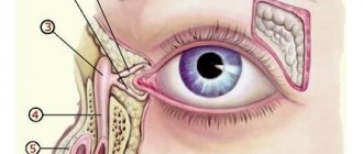
The tarsal or tarsal canal is a canal between the medial malleolus, talus and calcaneus, and the flexor tendon retinaculum, a connective tissue structure that runs from the medial malleolus to the calcaneus. The channel contains:
- tibialis posterior tendon;
- flexor digitorum longus tendon;
- posterior tibial artery and vein;
- tibial nerve;
- flexor tendon of the thumb.
The tibial nerve divides into two terminal branches—the medial and lateral plantar nerves—as it passes through the tarsal canal. The medial calcaneal nerve arises from the tibial nerve near or superior to the flexor retinaculum.
Tarsal tunnel syndrome (TTS) is a rare compressive neuropathy of the tibial nerve or one of its branches as it passes under the flexor retinaculum.
Friends, this and other questions will be discussed in detail at the Neurodynamics seminar. Find out more...
In the literature on TTS, the tibial nerve is also called the posterior tibial nerve, so TTS is also known as posterior tibial neuralgia. Some authors refer to compression of the deep branch of the peroneal nerve as “anterior tarsal tunnel syndrome.” This article focuses on TTS as a condition that results in compression of the tibial nerve or its branches.
Epidemiology/Etiology
The incidence is unknown. A higher prevalence of TCS has been reported among women than among men. Moreover, this occurs at any age. Causes of TCS include:
- Repetitive activities that strain this area, such as running, long walking, or standing.
- Injuries such as fractures, dislocations, or sprains.
- Varus or valgus heel.
- Fibrosis.
- Excess body weight.
- Pathologies occupying space in the tarsal canal area, such as tumors, edema, osteophytes or varicose veins.
- Tendinitis.
- Systemic diseases that cause inflammation of the ankle joint or disorders of its innervation (for example: diabetes, arthritis).
Many cases (20-40%) are idiopathic.
Characteristics/Clinical picture
Common symptoms of TTS include paresthesia (burning, numbness, or tingling) in the tibial, lateral, and/or medial plantar nerves. There may be burning, tingling, or pain along the medial ankle and plantar surface of the foot, as well as localized tenderness behind the medial malleolus. Symptoms usually worsen with forced eversion and dorsiflexion of the foot. When the medial plantar nerve is isolated, patients may experience stabbing pain in the midfoot, which is typically seen in middle-aged runners. If the condition is progressive or chronic, there may be muscle weakness of the abductors and flexors of the fingers. In more serious cases, muscle atrophy occurs. Patients may also experience night pain that reduces sleep quality, as well as severe pain when walking for long periods of time.
Diagnostics
Pain, numbness, pins and needles and disturbances in arm movement can be caused not only by compression of the nerves in these bone canals. There are also diseases of the brachial plexus, spinal hernias, muscle tightness and other conditions that cause pain in the arms. Therefore, the most reasonable thing is to immediately contact a specialist, and not try to figure it out on your own.
Diagnosis of pain in the arm may take 2-3 days. First of all, this is an examination by a neurologist, as well as conventional clinical methods (tests, X-rays, MRI) and special techniques that allow you to accurately determine the extent of nerve damage - electromyography and electroneurography.
These methods examine the electrical activity of muscles and the speed of transmission of nerve impulses. By combining the results of both methods, one can understand whether the nerve has retained its function or has degenerated and been replaced by connective tissue. The treatment method depends on the results of the examination.
Survey
It is important to take a thorough history. The physical therapist should learn about the following:
- Mechanism of injury – was there trauma, sprain or overuse?
- Duration and location of pain and paresthesia?
- Weakness or difficulty walking?
- Are back and buttock pain associated with distal symptoms?
- Does the pain get worse, stay the same, or get better?
Key symptoms
- Paresthesia or burning in the area of the distal branches of the tibial nerve.
- Prolonged walking or standing often worsens the patient's symptoms.
- Dysesthesia (an abnormal or unpleasant sensation) occurs at night and can interfere with sleep.
- Muscle weakness.
Observation
- Atrophy of the abductor pollicis may be noticeable.
- Assessment of the arches of the feet.
- Position of the talus and calcaneus.
Gait Analysis
- Examine for abnormalities (excessive pronation/supination, excessive inversion/eversion, antalgic gait, etc.).
Sensitivity assessment
- Testing surface sensitivity, sense of discrimination.
- Sensitivity will be impaired in the area of innervation of the tibial nerve.
Palpation
- Tenderness on palpation between the medial malleolus and the Achilles tendon (palpation is painful in 60-100% of patients).
Movement amplitude
- Focus on the range of motion of your ankle and toes.
Manual muscle testing
- Decreased strength usually occurs in the late stages of STS.
- First, the abductors of the fingers are turned off, and then the short flexors of the fingers.
Special tests
Tinnel's sign
- Percussion in the area of the tarsal canal leads to the spread of paresthesia in the distal direction (occurs in more than 50% of patients).
Dorsiflexion-eversion test
- Place the patient's foot in the dorsiflexion position and hold it in eversion for 5-10 seconds. This results in the patient becoming symptomatic.
Electromyography
- The presence of an isolated lesion of the tibial nerve in the tarsal canal is confirmed by measuring the velocity of impulses along sensory and motor fibers.
- Assessment of conduction along sensory fibers of the medial and lateral plantar nerves. This is best done by recording from the tibial nerve just above the flexor retinaculum and stimulating the ball of the foot. When surface electrodes are used, responses to stimulation are of low amplitude.
- Measuring conduction velocity along motor fibers by recording the distal latency of the abductor pollicis is a much simpler but less sensitive method. An important finding of electromyography is the detection of axonal damage when readings are recorded from the distal muscles innervated by the tibial nerve.
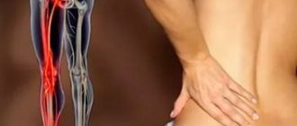
Signs and symptoms
The specific symptoms of tarsal tunnel syndrome can vary from person to person. In some cases, symptoms may develop suddenly, in others they may develop gradually. Some patients may experience sharp shooting pain along the tibial nerve. This nerve branches off from the sciatic nerve and runs down the leg to the ankle and then to the foot. The pain can be so severe that the person walks with a limp. Patients may experience radiating pain that is not localized to one place. In addition to or instead of pain, patients may experience numbness of the affected area, a burning or tingling sensation (paresthesia) that feels like a pin or needle.
For some people, symptoms may affect one location, such as the inside of the ankle. For others, symptoms affect the ankle, heel and foot. For example, pain may spread from the ankle to the heel or even the foot, depending on which part of the nerve is affected. Less commonly, the pain spreads from the ankle to the calf.
Symptoms of tarsal tunnel syndrome are often worsened by activities such as prolonged standing or walking. Consequently, the pain may worsen during an active day. Symptoms usually go away with rest. However, as the disease progressed, some patients reported pain during rest or at night when trying to sleep.
Rating scales
- Functionality Questionnaire of the Foot and Ankle (FAAM). The FAAM is a reliable, sensitive, and valid measure of physical function for people with a wide range of musculoskeletal disorders of the leg, ankle, and foot.
- Tarsal tunnel syndrome severity rating scale.
| Symptoms | None | In some ways | Present |
| Pain, spontaneous or with movement | 2 | 1 | 0 |
| Burning pain | 2 | 1 | 0 |
| Tinnel's sign | 2 | 1 | 0 |
| Sensory impairment | 2 | 1 | 0 |
| Muscle atrophy or weakness | 2 | 1 | 0 |
Disorders with similar symptoms
Symptoms of the following conditions may be similar to those of tarsal tunnel syndrome. Comparisons may be useful for differential diagnosis.
Plantar fasciitis (plantar fasciosis) is a common condition characterized by inflammation of the thick band of tissue that runs along the bottom of the foot (plantar fascia). The plantar fascia supports the arch of the foot and acts as a shock absorber. Plantar fasciitis is a common cause of heel pain, often described as sharp pain in the heel. The pain associated with plantar fasciitis tends to develop gradually and is worse in the morning and may improve throughout the day and with stretching exercises. Additionally, this pain is different from the shooting and tingling pain associated with tarsal tunnel syndrome.
Foot or ankle pain can cause a wide range of conditions, including diabetes (diabetic neuropathy), posterior tibial tendinosis, stress fractures, some rare disorders such as reflex sympathetic dystrophy, and certain disorders that affect nerves outside the central nervous system (peripheral neuropathy).
Treatment for tarsal tunnel syndrome
Medication support
To optimize recovery and reduce functional impairment in patients with TTS, pharmacological methods are used in combination with physical therapy. These include:
- NSAIDs.
- Corticosteroid injections.
Surgery
Surgery is indicated for patients who have not benefited from conservative treatment such as physical therapy and have symptoms that significantly affect their daily life. People with severe disorders tend not to respond to conservative treatment and often require surgery. Godges and Klingman identified several characteristics that were associated with successful response to surgery. These include young age, short disease duration, no history of ankle problems, early diagnosis, and specific etiology.
- Decompression of the tibial nerve.
- Cryosurgery.
Physical therapy
There is a lack of high-level evidence regarding physiotherapy treatment for tarsal tunnel syndrome. Further research is needed to identify specific rehabilitation exercises for patients with TCS. There are small randomized controlled trials that include analyzes of the effectiveness of specific treatments.
Ulnar nerve compression or cubital syndrome
MRI of the hand
- Cost: 7,000 rub.
More details
Occurs in those who often bend their arm at the elbow - cyclists, programmers and others. The ulnar nerve is often affected in very thin women. In this case, pain and numbness begins with the little finger, spreading to the entire hand. The muscles on the back of the hand between the thumb and index finger become thinner and begin to work less well. In damp, cold weather the pain becomes almost unbearable.
Conservative treatment
| Stages | Physical agents | Orthotics | Therapeutic exercises | Manual therapy |
| Acute stage Goal: reduce pain and swelling |
|
|
|
|
| Subacute stage Goal: Increase flexibility and improve straight leg test | See above | See above | Tibialis Posterior Stretch | See above |
| Recovery stage Goal: develop symmetrical flexibility, improve straight leg test and functional mobility | See above | See above | Tibialis Posterior Stretch | See above |
One of the mechanical causes of TCS is thought to be excessive calcaneal eversion, leading to collapse of the medial arch (overpronation), which exerts a traction effect on the tibial nerve and leads to compression under the flexor retinaculum. Scherer believes that custom orthoses can correct overpronation and therefore reduce stress on the tibial nerve. Although there are no clinical trial results to support the effectiveness of orthopedic treatment, it may be an important modality to consider when treating patients with TCS.
A number of articles listed the main methods of conservative treatment of STS as a guide for rehabilitation, but did not provide patient outcomes.
Postoperative treatment
| Phases | Period | Goals | Intervention |
| Phase I | 1-3 weeks |
|
|
| Phase II | 3-6 weeks |
|
|
| Phase III | 6-12 weeks |
|
|
Incidence (per 100,000 people)
| Men | Women | |||||||||||||
| Age, years | 0-1 | 1-3 | 3-14 | 14-25 | 25-40 | 40-60 | 60 + | 0-1 | 1-3 | 3-14 | 14-25 | 25-40 | 40-60 | 60 + |
| Number of sick people | 0 | 0 | 1 | 10 | 10 | 30 | 20 | 0 | 0 | 5 | 100 | 100 | 80 | 70 |
Case study
Romani et al reported their results in a 22-year-old lacrosse player with tarsal tunnel syndrome. The player suffered a mild eversion ankle sprain that was successfully treated with conservative treatment. Following a recurrent ankle sprain, the patient made the decision to compete in the NCAA Tournament, which led to an exacerbation of symptoms and, ultimately, to surgery. The 13-week rehabilitation program included: RICE, range of motion maintenance, balance exercises, therabend exercises, aquatic therapy and walking, which eventually progressed to running. At the end of week 13, the athlete returned to lacrosse, competing at the elite level.
Dr. Karen Hudes conducted a separate case study on the conservative approach to treating TTS. A 61-year-old patient diagnosed with tarsal tunnel syndrome reported pain and discomfort in the medial ankle area (verbal rating scale 9/10). Initial treatment included orthopedic techniques for the first ten weeks, after which the patient reported little change in symptoms. Following failure of orthopedic therapy, treatments such as transverse friction massage, high-velocity, low-amplitude manipulation of the talonavicular joint, and cuboid mobilization were used twice weekly. The patient's symptoms began to improve after 3 weeks, resolved by week 6 with occasional relapses of pain, and resolved completely by week 12. The patient reported no pain during the ten-month follow-up.
Causes
The exact causes of tarsal tunnel syndrome are still unknown. Usually, at the onset of the disease, patients report the occurrence of tissue swelling in the area of the tibial nerve. It is precisely because of this swelling, caused, for example, by a banal fall or blow, that making an accurate diagnosis and directly treating the disease can be delayed.
In addition, the likelihood of tarsal tunnel syndrome is influenced by a special mechanism of functioning of various systems within a person, called the “flexor retinaculum.” The tunnel, where the nerve endings of the tibia are concentrated, is a dense structure that can stretch and contract as necessary. This is what makes the flexor retinaculum work. Any pathological inflammation within this system compresses everything inside the structure.
So, for example, with a strong blow to the leg, swelling occurs and inflammation begins, against the background of which pressure on the tibial nerve increases. As a result, a disease such as tarsal tunnel syndrome may occur.
Symptoms
The symptoms of tarsal tunnel syndrome are quite varied, but the main symptom of this disease can be considered numbness of the foot in the heel and surface in the area of the leg injury.
In addition, almost every patient with this diagnosis reports unbearable pain in the plantar area of the foot. Such discomfort is associated with the functioning of the nervous system. After all, every nerve ending is a sensitive component, so when it is compressed, pain should appear instantly. The patient feels a tingling and burning sensation, especially if the leg is under stress such as:
- run;
- walking;
- standing in one place for a long time.
If the injured leg is in a calm, relaxed state (rest, sleep), the pain decreases significantly, but almost never disappears completely.
Diagnosis
When making a diagnosis, the medical expert first takes a history of the disease. During the examination, a podiatrist, a specialist in solving problems with the functioning of the foot, asks about symptoms, the nature of pain, and also prescribes some types of tests.
But how can one accurately determine whether a person has tarsal tunnel syndrome? For this, there is a special examination, within the framework of which the speed of nerve conduction is checked, in other words, the latter must find out how many impulses the nerve emits in a certain time.
So, if the speed of nerve conduction is slow, then in 90% of cases the patient is diagnosed with tarsal tunnel syndrome. Also, with this disease, patients experience a positive Tinel's symptom, which is characterized by a feeling of lumbago when tapping along the nerve in the compression zone.