If a person feels pain in the jaw, radiating to the ear and temple, we may be talking about arthritis of the mandibular joint. The pathology is not just painful, but also dangerous. If not diagnosed in a timely manner, it can lead to severe complications, including paralysis of the facial nerves. Therefore, the first thing to do when such symptoms appear is to consult a specialist. In 10-18% of cases, diagnosis and treatment of TMJ arthritis is the responsibility of dentists and occurs in people from 25 to 45 years old.
What is the temporomandibular joint - TMJ
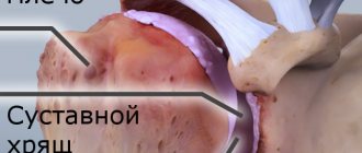
We use this joint while talking, chewing, yawning, laughing - very often. Around it are muscles and tendons that allow the jaw to move in different directions for different purposes.
TMJ problems occur for many reasons:
- violation of the dental system, for example, congenital anomalies of teeth, gums or bone tissue, malocclusions, injuries, poor-quality prosthetics;
- congenital anatomical abnormalities of this joint;
- increased or decreased tone of muscle fibers around the TMJ;
- active conversational load, for example, among actors, singers, speakers;
- central nervous system problems;
- disruptions in the functioning of the endocrine system;
- excessive tension of the jaw muscles, for example, due to the bad habit of biting nails.
What is the temporomandibular joint and why do problems arise with it? Detailed and clear - in the video:
Actors and singers with active oratorical articulation are at risk for TMJ arthrosis
Cellular space
In the temple area there are two fascia: superficial (in the form of a thin plate running across the entire temple) and proper. When they split, they form an interfascial fiber gap filled with fatty tissue, penetrated by many fibrous bridges.
The cellular spaces of the temporal region consist of the following structures:
- Subcutaneous tissue lying between the skin and the superficial fascia. This is a kind of continuation to the temple area. It contains blood vessels, nerve and muscle fibers.
- Interponeurotic tissue located between the superficial and deep plates of the temporal fascia.
- Subgaleal tissue enclosed between the temporal fascia and musculus temporalis. It contains fat and veins.
The last, deep layer is located between the temporalis muscle and the periosteum. It runs along the blood vessels and nerves that supply muscle tissue.
How to understand what is bothering you with TMJ: 4 obvious signs
If you feel discomfort while talking, chewing, or yawning, look out for these signs:
- Are there any clicks or popping sounds during jaw operation (only 20% of people who notice this symptom complain of pain, however, this is a reason to suspect arthrosis of the temporomandibular joint and go for a diagnosis);
- Is there a feeling of jamming of the joint (if the fossa and the articular head are not in perfect contact, you have to open your mouth as much as possible, trying to find the right point);
- How severe is the pain (can occur when chewing, radiate to the temples, under the tongue, ears, neck, sternoclavicular region);
- Has your general health changed (headache and dizziness, insomnia, hearing impairment, depression and other uncharacteristic manifestations are possible).
With TMJ dysfunction, some patients develop snoring
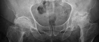
CT scan of the temporal bones: when is this study needed?
Computed tomography of the skull bones is a fairly common study. However, some diseases may require targeted images of individual bones of the skull.
We talk about the features of one of these studies - computed tomography of the temporal bones - with Roman Aleksandrovich Strokov, a radiologist at the Expert Clinic Kursk.
— Roman Aleksandrovich, where are the temporal bones located and what is their function?
- They are located in the base of the skull and participate in the formation of its lateral part. The temporal bones contain the organ of hearing and balance. In addition, the bone canals of the temporal bones contain important nerve structures such as the facial and vestibular-cochlear nerves, and the trigeminal ganglion is located.
— What can be a reason for a CT scan of the temporal bones? For what diseases is this diagnosis effective?
— Computed tomography of the temporal bones is a technique that is carried out according to a special protocol. This helps to clearly detail all the anatomical formations that are located in the temporal bone. Therefore, doctors refer patients with damage to the organ of hearing and balance, or with traumatic brain injury (TBI), when, in particular, a fracture of the base of the skull is suspected, for a CT scan of the temporal bones.
The study is also recommended for patients with neurological symptoms that correspond to damage to the nerves located in the temporal bones. A CT scan of the temporal bones may also be needed when planning surgical treatment, for example, for a disease such as otosclerosis. This is done to evaluate the anatomical features of the temporal bone in a particular patient and to select the appropriate cochlear prosthesis (prosthesis of the cochlea of the inner ear).
CT scan of the temporal bones is also informative for inflammatory processes of the ear (acute and chronic otitis media), injuries, and tumors of this area.
Read materials on the topic:
Is there noise in your ear? Rule out otosclerosis What is otitis media and how to avoid it?
— What does a CT scan of the temporal bones show?
— On a CT image we observe the structures of the outer, middle and inner ear (auditory ossicles, configuration of the ear canal). In addition, it is possible to evaluate the bone elements, as well as the air cavities of the temporal bone for violations of their pneumatization (filling with air). Based on these data, we conclude whether the identified changes are the cause of the illness that worries the patient.
— Roman Aleksandrovich, please tell us how a CT scan of the temporal bones is done
— A standard examination of the temporal bones is performed without contrast (that is, without the use of a contrast agent). The patient enters the office, undergoes instructions, undresses, lies down on the tomograph table and lies motionless throughout the entire procedure. It usually takes a few minutes.
The tomograph is equipped with means of communication with personnel. If during the study the patient experiences discomfort or deteriorates in health, he can report this to the staff.
At the end of the scan, the radiologist checks the quality of the images obtained and their information content. The patient receives pictures on film, a disk with a recording of the procedure and a doctor’s report.
— Do I need to prepare for this procedure?
— There is no special preparation for CT scanning of the temporal bones. The only condition is that the patient must remove all metal-containing jewelry from the outer ear.
— What are the contraindications to this research method?
— CT scanning of the temporal bones, like computed tomography of any other area, is not recommended for pregnant women and children due to ionizing radiation.
— Are there cases in which this study can be performed on pregnant women or children?
— For children, it is performed according to the necessary indications and when other diagnostic methods and clinical examination data do not provide sufficient information to make a diagnosis. Most often, the indications for doing a CT scan of the temporal bones are:
- inflammatory diseases of the ear, congenital anomalies of its development;
- traumatic brain injury;
- planning of surgical treatment.
For pregnant women, this study is carried out strictly according to indications if there is a life-threatening condition.
— Do I need a referral to take this test?
— I believe that it is always advisable to have a referral for a computed tomography scan, not only for children, but also for adults. Why? Because in the referral, the attending physician (otorhinolaryngologist, audiologist, therapist, traumatologist) indicates a preliminary diagnosis made on the basis of the examination. And to confirm or refute his opinion, he sends him for a computed tomography scan. For example, a doctor suspects a patient has an inflammatory process in the middle or inner ear and points this out. That is, the radiologist is asked whether there is inflammation or not. Accordingly, the radiologist must answer this question, specifically describing changes in the structures of interest to the attending physician.
In the vast majority of cases, patients come to our clinic for a CT scan of the temporal bones with a referral. If the patient comes on his own, it is difficult for the radiologist to give a clear answer. Often such a subsequent study is not informative enough for the attending physician.
Interviewed by Sevilya Ibraimova
You can sign up for a CT scan of the temporal bones here. ATTENTION: the service is not available in all cities
The editors recommend:
When is CT indispensable? Why can't my ear hear?
For reference:
Strokov Roman Alexandrovich
Graduate of the medical faculty of the Tashkent State Medical Academy in 2011. In 2012, he completed his internship in the field of Radiology. He currently holds the position of radiologist at Clinic Expert Kursk. Receives at the address: st. Karl Liebknechta, 7
Treatment tactics
If there is no arthrosis, but there is dysfunction, the tactics are as follows:
- determine the root cause and eliminate it, for example, grind teeth and fillings that impede the functioning of the joint, correct prosthetic errors, and correct the bite;
- reduce the load on the joint - the patient is recommended to switch to soft foods, massage the facial muscles and exercises to relax the jaws;
- if the defects are serious, surgical treatment is performed - bone or muscle plastic surgery of the area of dysfunction.
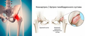
If the doctor determines degenerative changes in the cartilage tissue, treatment for osteoarthritis will be prescribed. With the help of painkillers, they relieve pain. If necessary, a complex of physical therapy is prescribed, for example, laser or ultrasound. Good results are achieved by intra-articular injections of the Noltrex synovial fluid prosthesis, which restore the functionality of the joint by replenishing the missing lubrication.
Arthrosis of the TMJ is successfully treated with intra-articular injections of Noltrex
Functional meaning
The temple, like the entire cranial vault, performs a protective function, preventing mechanical damage to the brain. This area contains brain regions responsible for the following functions:
- visual and long-term memory;
- processing of sensory information entering the central nervous system;
- acoustic perception with receiving and processing of sound signals;
- visual perception.
The temporal region is penetrated by the most important nerves and blood vessels that supply the brain, organs of hearing and vision.
What symptoms should you see a doctor for?
The earlier the diagnosis is made, the more favorable the prognosis. So seek help if:
- you experience pain in the lower jaw - intense or mild;
- you cannot fully move your jaw, you feel limited;
- hear a painful clicking or crunching sound when opening and closing your mouth;
- pain and noise in the ears appeared (especially if the otolaryngologist did not find any pathologies);
- There was a feeling of incomplete bite.
Arthrosis of the TMJ is not a very common phenomenon, but quite dangerous. Our usual comfort during conversation, eating, and other household activities is at risk. To avoid this, do not attribute alarming symptoms to fatigue and do not rely on “it will go away on its own”: be sure to find time for yourself and your health!
Temporal lobe
Many researchers have found a decrease in neuronal density or a decrease in the volume of nerve cells in the temporal lobe in schizophrenia.
The results of a number of studies indicate an asymmetry in the size of the right and left temporal lobes in schizophrenia, with the former being larger than the latter.
As the disease progresses, the temporal lobe of the brain gradually shrinks in size. If in the early 90s only 50% of researchers adhered to this point of view, then already in 2001, with the expansion of the capabilities of neuroimaging methods for studying the brain, data on a reduction in the temporal lobe by an average of 3% in schizophrenia were confirmed in the works of more than 60% of scientists .
A decrease in the volume of the temporal lobes is less frequently recorded in patients with the first episode of schizophrenia compared to patients who have suffered from this disease for a long time. It was found that the volumes of the medial parts of the temporal lobes in patients with schizophrenia are always reduced compared to the volumes of these brain regions of healthy people.
Temporal lobe changes in schizophrenia
- Increased volume of the temporal lobe in childhood schizophrenia
- Reduction in the amount of gray matter in the superior temporal gyrus of both hemispheres
- Reduction in the volume of the left anterior superior temporal gyrus
- Reduction in the volume of the medial temporal lobes (amygdala)
- Reduction in the size of the fusiform gyrus
- Increased activity in the right temporal lobe and decreased activity in the left temporal lobe
- Increased activity in Wernicke's and Broca's areas
- Disruption of connections between neurons of the temporal and frontal cortex
- Disruption of connections between neurons of the temporal cortex
In schizophrenia, the activity of certain parts of the temporal lobe of the brain is increased, for example, Wernicke's and Broca's areas , and this activity is usually observed in those patients who experience auditory hallucinations and mistakenly perceive their thoughts as voices addressed to them.
| | MRI allows you to diagnose changes in the size of the temporal lobe, and you can track brain activity in these areas using EEG |
Clinically, damage to the temporal lobe of the brain is relatively often manifested by auditory hallucinations and various disorders of thinking and speech. It is known that the superior temporal gyrus of the left hemisphere of the brain is the anatomical substrate for speech and communication. In Heschl's transverse gyrus there are 41 primary auditory cortexes associated with Brodmann's area. This area of the cortex is responsible for the non-interpretive awareness of auditory impulses that come from the inner ear and through the lemnicus lateralis, corpus geniculate mediale of the thalamus, reach the anterior transverse gyrus in the gyrus transversi. Interpretation of perceived sounds, previously distinguished only as sounds of different frequencies, occurs in the secondary auditory area of the cerebral cortex, which also serves as the sensory center for speech. Localized in Brodmann's areas 42 and 22 and anatomically corresponding to the planum temporale, it borders on the primary auditory cortex, located as if along its periphery. As a result of integration processes occurring in this area, sounds begin to be recognized as noises, words and melodies.
The auditory zone of the cortex is finely differentiated, each frequency has its own ending point, high frequencies are localized to a greater extent in the posteromedial area of the cortex, low frequencies, on the contrary, show anterolateral localization.
In patients with schizophrenia, the volume of the superior temporal gyrus is reduced compared to the volume of this region of the brain in healthy people. More noticeable is the change in the size of the left anterior superior and posterior superior parts of the temporal lobe. The fusiform gyrus in schizophrenia is also somewhat reduced in size.
Data were obtained indicating a correlation between thought disorder and the volume of the left anterior superior temporal gyrus. Moreover, the severity of thinking disorders correlated with the degree of reduction in the volume of this area of the brain (Shenton M. et al., 1992).
According to some authors, enlargement of the right superior temporal gyrus in childhood may be a biological risk marker (phenotypic risk indicator) for the onset of schizophrenia.
The most pronounced changes and, in particular, a decrease in size in schizophrenia are found in the gray matter of the superior temporal gyrus. However, when assessing the total size of the gray and white matter of the temporal lobe of the brain, the differences become less distinct.
A number of authors suggest that the pathological process in schizophrenia sequentially spreads from the deep structures of the brain, the parietal lobe to the frontal and then the temporal lobes, noticeably manifesting itself in the area of the superior gyrus of the right temporal lobe (gyrus temporalis superior).
The loss of cortical gray matter in schizophrenia is characterized by its dynamics and occurs in a specific order: starting from the parietal lobe, through the frontal lobe and the dorsolateral prefrontal cortex to the temporal lobe. At the same time, the temporal lobe of the brain and, especially, the superior temporal gyrus undergo structural changes much later than other brain structures. Note that the superior temporal gyrus is distinguished by the expression of highly differentiated dopamine receptors (D2), however, the density of these receptors here is still less than in the striatum.
Some studies have demonstrated a relationship between the severity of positive symptoms of schizophrenia and the degree of reduction in the gray matter of the superior temporal gyrus, and the intensity of auditory hallucinations correlated with a decrease in its anterior part, which includes Heschle's gyrus, and the size of the posterior part of the superior temporal gyrus (planum temporale) correlated with the severity thinking disorders. At the same time, according to some authors, noticeable cortical atrophy was not observed in patients with paranoid syndrome.
Some authors explained the decrease in the volume of gray matter in the temporal lobe and the increase in the volume of the basal ganglia with antipsychotic therapy. This process typically began following an increase in cortico-subcortical glutamate expression. In those patients with schizophrenia who did not receive antipsychotics, on the contrary, there was a decrease in the volume of the basal ganglia.
In schizotypal disorder, changes in the superior temporal gyrus have also been noted.
In childhood schizophrenia, in contrast to the decrease in the size of the temporal lobe recorded in adult patients with schizophrenia, on the contrary, an increase in the volume of this lobe of the brain was noted. The increase in the volume of the temporal lobe in childhood schizophrenia is explained as a secondary reaction to primary hypoplasia of adjacent brain structures. It can also be assumed that the temporal lobe of the brain is initially excluded from the destructive processes associated with the development of schizophrenia .
As childhood schizophrenia progresses, the right temporal lobe begins to shrink in size at a faster rate than the left. Thus, schizophrenia manifests itself in a progressive disorder of the structures of the temporal lobe of the brain (“neuronal developmental disorder”), in which early and late periods of change can be distinguished.
In schizophrenia in childhood and adolescence, some researchers have found an increase in the medial structures of the temporal lobe of the brain, in particular, the size of the amygdala.
For childhood schizophrenia, most researchers consider the presence of a defect in the genes responsible for the synthesis of factors involved in the development of certain brain structures to be significant. This defect also explains the disruption of neuronal connections, manifested in the form of speech deficits.
An MRI study of patients with schizophrenia revealed an association between changes in the superior temporal gyrus with cognitive deficits and hallucinations. Speech deficits in the premorbid period suggest a neuromorphological deficiency in this area of the brain long before the manifestation of clear clinical symptoms of schizophrenia. It is likely that a significant change in the structure of the temporal lobe of the brain in adolescence is a consequence not only of genetic predisposition, but also the result of developed psychosis, since clear correlations of structural changes in the temporal lobe of the brain with the severity of symptoms of psychosis have been shown in a number of studies.
In schizophrenia, the activity of the right temporal region, responsible for processing sound information, increases, and the activity of the left temporal region, associated with one's own speech, decreases. It is known that there is a certain connection between the left parietal-temporal region and the ability to understand mental states (Ferrari P., 2006).
Other studies have demonstrated abnormalities in the activity of brain regions associated with the perception of faces (fusiform gyrus), recognition of emotions (amygdala), and perception of human voices (superior temporal region).
At the neurophysiological level , with mental automatism syndrome, one can register increased activity in the temporal and parietal areas of the cerebral cortex , as well as in those structures that are responsible for the perception of one’s own actions and their consequences.
Experimental studies have shown that the more pronounced the hallucinations, the worse the connection between the temporal cortex and its frontal regions . Note that the auditory association cortex projects widely to other brain structures, including the amygdala and hippocampus.
It is a fair assumption that hallucinations can be a consequence of a “break” in any part of the “neural network” connecting these structures to each other.
Magnetic resonance spectroscopy using phosphorus (31P) revealed a significant decrease in the indicator characterizing the NAA/Cre ratio (N-acetylaspartate/creatinine) in the superior temporal gyrus in patients with schizophrenia who had suffered a first psychotic episode.
All multivoxel studies of patients with schizophrenia and their siblings found a decrease in the NAA/Cre index in the hippocampus and medial temporal regions. This fact indicates a genetic predisposition to schizophrenia, initially expressed in the inferiority of these parts of the brain. However, in the literature one can find studies whose results refute such conclusions.
Return to Contents