A tenth of all injuries are fractures of the lower leg bones. Most of the latter are: fractures of the outer and inner ankle. Such injuries are possible when exposed to intense impact force that occurs during road traffic accidents, unfortunate accidental falls in combination with an unnatural position of the limb or foot. Considering that large bones are damaged, surrounding soft tissues and blood vessels may be involved. Such injuries are accompanied by severe pain and significant blood loss.
Anatomy of the lower leg bones
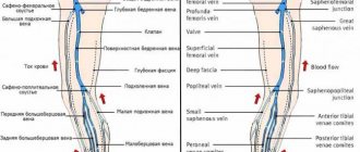
Anatomically, the free lower limb is divided into the thigh, lower leg and foot. The proximal (upper) part of the tibia is fixed to the thigh, forming the knee joint, and in the distal (lower) part the ankle joint. The bone structures of the lower leg are represented by large tubular bones:
- tibia, located medially (closer to the median plane);
- fibula, located laterally (further from the median plane).
Content:
- Anatomy of the lower leg bones
- Fractures
- Symptoms of shin bone fractures
- Help with fractures of part of the lower limb
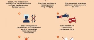
- What not to do
- Conclusion
The distal epiphyses (lower ends) of both bones expand downward, forming the so-called medial (inner) and lateral (outer) ankles, respectively. These bones are connected to each other by fibrous joints, tibiofibular syndesmosis, interosseous membrane, and joints. The lower leg bones are often damaged in lower extremity injuries.
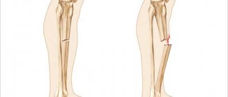
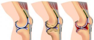
Open and closed fracture of the tibia
Closed fracture of the tibia
A closed fracture of the tibia is a very serious injury. It is characterized by the fact that damage to tissues located far from the bones does not occur, just as there is no contact of the damaged area with the external environment.
With closed fractures, the bones of the ankle and the condyles of the tibia may be damaged, its tuberosity may be torn off, the head of the fibula or the diaphysis of both bones may be damaged. If the far end of the tibia is damaged, the fracture can be either intra-articular or periarticular.
Symptoms of a closed fracture are as follows:
- Sharp limitation of limb mobility. The person will simply not be able to lift his leg up.
- If an attempt is made to raise the shin slightly, the end of the tibia (proximal) will protrude under the skin.
- If, upon palpation, crepitations are heard, that is, characteristic crunching sounds, then this symptom clearly indicates the presence of a closed type fracture. In this case, you should not cause the sound on purpose, since such manipulations can lead to the displacement of fragments that may be present under the skin.
If in patients who have not reached retirement age, closed fractures are more often split, since the bones have a solid structure, then in older people the injuries are depressed, which occurs due to the high porosity of the bone tissue.
Diagnosis, as a rule, is not difficult for an experienced doctor and palpation examination is enough for him to determine a closed fracture. This is explained by the fact that the bones of the lower leg are located close to the skin and are not covered with a thick layer of muscle. However, it is impossible to do without X-ray examination. It will reveal the characteristic features of the fracture and possible displacement of fragments. Pictures must be taken in several projections, most often in two.
Treatment is carried out in a hospital setting. The tasks that doctors face are restoring bone integrity, relieving pain, returning the patient to normal life activities, which will proceed without restriction of movements.
The following methods are used:
- Extension, which involves stretching the damaged bone. It can be skeletal or adhesive.
- Fixation method. It is implemented by applying a certain type of plaster cast.
- An operative method that involves intraosseous fixation using either metal plates, or metal knitting needles, or metal rods, or metal wire.
But, naturally, before one or another method of limb fixation is applied, it is necessary to reposition the fragments, if any. A Delbe bandage is often used to immobilize the fracture site. It has a number of advantages over conventional plaster, since after its application a person can move the knee and ankle joints if they are not damaged. This bandage makes it possible for outpatient treatment, without tying the patient to a hospital bed.
Open fracture of the tibia
If open fractures of other bones of the human skeleton are a relatively rare phenomenon, then with a fracture of the tibia they occur much more often, which is quite explainable by its anatomical features. The tibia itself is located directly under the skin, so it often breaks through it with sharp edges, which leads to an open fracture. In addition, if the injury was received as a result of an accident, then it can be heavily contaminated. This circumstance significantly complicates her character.
The main symptoms of open fractures of the tibia include:
- Bleeding.
- A gaping wound with bones that have broken through the skin and soft tissue.
- Traumatic shock.
- Limitation of mobility.
- Sharp pain.
- Weakness and dizziness, up to loss of consciousness.
Open injuries with the formation of fragments are the most difficult to treat. This occurs because nearby nerves and blood vessels are damaged. Sometimes the question may even arise about the need to amputate a limb.
The deciding factors are:
- How extensive is the area of tissue damage?
- What is the degree of disruption of the blood supply to the foot and leg.
- No pulsation.
- Area of damage to the skin. If it is very extensive and it is not possible to replace it, then this will be a decisive factor speaking in favor of amputation of the limb.
The longer it takes to decide to amputate, the greater the risk that gangrene may develop. Any open fracture must be treated as quickly as possible. After this, the introduction of several drainages is indicated. It is more expedient to pass them through the holes made for this purpose. The wound should be fixed with a rare suture.
When an open wound does not form immediately, but as a result of a puncture by the edge of a fragment and is secondary, then after treatment with antibacterial compounds, sutures are immediately applied, and there is no need to insert drainage.
When a secondary wound is accompanied by large-scale damage to the skin, its transplantation is necessary. It cannot be separated from the fabric for the purpose of stretching. Another important feature in case of an open fracture is that the fragments must be set immediately after treatment with disinfecting compounds, but in no case vice versa. Because this can lead to blood poisoning. In modern medical practice, the use of osteosynthesis is increasingly indicated, which is also carried out after disinfection of an open injury.
If the fracture is transverse, then one reduction will be sufficient; as a rule, the fragments are held securely. If the fracture is oblique or helical, then up to two sutures should be placed with fixation on the wire.
Also, when treating open fractures of the tibia, there is a tendency to insert a special metal rod into the bone. It is empty inside and has holes on the sides. Through it, special medicinal substances, including antibiotics, will be supplied to the bone marrow canal. After its installation, plaster application is indicated.
The prognosis for limb recovery after an open fracture of the tibia largely depends on how well the initial antiseptic and antibacterial treatment was carried out. Proper immobilization of the limb plays a significant role. Treatment after applying a cast is carried out similarly to therapy for a closed fracture, but it is natural that open injuries heal over a longer period of time.
Fractures
A fracture is a violation of the integrity of the bone structure. Fractures of the lower leg bones, depending on the location, are divided into injuries in the area:
- proximal (upper) segment:
- diaphyseal (middle) segment;
- distal (lower).
Damage to the ankle segment (in the ankle area) is considered separately.
Fractures are also divided depending on the damage to the joint:
- periarticular;
- intra-articular.
Based on damage to the diameter of the bone, fractures are divided into: complete and incomplete.
Based on the presence of splinters and bone fragments, there are fractures:
- simple;
- complex (with the presence of fragments).
Damage to the shin bones is a common and quite serious injury. Therefore, it is important to properly provide assistance to a victim with such an injury.
Stages of treatment of open fractures in the Medicenter clinic network
Immediately after an open fracture, the main danger for the patient is pain shock and blood loss, so first of all we administer painkillers and correct the patient’s general condition.
Next, you need to determine the severity of the damage. In order to establish the location of bone fragments, patients need an x-ray examination. The X-ray room at the Medical Center is open from 09:00 to 21:00.
Then we perform surgical treatment of the wound. We thoroughly rinse with an antiseptic solution and remove necrotic tissue. It is optimal if this is done in the first hours after injury, so we emphasize once again that it is very important to immediately take the patient to the clinic. If there is suspicion of damage to nerve endings and blood vessels, we call our consultants in the field of neurosurgery and vascular surgery. Given the risks of infection, our surgeons may recommend not suturing the wound immediately. It is left open so that regular processing and monitoring of its condition can be carried out. If the first signs of infection appear, we immediately begin the entire range of measures aimed at treating the wound infection.
The next stage is the direct healing of the fractures. Since the initial contamination of the wound prevents immediate reduction (comparison of bone fragments) and osteosynthesis (operations for fusion of bones using metal structures), we treat such fractures using skeletal traction, open orthrodesis (such as a plaster or polymer bandage) or compression-distraction devices.
The final stage of treatment is rehabilitation. For rehabilitation, we use an integrated approach, including physical therapy, physiotherapy and massage.
Symptoms of shin bone fractures
Similar to any bone fracture, the main signs are:
- intense pain at the site of injury, sharply increasing with movement;
- inability to support the leg;
- deformation, unnatural position of the limb;
- redness or cyanosis of the skin, and if the integrity of the skin is damaged, there may be a wound, then they speak of an open fracture;
- local swelling, swelling of soft tissues;
- a feeling of crepitus (crunching) at the site of injury;
- lack of foot movement in case of a tibia fracture and injury to the main nerves of the lower limb (usually the peroneal nerve).
Displaced tibia fracture
A displaced fracture of the tibia is most often formed as a result of a direct blow in the transverse direction. In this case, fragments are formed that can move in different directions. The displacement can be lateral, peripheral, angular, with divergence, wedging, and insertion of broken parts.
This type of injury is characterized by the following symptoms:
- The length of the leg will become shorter compared to a healthy limb. Most often, this does not even require additional measurements. The difference will be visible to the naked eye.
- The movement of the lower leg can be carried out in an unnatural direction for it.
- Sometimes the fragments can shift so much that they break through the soft tissue and skin.
- Sometimes a depression or depression forms in the place where the movement of the fragments occurred.
- Pain is a constant accompaniment of any fracture, as well as a crunch during injury.
- At the site of the fracture, bruising and swelling form, with a pronounced impairment of the motor function of the limb.
Most often, the condition of a person who has suffered a displaced fracture of the tibia is still satisfactory, but sometimes traumatic shock can occur.
Treatment will begin with the mandatory comparison of the resulting fragments. This is necessary to give the limb the correct shape and its subsequent normal fusion. Reposition is carried out either manually or using special tools. To do this, the victim must be placed on his back and anesthetized with appropriate drugs. After this, one doctor holds the patient by the thigh, and the other grabs the leg so that one hand firmly holds the heel and the other the back of the foot. Then a slow and systematic stretching of the muscles pulled to the site of the fracture is carried out, and with the help of palpation the position of the fragments that have been displaced is determined. After the reduction is completed, the doctor will definitely check the length of the limb and compare it with the length of the healthy leg. If the parameters converge, then you can begin to apply a plaster cast.
For control, the patient will have to undergo an x-ray again after 10 days so that the doctor can make sure that the fusion of the leg bones is happening normally. Sometimes the skeletal traction method can be used. An operation is required when closed reduction cannot be performed due to the fact that fixation of fragments requires the use of metal structures.
Features of the treatment of elderly people, as well as young patients who have received a displaced leg injury, are that they must be left immobilized for as short a period as possible. That is why you should choose the least traumatic method of treatment.
Help with fractures of part of the lower limb
First aid for a broken leg, main directions of action:
- Stop bleeding if present;
- if there are signs of arterial bleeding (scarlet blood may flow in a stream), a tourniquet is applied above the injury, taking into account the possibility of its removal during immobilization (applying a splint) and with a time stamp of application;
- treatment around the wound with an antiseptic, a sterile pressure bandage.
- Anesthesia
- the administration of analgesics is carried out intramuscularly, intravenously by specially trained people;
- local cold, apply ice, cold water bottles wrapped in cloth.
- Immobilization of a limb with fixation of at least two joints (knee and ankle):
- the injured leg is tied to the healthy one above and below the injury;
- splinting with improvised materials (boards, cardboard) wrapped in soft material. The main splint should run along the back of the leg.
First aid
Home / Articles / First medical aid
FIRST MEDICAL AID FOR WOUNDS, INJURIES AND ACCIDENTS.
Wounds can be through or blind. With a perforating wound, the wounding object passes through the wound area. With such a wound, there are two openings on the skin - an entrance and an exit. In blind wounds, the wounding object becomes stuck in the tissues of the body. Injuries can also be tangential. In such cases, the wounds are a tissue defect in the form of a groove, the bottom of which is the deeper tissue.
The most dangerous complications of wounds are bleeding. . associated with injury to large blood vessels and shock, usually developing with extensive soft tissue injuries and fractures of the long tubular bones of the femur, shoulder, and lower leg. At a later date, the wound may fester and tetanus and gas gangrene may develop. Every wound is fraught with the risk of late complications. In this regard, when providing primary care, it is first of all necessary to stop the bleeding, cover the wound with a bandage in order to protect against further infection, _ and in case of bone fractures, apply a splint. which significantly reduces the danger and often prevents the wounded from going into shock.
STOP THE BLEEDING.
Depending on the type of damaged vessel, arterial, venous and capillary bleeding are distinguished. Bleeding can be external, when blood pours out from a damaged vessel, and internal, when blood pours into any body cavity - stomach, chest, skull.
The most dangerous is arterial bleeding, in which blood from a damaged vessel is ejected under high pressure in a pulsating fountain, and in a short time blood loss can reach life-threatening proportions. It is believed that a loss of more than 40% of blood, about 2.2 - 2.5 liters, is fatal.
For venous bleeding. blood flows abundantly and evenly through the edges of the wound.
Capillary bleeding accompanies any wound. Such bleeding stops under the bandage on its own. Arterial and venous bleeding should be stopped immediately at the site of injury.
Bleeding can be temporarily stopped by pressing the damaged vessel, applying a pressure bandage, tourniquet or twist.
Pressure points of the most important arteries: temporal, occipital, mandibular, right common carotid, left common carotid, axillary, brachial, radial. ulnar, femoral, posterior tibial, dorsalis pedis artery.
Pressing the artery along its length with a finger is an accessible way to stop bleeding under any conditions.
However, it can only be used for a short time before applying a tourniquet or twisting.
To stop bleeding on the neck, head or face. the common carotid artery should be pressed against the transverse processes of the cervical vertebrae at the level of the middle of the anterior edge of the sternomaxillary muscle.
For bleeding in the shoulder area. and the shoulder girdle, the subclavian artery should be pressed against the first rib in the subclavian fossa or the brachial artery against the humerus in the area of the subclavian fossa.
When bleeding from the thigh. you need to press the femoral artery to the pubic bone in the groin area or the femur on the inside of the thigh.
Arterial vessels must be squeezed with considerable effort with 2-4 fingers.
To stop bleeding from small veins and capillaries. It is enough to apply a sterile pressure bandage to the wound. In these cases, the pad of an individual dressing bag or sterile gauze pads should be applied in a thick layer to the wound area and secured tightly with a bandage.
To temporarily stop arterial bleeding from large blood vessels, mainly in the extremities, a rubber elastic tourniquet is used. The tourniquet is a rubber band 125 cm long, about 2.5 cm wide and 4 mm thick. At one end there is a metal hook, and at the other a chain, with the help of which the ends of the tourniquet are secured after it is applied.
The tourniquet is applied above the wound and as close to it as possible. . You should not apply a tourniquet in the middle third of the shoulder, as this can lead to compression of the radial nerve and subsequent development of paralysis. The place where the tourniquet is supposed to be applied must be protected with a cotton-gauze pad or a clothing pad.
To apply a tourniquet, you need to stretch it strongly, make several rings around the limb and secure the ends with a hook and chain. After applying the tourniquet, you should check whether it has sufficiently compressed the soft tissues and blood vessels. The absence of a pulse in the vessels below the wound site indicates sufficient compression of the vessels by the tourniquet. Do not tighten the tourniquet too much. If the tourniquet is tightly tightened, paralysis may develop.
In the absence of a tourniquet, a twist is used to stop bleeding. It can be made from any sufficiently durable fabric, belt, braid, rope. You cannot use thin cords (for example, a telephone cable) for twisting, as they can damage soft tissues.
As when applying a tourniquet, the place where the twist is applied must be protected with a gasket. The twist is wrapped around the limb and the ends are tied in a knot. A stick is inserted into the resulting ring and twisted until the bleeding stops. The end of the stick is fixed with a bandage or other method.
The time for applying a tourniquet or twisting must be indicated on the tourniquet, bandage or in a note.
In summer, the tourniquet can be left on continuously for no more than an hour, and in winter – for half an hour. At these intervals it should be loosened for a few minutes and then tightened again. In total, the tourniquet should not lie for more than 2 hours. In cold weather, the limb with the tourniquet should be well insulated.
A wounded person with vascular damage must be given rest, and the damaged area of the body must be given an elevated position. This achieves a slight reduction in bleeding of the wound area, improves venous outflow and creates more favorable conditions for spontaneous blockage of the vessel with coagulated blood/thrombus/. The final stop of bleeding is carried out in medical institutions.
FIRST AID FOR BONE FRACTURES.
There are closed and open bone fractures. With closed fractures, the integrity of the skin is not compromised and the fracture site is protected from infection. With open fractures, the skin over the fracture site is broken, and bone fragments can come out of the wound. Gunshot fractures are always open.
The most characteristic signs of fractures are: change in the shape of the limb, mobility in an atypical place, inability to use the damaged limb due to severe pain in the fracture area, and in open fractures - the presence of bone fragments in the wound. These signs, however, are not always visible. Therefore, if a fracture is suspected, especially a closed one, first aid should be provided, as in the presence of a fracture. In this case, it is necessary to do immobilization.
Immobilization /IMB/. - / ensuring immobility at the fracture site / reduces pain, protects against the spread of infection, as well as secondary bleeding that can occur due to damage to nearby blood vessels by bone fragments. IMB is given to all wounded people with bone fractures, extensive soft tissue injuries, as well as to wounded people with a hemostatic tourniquet applied. It is done at the site of the injury. If there is bleeding, before IMB it is necessary to stop it and cover the wound with a sterile bandage.
To achieve immobility of the bones at the fracture site, at least 2 joints closest to the fracture site must be fixed during IMB. For MPI, improvised materials or other available techniques are used. For fractures of the bones of the shoulder girdle. /collarbone, scapula, humerus/ the wounded arm can be bandaged to the body, or better yet, on the area of the shoulder and forearm, a splint can be applied using improvised means/branches, boards, plywood/. Then hang your hand on a scarf, a belt, using the hem of a tunic, or bandage it to the body.
For fractures of the bones of the hand and forearm. the splint is applied to the dorsal palmar surface. It should start at the elbow joint and extend slightly beyond the fingers. To ensure a half-bent position of the fingers, a soft object is placed in the hand, and the hand should be suspended.
It is most difficult to achieve MPI for femoral fractures. In these cases, 3 joints should be fixed: hip, knee and ankle. For IMB of hip fractures, 2 boards can be used. In this case, the outer board should rest against the armpit with one cone, and protrude slightly beyond the foot with the other. The inner board, which is shorter, should reach the perineum with one end, and also protrude beyond the foot with the other end. The boards are bandaged to the limb, and the upper part of the outer board is bandaged to the body using bandages, belts, scarves, etc. In the absence of materials that can be used for the splint, the damaged limb is bandaged to the healthy one.
In case of fractures of the leg bones, as well as in case of hip fractures, the wounded limb can be bandaged to the healthy one. If it is possible to apply a splint, it should start at the level of the upper third of the thigh and protrude slightly beyond the foot.
The MPI for fractures of the lower jaw bones is achieved. by pressing it with a bandage to the upper jaw.
If there is a gunshot fracture of the spinal bones, the wounded person should be placed on a wooden shield. Place a rolled up overcoat or duffel bag under your feet in the area of the knee joints. This significantly limits the mobility of the spine and relaxes the thigh muscles. When applying a splint, the protrusions in the joint area must be protected with a soft pad from uniform items, a towel or other means.
FIRST AID FOR BRUISES.
Bruises are accompanied by damage to soft tissues and blood vessels. Blood pouring into the tissue forms a bruise, and with significant hemorrhages, a cavity filled with blood or a hematoma. The outer skin is usually not damaged.
Signs of a bruise include pain, swelling in the area of the bruise, and dysfunction. . The pain is especially severe in the first time after a bruise. It is associated with compression of the nerve endings by the blood pouring into the tissue. Hemorrhage at the site of the bruise is visible only in cases where it occurs directly under the skin. After some time, a dark purple spot appears on the skin where there is hemorrhage - a bruise.
When providing primary care for bruises. It is necessary to apply cold to the bruised area, and then apply a pressure bandage and create maximum rest for the bruised area. Cold constricts blood vessels and reduces bleeding into the tissue.
A bruise on a large surface of the body often causes damage to the entire body - contusion. With mild concussion, there may be a short-term loss of consciousness, vomiting, slow pulse, hearing loss, memory loss and dizziness. In cases of severe concussion, loss of consciousness can be prolonged, and all other phenomena are more pronounced. When providing primary care to shell-shocked people, it is necessary to administer an anesthetic, create rest and send them to the nearest emergency room.