The lower jaw is subject to traumatic damage much more often than other bones of the facial part of the skull. Although it is one of the strongest bones, its mobility and protruding position make it susceptible to fractures. Up to 85% of all injuries to facial bones occur due to fractures of the lower jaw. These figures include both isolated fractures of the lower jaw and fractures with simultaneous damage to other bones of the facial skeleton.
Experts predict that the number of injuries to the lower jaw will only increase, as well as the nature of such injuries. This is facilitated by an increase in the number of vehicles, an increase in their speed limits, as well as complex technical equipment in production.
Types and localization of mandibular fractures
Non-gunshot, most often linear fractures of the mandible occur in the area of the condylar process, the angle of the mandible, the central incisors, the canine and the mental foramen. These places are usually called “places of weakness.”
Direct fractures of the mandible occur at the site of application of force.
Reflected fractures are localized due to the direction of impact and the area of damage. For example, as a result of a lateral impact, a reflected unilateral fracture of the neck of the lower jaw often occurs. The maximum tension in the bone tissue in the midline area is created due to bilateral compression of the mandibular bone in the region of the molars (sixth, seventh and eighth teeth).
How the fragments will be located during a fracture of the lower jaw depends on several factors. These include: the strength of the damaging factor, the volume of the injured area, as well as muscle groups attached to the surface of the injured area.
Causes of mandibular fractures
Mandibular injuries are divided into four types:
1. Domestic injuries that arise from conflicts in everyday life or doing housework.
2. Street injuries resulting from the use of vehicles, and street injuries not related to damage in transport. This could be a fall while walking due to ice, other bad weather, or poor health.
3. Sports injuries received while playing sports.
4. Industrial injuries sustained in industrial production in agriculture.
Symptoms of a mandibular fracture
With a fracture of the lower jaw, patients may complain of impaired or limited mobility of the lower jaw, pain that may intensify with movement, biting or chewing, malocclusion, changes in the sensitivity of the skin on the lower lip or chin, bleeding from the oral cavity - a symptom characteristic of ruptures of the mucous membrane .
The range of complaints that the patient presents helps the doctor understand what kind of damage is being discussed and suggest the location of the fracture.
Diagnosis of mandibular fractures
Diagnosis is based on examination data and collection of patient complaints.
During the examination, the doctor pays attention to the condition of the skin, notes whether the patient has bruises, abrasions or wounds, and records the presence of hyperemia, cyanosis and bruising. Notes the presence of asymmetry on the damaged side of the face.
If a fracture of the lower jaw is suspected, the doctor first feels the undamaged area with his fingertips, and then the damaged one. He marks the places of greatest pain, unevenness, violation of integrity, determines the amplitude of movement of the head of the lower jaw in the articular cavity - for this, the doctor places the tip of his finger in the external auditory canal. The doctor determines the condition of the head of the lower jaw or its displacement by palpating the surface in front of the tragus of the ear both in motion and at rest.
Violation of the integrity of the lower jaw confirms a number of symptoms associated with pain in the fracture area:
Symptom of referred pain
(indirect load), in which pain in the fracture area occurs in response to finger pressure on the chin.
Spatula symptom
provokes pain at the site of the jaw fracture, both lower and upper. To do this, a wooden spatula is placed between the patient's teeth, after which the teeth are closed, and the doctor makes a small blow to the outer part of the spatula.
The occurrence of pain with simultaneous pressure and bringing together the angles of the lower jaw may indicate a fracture of the chin.
Impaired pain and tactile sensitivity of the skin of the lower lip and chin may indicate damage to the lower alveolar nerve.
Fractures of the lower jaw are characterized by a change in bite (the teeth of the upper and lower jaws do not close together correctly), which depends on the nature and location of the fracture.
Thus, with a unilateral fracture in the area of the body or angle of the lower jaw, the closure of the teeth will occur on a small fragment.
A unilateral fracture with displacement of the condylar processes is characterized by closure of the molars only on the damaged side, and no contact occurs on the undamaged side.
With a bilateral fracture of the angles or body of the lower jaw, contact is possible between the molars, while the front teeth cannot close - the patient has an open bite.
For a fracture in the central part of the lower jaw, malocclusion may not occur. But if there is displacement of the fragments, the patient will have a tilt of the chewing teeth towards the tongue (tubercular contact).
A change in bite may also be indicated by a shift in the midline between the central incisors on the upper and lower jaws, and a mismatch in the position of the frenulum of the upper and lower lips.
Examination of the oral cavity helps to find breaks in the mucous membrane of the alveolar process, as well as to detect hemorrhages in the area of the transitional fold (often with exposure of bone). Palpation allows you to determine the location of sharp edges under the mucous membrane and confirms abnormal mobility of the lower jaw. If there are displaced fragments, the doctor visually fixes the neck or root of the tooth in the fracture line.
After examination, the nature and location of the fracture is confirmed using X-rays (pictures are taken in frontal and lateral projections) or using multispiral computed tomography. Orthopantomography does not always allow one image to see all the changes that occur due to trauma to the lower jaw. It should be noted that for fractures in the area of the angle, if the fragments are displaced outward, the orthopantomogram may not show the displacement. It should be considered as two lateral radiographs, to which it is necessary to additionally take a radiograph in a frontal projection.
Pictures obtained by orthopantomography can be used as lateral projections, but always require an additional AP picture obtained using radiography.
The x-ray image better shows the violation of the integrity of the bone tissue, the presence of a tooth root in the fracture gap.
After an imaging test, the doctor makes a final diagnosis and determines a treatment plan for the patient.
How to take care of your mouth and brush your teeth while wearing a splint?
Often patients are given a splint that tightens the upper and lower rows of teeth. Therefore, difficulties arise with daily care of teeth and gums. However, it is necessary to maintain hygiene, because a number of studies1 show that after wearing splints, many patients experience periodontal inflammation, as well as caries if hygiene was lacking.
Hygiene can be carried out using antiseptics and rinses that are used to rinse the mouth. To clean teeth and dental structures, you need to buy a brush with soft bristles and a brush. It is also recommended to purchase an irrigator that will help thoroughly rinse the mouth from food debris. True, you can use it no earlier than 10 days after the operation, and only on the most gentle regimen. It is necessary to rinse and irrigate up to 8–10 times a day.
First aid for fractures of the lower jaw
It can be carried out either at the scene of the incident or in an ambulance. It can be provided by both medical and non-medical workers in the form of mutual assistance.
The victim’s damaged jaw is temporarily (for several hours) fixed (the upper jaw is pressed to the lower) using bandages or other devices in order to be able to deliver him to a medical facility.
For fixation, the following can be used: a circular bandage parietal-mental bandage, a soft chin sling (Pomerantseva - Urbanskaya), an Entin splint (standard transport rigid bandage), as well as various types of intermaxillary ligature binding. If the patient has a traumatic brain injury, ligature binding is used with extreme caution - fixed jaws do not allow opening the mouth, and can lead to aspiration of vomit or blood.
What needs to be done during the rehabilitation period?
- After the operation, it is allowed to eat exclusively liquid food and broths: it is recommended to eat food through a straw. Later, when the splint is removed (after about 21 days), you are allowed to eat food that has been blended and has a pureed consistency, as well as ground meat. A gentle diet must be followed for at least another month from the moment the tires are removed,
- you cannot open your mouth and strain your jaw: you will have to say “no” to meeting friends, communicating with colleagues, talking and laughing,
- it is necessary to follow the recommendations of doctors and take prescribed medications and drugs,
- It is important to undergo a course of physiotherapy procedures that promote accelerated tissue restoration: they can be carried out no earlier than 3–5 days after surgery. This could be magnetic therapy, electrophoresis,
- physical therapy: it is done after removing the fixing splints,
- undergoing routine examinations: they are carried out at least once a week until complete recovery.
Do not open your mouth or load your jaw
Permanent fixation of fragments
Fragments of the lower jaw can be fixed using conservative and surgical methods.
Permanent fixation can be carried out using: steel standard tape splints (Vasiliev splints), Tigerstedt dental wire splints, dental dentogingival and supragingival plastic splints, which are made in dental laboratories (they are rarely used nowadays).
In recent years, some clinics have begun to use orthodontic screws, which are inserted through the mucosa into the alveolar processes of the jaws, 3 pieces on each side, after which a rubber rod is put on the heads of the screws, providing intermaxillary fixation. This avoids the time-consuming procedure of double-jaw splinting. On the other hand, when inserting screws, the roots of the teeth can be damaged; in addition, there are often situations when the screw, under the action of a rubber rod, becomes mobile, which leads to inadequate intermaxillary fixation and removal of the screw.
If conservative treatment methods do not produce an effect, osteosynthesis is used - this is a surgical method that, with the help of devices, allows you to fix the fragments and eliminate their mobility. Osteosynthesis is performed if the patient has comminuted fractures of the lower jaw, or the displacement of the fragments is so pronounced that they do not allow closed reduction. Osteosynthesis is also indicated in the absence or insufficient number of teeth in the patient.
Approximately 30% of patients with mandibular fractures require surgical treatment. Currently, osteosynthesis is one of the leading methods of surgical care, and is used much more often than in the past.
This is due both to an increase in the arsenal of technical means for performing surgery (the presence of physiodispensers, titanium miniplates), which leads to improved surgical techniques, greater predictability of results, and to the requests of patients, which are aimed at a more comfortable course of the post-traumatic period, reducing the time of intermaxillary fixation, and in some cases, complete abandonment of it.
Methods for treating fractures using surgical techniques make it possible to compare and fix mobile bone fragments in a normal anatomical position. This helps reduce treatment time and achieve early restoration of lower jaw function. However, according to various data from Russian and foreign clinics, there is also a negative aspect of using osteosynthesis - about 30% of patients experience complications after using this technique. This is due to the use of materials to hold fragments: steel, titanium, etc. Even the most bioinert alloys are not ideal. Being in bone tissue, they undergo corrosion and cause galvanosis, which negatively affects the recovery process and can cause purulent-inflammatory complications and pain reactions.
Can the upper jaw break?
Maybe, but doctors admit that this is a very rare situation. Due to anatomical features and lack of mobility, even in the most complex accidents, the upper jaw often remains intact. However, experts also emphasize that when the upper jaw is damaged, the symptoms and consequences are much stronger, they more often lead to death.
A fracture of the upper jaw is much more serious and dangerous than the lower jaw
The mandibular bone is massive and mobile, protrudes forward, therefore, with any external influence on it, it “suffers” first of all. According to statistics, it is damaged in 80% of cases of the total number of injuries to the bones of the facial skeleton.
Osteosynthesis methods
There are direct and indirect methods of osteosynthesis.
Direct methods:
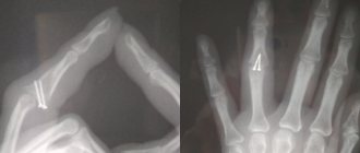
— intraosseous osteosynthesis using knitting needles, rods, pins and screws,
— bone osteosynthesis using bone plates, staples, circular ligatures, frames,
— intraosseous osteosynthesis based on bone sutures or their combination with knitting needles and staples.
Indirect osteosynthesis is based on the use of various fixation devices, both extraosseous (bone clamps and clamps) and intraosseous (rods, pins, screws and knitting needles).
Osteosynthesis can be performed both under local anesthesia in combination with neuroleptanalgesia and ataralanalgesia, and under general anesthesia.
The nature of the access (extraoral or intraoral) is determined by the doctor.
If osteosynthesis is performed using an extraoral approach, then in case of a fracture in the area of the angle and body of the lower jaw, the doctor makes an incision in the submandibular region. If the fracture occurred in the frontal area, the incision will be in the submental area. For fractures in the area of the condylar process - in the retromandibular region. During the operation, the doctor exposes bone fragments, removes small fragments, and fixes large fragments and fragments in a normal anatomical position using the selected design.
If the doctor uses intraoral access to perform osteosynthesis, he makes an incision in the mucosa along with the periosteum, then compares the fragments and performs osteosynthesis.
Bone or wire suture
In 1825, surgeon Rogers from Dublin performed the world's first operation using silver wire. With its help, he connected the fragments of the lower jaw. Later, in 1863, Russian surgeon Yu.K. Szymanowski successfully used a bone suture. From this point on, the bone suture was successfully used for osteosynthesis for many years. The main material was initially stainless steel, later it was replaced by titanium, nichrome, tantalum, etc.
There are various modifications of the bone suture (loop-shaped, cruciform, figure-of-eight, trapezoidal, double, etc.). The choice depends on the nature and location of the fracture.
The application of a bone suture occurs according to certain rules. It is important to make holes for the material in those areas where damage to the mandibular canal and tooth roots is excluded, and no closer than 1.0 cm from the fracture line. Ideally, the suture should cross the fracture line in the middle of the distance between the edge of the mandible and the base of the alveolar process.
Is it possible to do without splinting?
Even if the case is not severe - the fracture is one-sided, closed and without displacement - it is imperative to take measures to prevent the development of such unpleasant complications as:
- accidental displacement of fragments,
- re-injury
- development of inflammation of soft tissues,
- infection of the fracture site.
To do this, it is necessary to immobilize the jaw by any available method. This can be a sling bandage, but it is much more convenient and effective to use a splint. In case of a complicated fracture, splinting is absolutely indispensable, regardless of the location of the injury.
Metal Kirschner spokes
This type of wire was first used to treat fractures of the lower jaw in 1933. Intraosseous insertion of these wires can be carried out both percutaneously (without incisions) and with soft tissue incisions.
In 1975 V.V. Donskoy used an original technique, with which he inserted a wire into the branch of the lower jaw through the mucosa without an incision, then carried out a reposition, and fixed it like a splint to the teeth or to a splint. Later, in 1988, Deryabin E.I. and Osipov V.Yu., and Yu.G. Kononenko and G.P. Ruzin proposed modifications of this method in 1991. Today there are many techniques where the wire suture is combined with knitting needles, staples, surrounding wire ligatures, etc.
Treatment tactics for displaced fractures
In such cases, before applying a splint, it is necessary to compare the jaw fragments, for which reduction orthopedic devices are used. A broken upper jaw requires traction using special dental splints.
Such injuries are very dangerous because they can cause asphyxia. But correctly provided first aid will prevent suffocation. Clear the oral cavity of foreign bodies or blood, lay the victim face down, placing a cushion rolled up from clothes, blankets, etc. up to the chest.
International standards
A real revolution in maxillofacial surgery occurred in 1958, when M. Muller, M. Allgower, R. Schneider, H. Willenegger organized the International Association for the Study of Internal Fixation (AO/ASIF - Ardeitsgemeinschaft fur Osteosynthesefragen/Association for the Study of Internal Fixation – working association for the study of osteosynthesis/association for the study of internal fixation).
According to the postulates of AO/ASIF, the osteosynthesis technique implies that:
1. the structures used must be made of bioinert metal alloys;
2. bone fragments must be anatomically accurately compared and fixed;
3. the use of gentle surgical techniques ensures the preservation of blood supply to bone fragments and surrounding soft tissues;
4. stable fixation of fragments is ensured by interfragmentary compression;
5. early application of functional load is indicated;
6. restoration of contractile activity of muscles and movement in the joint.
AO/ASIF staff also developed and implemented metal plate systems for mandibular osteosynthesis:
· dynamic compression plates;
· reconstructive (blocking) plates (Locking reconstruction plates);
· blocking (locking) plates (Locking plates 2.0 mm);
· universal plates (Universal fracture plates);
· mandibular plates (Mandible (Mandible plates 2.0 mm).
They developed dynamic compression plates (DCP), through which it was possible to create compression between fragments for their primary fusion. The design of these plates includes oval holes with beveled walls, which allows the fragments to be brought closer together when the screws are tightened. The use of dynamic compression plates made it possible to achieve stable internal immobilization, reduced the number of cases of delayed fusion of fragments, and eliminated the need for additional fixation. But their use still does not eliminate the risk of microcracks in the area of the fracture line and the development of osteoporosis in the bone at the site of contact with the plate.
The locking plate/screw system, with threaded plate holes and locking screw heads, was developed to prevent bone necrosis under the plate. The system provides rigid fixation of bone fragments using a plate and a plate and screws to each other - this helps prevent the screws from unscrewing and avoid possible displacement of the fragments while tightening the screws in the hole of the plate. The plate itself is located at a certain distance from the surface of the bone, which prevents the development of lysis.
Foreign studies have not revealed significant differences in the effectiveness and possible development of postoperative complications during osteosynthesis with locking plates with screws and non-locking plates.
Another system developed by AO/ASIF experts is the LCP (locking compression plate system with angular stability) is a design of multi-cell plates with numerous holes and consists of two parts: a threaded one for fixing the head of a locking screw and a hole for creating dynamic compression by eccentric insertion of standard cortical or cancellous screws.
Installation of the plate requires special tools and is carried out according to clearly established technology.
If the adjustment of the LCP to the shape and relief of the outer surface of the lower jaw bone is carried out in accordance with the established requirements, then this creates ideal conditions for the fusion of fragments during osteosynthesis of multiple comminuted fractures of the lower jaw of various locations, in case of suppuration of a bone wound, traumatic osteomyelitis, in case of a fracture with the occurrence of a bone defect tissue, fracture of toothless jaws. Limited contact of LCP with bone helps prevent the development of bone necrosis under the plate.
First aid
In a situation with a serious injury, the people around him must provide first aid to the victim. Medical recommendations exclude independent adjustment of individual parts of the skull or contact with open wounds. The main tasks are to create conditions of complete rest, eliminate re-injury, try to stop the bleeding and call an ambulance team.
In case of acute pain, an injection of an anesthetic is allowed. To stop continuous bleeding, it is necessary to press down the artery. In cases where the wound is small, you can treat the area with hydrogen peroxide, as well as ensure careful application of cold.
Recommendations
Treatment of mandibular fractures involves long-term immobilization of the jaws, which becomes a significant psychological problem for the patient. The success of treatment directly depends on how closely both the doctor and the patient act together and take the process seriously.
For the entire period of healing of the fracture, the patient is prescribed a diet (maxillary table No. 1 and No. 2); food is allowed to be taken only in a creamy consistency (well boiled and passed through a blender). In most cases, opening the mouth is impossible, because... The patient has an intermaxillary rubber traction device installed. Food is supplied through tubes, tubes and sippy cups.
From the moment of double-jaw splinting or osteosynthesis, the splints are left in place for an average of 21–30 days. If the doctor has confidence in the favorable course of the recovery process, then it is possible to reduce the period of wearing the intermaxillary rubber traction.
Even after removing the splints, the patient cannot fully open his mouth for 1-2 weeks. To restore the function of the chewing and facial muscles, he is prescribed myogymnastics.
How long will it take for the injury to heal?
When asked how long it takes for a jaw fracture to heal, doctors answer that on average rehabilitation lasts up to 3 months. The trauma is significant, and only after the first month of treatment do patients begin to feel relief. Much of the relief is due to the fact that 21 days after the operation the fragments heal and doctors remove the splint, but sometimes this happens later, only on days 30–40.
On average, rehabilitation lasts up to 3 months
If you do not follow the doctor’s recommendations, in case of chronic diseases that disrupt tissue trophism (for example, diabetes), the healing process may take 2–4 weeks longer.
“I suffered a broken jaw in an accident. How many tears I shed while being treated!!! This whole thing heals quite slowly, you have to wear a splint, and it’s generally difficult to eat normally. If you want to lose weight and get rid of extra pounds, break your jaw. Black humor! This splint also constantly rubbed the mucous membrane on the cheeks and lips. All this time there was pain. After the splint was removed, there were sores and inflammation in my mouth. Then she replaced the teeth, since some were knocked out during the accident. In total, it took six months to recover. Now, thank God, the pain is gone, but it pulls and aches for any reason, for example, if the weather changes...”
Mi, fragment of review from otzovik.com