Total information
Perichondritis of the costal cartilages is not observed as often as other lesions of the chest wall. It should also be borne in mind that inflammation of the perichondrium is generally less common than inflammatory lesions of other tissues.
Damage to the perichondritis of the costal cartilages is more common than other perichondritis - together with the same lesion to the cartilage of the larynx and auricle, it makes up the top 3 perichondritis in terms of frequency of occurrence. At the same time, perichondritis of the costal cartilages is more often observed due to trauma than similar damage to other locations. Thus, inflammation of the connective tissue membrane of the ear cartilages mainly develops during purulent processes in the area of the outer and middle ear, and perichondritis of the laryngeal cartilages is often a complication of intubation or radiation therapy for cancer of the larynx.
note
In comparison with other perichondritis, the described disease develops against the background of an infectious lesion much less frequently than against the background of trauma.
All perichondritis of the costal cartilages are divided into:
- aseptic - without the participation of an infectious agent;
- purulent - with the addition of microbial pathogens.
Anatomy of the ears
The outer ear consists of the pinna and the auditory canal. The pinna directs sound waves from the environment through the ear canal to the eardrum. This sinuous, shell-like, flexible structure is functionally important. Abnormalities or deviations in the position, shape, size or architecture of the auricle are noticeable and undesirable. Careful surgical correction or reconstruction can restore the beauty of the ear and its harmonious appearance with the face.
The technique of aesthetic otoplasty is elegant and often complex and is based on a thorough understanding of anatomy. Along with knowledge of the muscles, nerves and blood vessels that supply the pinna, every fold, concavity and convexity of the ear must be clearly imprinted in the mind of any plastic surgeon embarking on aesthetic otoplasty and pinna reconstruction.
Auricle
The pinna is an irregularly shaped part of the outer ear that protrudes laterally at the level of the temporal bone.
The auricle consists of an elastic, sinuous cartilaginous plate covered with skin. The pinna is inclined slightly posteriorly, so that the long axis creates an angle of about 8° with the retromandibular line. The upper part of the auricle is located in a vertical plane at the same level as the eyebrow. The normal distance (projection) of the auricle from the mastoid process is from 10 to 17–20 mm (from the mastoid process to the free edge of the helix).
In children, the auricle is wider and shorter. The ratio of height to width increases with ear growth, reaching about 7:4 in adulthood. Depending on the position of the pinna, its surface can be described as anterior or lateral, as well as posterior or medial. From the front, the helix should be visible 2–5 mm in the transverse direction, located behind the antihelix.
Anterior (lateral) ear surface
The outline or edge of the pinna is formed by a whorl, an incompletely coiled tube, which forms a groove or pit of the concha along most of its length. The helix begins at the front and emerges from the cavity of the concha of the ear in the form of a helix stalk, ultimately forming a rounded arch. There is often a bump or bump on the helix, known as Darwin's tubercle.
Inside the helix is the antihelix, which forms part of the bottom of the auricle and borders the concha. On top of the antihelix there are two legs: upper and lower, between which lies a triangular fossa. From below, the antihelix is attached to the antitragus.
Both the helix and antihelix are visible from the front, but the antihelix projects more as it extends from top to bottom, creating an angle of approximately 20° between the planes of the helix and antihelix. The lower crus of the antihelix is oriented higher in tall people with long ears. In children, the lower leg of the antihelix is oriented more horizontally.
The concha is a partial cavity that leads into the ear canal. The sharp upper edge of the concha is formed by the lower leg of the antihelix, and the lower edge is formed by the sharp edges of the tragus and antitragus, which hang over the concha. In contrast, the lateral margin of the shell is smooth, rolled, and formed by an antihelix.
Between the tragus and antitragus there is a noticeable intertragus fossa with sharp edges. In older patients, a tuft of hair often protrudes from this fossa.
The lobule is the only part of the auricle without a cartilaginous frame. About a third of its perimeter is adjacent to the cheek, and the rest hangs freely.
The skin of the anterior surface of the auricle fits tightly to the underlying perichondrium and there is almost no fat between them. The vessels and nerves lie in a strip of fascia that separates the skin and the perichondrium.
Sebaceous and sweat glands are present in the triangular fossa and around the external auditory canal, respectively.
The skin on the back surface is very different from the tightly knit, fat-free skin on the front surface of the auricle. The posterior surface is covered with skin, which can wrinkle and slide over the underlying cartilage. This happens thanks to 2 layers of fat, dense on the surface and loose on the deep. The layer of fascia separating these layers of fatty tissue contains nerves, blood vessels and lymphatic vessels.
Posterior (medial) ear surface
The posterior surface of the pinna is largely hidden from view as the pinna protrudes only 10 mm from the mastoid process. The surface is characterized by projections and grooves, the opposite of those visible on the anterior surface of the auricle.
The main elevations are formed by a rook, a shell and a triangular fossa. A triangular fossa lies between these elevations.
Ear cartilage
The auricle gets its shape, appearance and flexibility from the underlying convoluted cartilaginous plate. The entire auricle, except for the lobule, has cartilage as a base.
The unique structure of the auricle, including the ridges and folds in the cartilage, allows it to be bent, folded and squeezed without discomfort or consequences. A thin, very dense layer of perichondrium covers the cartilage of the auricle.
The anterior/lateral surface of the cartilage closely resembles the pinna itself in that it is covered only by a thin layer of skin.
The helix curves in the anteroposterior direction from the helix stalk and then goes back, forming the cartilaginous edge of the auricle. The scaphoid fossa is located between the helix and the antihelix.
The antitragus and tragus are separated by the intertragus fossa. The concha and tragus continue medially, forming the cartilaginous part of the external auditory canal. This cartilaginous part enters the bony part of the external auditory canal and merges with it.
Muscles of the auricle
Small external muscles and ligaments of the ear provide attachment and support to the pinna, holding it firmly to the bony skull. The tone of the tiny vestigial intrinsic muscles provides structural support to the pinna and contributes to its characteristic tortuous and folded configuration
The extrinsic muscles consist of the posterior, superior and anterior auricular muscles.
The posterior muscle consists of a superior and inferior bundle, supported by the posterior auricular ligament. The muscle originates from the periosteum of the mastoid process and is attached to the cartilage in the area of the lower part of the conchal eminence. The posterior auricular ligament strengthens the posterior auricular muscle. These muscles are innervated by the posterior auricular branch of the facial nerve and supplied by the posterior auricular vessels.
The superior auricularis muscle is a short but strong muscle that originates from the supracranial aponeurosis and is inserted into the eminence of the triangular fossa on the posterior surface of the auricle. It is covered by layers of superficial temporal fascia and receives innervation from the temporal branch of the facial nerve. The superior auricular muscle receives its blood supply from small branches of the superficial temporal vessels.
The anterior auricularis muscle provides further support to the pinna from the front. It starts from the zygomatic arch and the supracranial aponeurosis and attaches in the region of the helix. It is supplied by branches of the superficial temporal vessels and receives innervation from the auriculotemporal nerve and the temporal branch of the facial nerve. The anterior auricular muscle is supported by the ligament of the same name.
The intrinsic muscles consist of tiny, anteriorly and posteriorly displaced vestigial muscles that connect and overlap the convexities and concavities of the auricle. In the anterior part around the conch are the muscles helicis major, helicis minor, tragicus and antitragicus. Posteriorly there are four main internal ligaments, as well as vertically and horizontally located muscle fibers that connect and support the cartilaginous convolutions of the auricle.
Innervation of the auricle
The auricle receives innervation from branches of the greater auricular nerve, lesser occipital nerve, auriculotemporal nerve, facial nerve and vagus nerve.
Most are sensory, with some vasomotor and secretory fibers arising from the facial nerve.
The greater auricular nerve provides the most significant sensory innervation to the auricle. It arises from the fibers of the second and third cervical nerves. The nerve at the ear is divided into anterior and posterior branches. The greater auricular nerve also innervates the anterior and superior walls of the external auditory canal.
The lesser occipital nerve also arises from fibers of the second, and sometimes third, cervical nerves. Includes the scalp, as well as the upper part of the cranial surface of the auricle.
The auriculotemporal nerve is a branch of the posterior trunk of the mandibular branch of the trigeminal nerve. Although it is primarily a sensory nerve, it also carries sympathetic and parasympathetic fibers and ganglion branches that communicate with the otic ganglion. The auriculotemporal nerve passes predominantly with the superficial temporal vessels along the zygomatic arch to the temple. Sensory branches from the auriculotemporal nerve innervate the tragus, the inferior part of the antihelix, the anterosuperior part of the scaphoid fossa, and the helix and pedicle of the helix. It also innervates the posterior wall of the auditory canal and the tympanic membrane.
The motor branches of the facial nerve innervate the external and internal auricular muscles. Autonomic sympathetic fibers passing with the facial nerve provide vasomotor and secretory function of the vessels and glands of the auricle.
Blood supply to the auricle
The auricle receives external blood supply through the branches of the external temporal artery in front and the posterior auricular artery in the back.
Causes
All the main causes of the development of perichondritis of the costal cartilages can be divided into groups:
- physical;
- chemical;
- infectious.
The physical reasons for the formation of this pathological process are:
- mechanical;
- thermal;
- radioactive.
Mechanical reasons that can provoke the development of perichondritis of the costal cartilages include injuries:
- non-penetrating;
- penetrating.
Non-penetrating injuries are bruises of the chest in the area of the costal cartilages. The chest wall is often injured, this explains the “leadership” of injuries among other factors that cause an inflammatory process in the perichondrium of the costal cartilages. Injury can happen:
- household;
- production;
- sports;
- criminal.
A separate mechanical cause of the occurrence of perichondritis of the costal cartilages is violation of the integrity of the periosteum during operations on the chest.
Everyday life is a “storehouse” of conditions for trauma to the chest with the subsequent development of perichondritis of the costal cartilages. Bruises can be easily caused by:
- due to inappropriate living conditions - in particular, cramped rooms filled with furniture;
- when bulky objects (mezzanines, shelves) fall;
- as a result of noisy children's games or capricious behavior of a child, who can purposefully or unintentionally hit the mother in the chest area.
Injury in industrial conditions, which can lead to the development of perichondritis of the costal cartilages, occurs in most cases when the principles of labor protection are not observed at work or when safety rules are ignored. By production we mean conditions not only in a factory or plant, but also in agriculture.
Thus, the occurrence of the described disease is quite often observed in workers who care for farm animals - a horse can hit the chest with its hoof, a cow can butt it with its horn, and so on.
Sports injury is also one of the most common causes of perichondritis of the costal cartilages. Athletes who engage in strength and team sports are mainly exposed to traumatic danger - these are:
- wrestlers;
- boxers;
- hockey players;
- basketball players;
- football players
and so on.
Also often injured:
- skiers;
- cyclists;
- jumpers.
Somewhat less frequently, perichondritis of the costal cartilages is diagnosed in people who have been subjected to physical violence (beating):
- in family;
- in a criminal environment.
Penetrating wounds of the chest, which can lead to the development of the described pathology, are puncture, cut, lacerations, chopped, and gunshot wounds.
The thermal factor that can provoke the development of perichondritis of the costal cartilages is mainly low temperatures. Banal ignorance of clothing according to the season can provoke the occurrence of this pathology. It develops especially often against the background of frostbite.
But critically elevated temperatures can also cause the formation of this disorder - for example, inflammatory processes in the perichondrium can occur during burns.
The radioactive factor leads to the development of perichondritis of the costal cartilages less often than other physical factors. Most often this happens due to:
- frequent chest X-ray examinations;
- radiation therapy prescribed for malignant tumors of the chest organs;
- contact with radioactive substances or equipment due to professional activities - often due to the same personal failure to comply with safety regulations or inadequate labor protection conditions at work.
Chemical factors that can lead to the development of perichondritis of the costal cartilages include chemicals that entered the perichondrium tissue by contact, through the blood or lymph flow, and provoked the occurrence of an inflammatory process in it.
Such toxic compounds are:
- exogenous;
- endogenous.
Exogenous provoking toxins include chemicals that are used in everyday life, in production and in agriculture.
Endogenous toxins are those produced in the human body. Most often their synthesis occurs:
- directly in human tissues due to a pathological process - most often suppuration and necrosis (death);
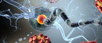
- during the development of an infectious process - such toxic substances are toxins of microorganisms that are released during their life activity or are formed as decay products of dead microbial bodies.
In the latter case, damage to the perichondrium develops against the background of a general infectious disease:
- malaria;
- flu
Less commonly, perichondritis of the costal cartilages is formed due to a local infectious lesion.
As for the specifics of the pathogen, the disease described is mainly caused by damage to a nonspecific infectious agent - this is:
- staphylococci;
- streptococci;
- Proteus;
- coli
and others.
Very rarely, specific tuberculous and syphilitic perichondritis of the costal cartilages are formed, but the possibility of their development should also be remembered.
Purulent perichondritis of the ribs often appears due to reasons such as:
- open chest injury accompanied by damage to the costal cartilages;
- contact spread of infection.
The latter can occur with diseases such as:
- mediastinitis – inflammation of the mediastinum (organs located between the lungs);
- pleural empyema – diffuse purulent lesion of the pleura;
- osteomyelitis of the sternum and ribs (for example, post-traumatic) - purulent melting of their bone tissue, which leads to the formation of fistulas (pathological tracts).
Correction of protruding ears in adults and children
Correcting protruding ears is one of the most common reasons for visiting a plastic surgeon. Prominent ears are not considered a medical pathology, so ear plastic surgery in this case is purely aesthetic.
Severe protruding ears can become the basis of psychological trauma and the cause of ridicule from others. An important feature of plastic surgery for protruding ears is the minimal level of intervention during surgery. The guaranteed safety of the operation allows otoplasty to be performed on a child.
Ear plastic surgery in children at EMC is performed from the age of 7, when the auricles are almost completely formed. The healing process goes quite quickly, the ears after otoplasty look natural and do not bother the child, after the healing process is completed the child will not even remember about the operation. Even upon closer inspection, no one will notice the small strip of cosmetic seam behind the auricle.
Development of pathology
The perichondrium is similar in its functions to the periosteum (the connective tissue membrane covering bone structures) - first of all, it:
- provides blood supply to cartilage (in this case, costal cartilage);
- protects it from the influence of various harmful factors.
On the other hand, pathological processes in the periosteum and perichondrium occur differently. In addition, their consequences are also different - this difference is explained by the different structure and nutrition of bone and cartilage. Bone structures have their own blood vessels and receive nutrients not only from the outside (from the periosteum), but also from the inside (from the bone marrow). Cartilaginous structures do not have blood vessels, so the perichondrium acts as the only source of nutrients. The result is that with periostitis, bone necrosis does not always develop, but destruction or detachment of the perichondrium in any case provokes cartilage necrosis. This state of affairs emphasizes the importance of the perichondrium and the fact that any damage to it (in this case, perichondritis) can lead to critical consequences.
Unlike the periosteum, the perichondrium does not have pronounced proliferative properties (the ability to grow). Therefore, when the inflammatory process develops in it, excess cartilage is not formed. As a result, the only forms of perichondritis (in this case, costal cartilage) that matter are the aseptic and purulent forms of the lesion.
Acute inflammatory changes during perichondritis of the costal cartilages can develop over a long period of time – up to 3 months. During this period, foci of destruction (destruction) form in the perichondrium, from which the infectious pathogen penetrates into the central part of the cartilage. Chondritis gradually forms, which spreads beyond the primary purulent focus, and from the affected central areas of the cartilage, provoking microorganisms enter the unchanged perichondrium. Such features of the spread of the purulent process lead to damage to large areas of cartilage. After 3 months, the inflammation of the perichondrium gradually regresses, tissue regeneration (restoration) is observed, but necrosis of the cartilage tissue may continue.
The cartilage destroyed due to perichondritis is replaced:
- more often - scar tissue;
- less often - bone.
Complete restoration of cartilage occurs very rarely.
Ear surgery after injury
Reconstructive ear surgery is necessary in cases where most of the tissue has been lost as a result of injury. As with microtia, reconstructive otoplasty can restore the normal shape and size of the ear after injury.
Earlobe surgery successfully solves the problem of earlobe correction after wearing so-called “tunnels” and heavy massive jewelry. As a result of wearing such accessories, the lobe becomes deformed, sags, and a fairly large gap remains at the place of the decoration.
Otoplasty allows you to correct this aesthetic defect: correction of the lobe in this case involves suturing the resulting cavities and holes with microscopic cosmetic sutures. Earlobe plastic surgery allows you to get the optimal result - the scar is practically invisible, and the earlobe will look as if it has never been pierced.
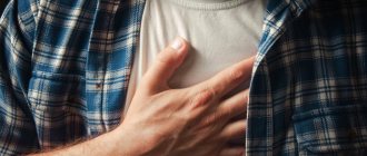
Symptoms of rib perichondritis
The clinical picture of perichondritis of the costal cartilages largely depends on its shape.
Important
With aseptic perichondritis, symptoms, having arisen, will increase, then a gradual regression of symptoms is noted. At the same time, the symptoms of the purulent form of the described pathology can only increase over time - this state of affairs is explained by the destruction (destruction) of cartilage and the formation of fistulas.
The clinical picture of aseptic perichondritis is manifested by local signs - these are:
- pain;
- breathing disorder.
Characteristics of pain syndrome:
- by localization - in the area of the affected costal cartilages;
- in terms of distribution - there is no irradiation as such, but in some cases pain can spread to the bone fragment of the affected rib;
- by nature - the “range” of pain sensations is different, it can be stabbing, aching, tugging pain;
- by severity - intensity depends on the degree of damage, as well as the pain threshold of the patients. Often, with minor perichondritis of the costal cartilages, patients complain that pain prevents them from working, sleeping and generally living as usual;
- by occurrence - they appear almost with the beginning of the development of the pathological process in the perichondrium, and as it progresses they intensify. Also, increased pain is observed with movements and deep breathing.
With the aseptic form of the described pathology, the general condition often does not worsen.
The purulent form of perichondritis of the costal cartilages manifests itself:
- local signs;
- violation of the general condition of the body.
Local symptoms are most indicative. When visiting a doctor, the patient complains of typical signs of suppuration, which successively follow each other. First, an infiltrate forms in the affected area - tissue compaction. After a certain period of time (on average it can last from 3-4 to 7 days), the dense lesion softens. In severe cases, inflammation can spread to the entire lower chest and upper part of the anterior abdominal wall. The formed abscess is opened - often through the skin (in this case the patient complains of purulent discharge from the “hole”), less often through the posterior perichondrium (pain in the chest will bother you). In the first case, a fistula is formed, in the second - purulent leaks in the soft tissues.
The characteristics of pain with purulent perichondritis of the costal cartilages are as follows:
- by localization - in the area of the affected costal cartilages;
- by distribution - along the ribs on the affected side;
- by nature - at first vague aching, then twitching, with a large abscess - pulsating;
- in terms of intensity - in the early stages the disease is insignificant, as the abscess matures it increases and can be very strong;
- by occurrence - appear when the first inflammatory changes appear in the perichondrium, intensify with any activity in the chest area on the affected side.
Signs of a general condition disorder are:
- hyperthermia – increased body temperature. Can reach 38.5-39.0 degrees Celsius;
- general weakness caused by the products of the purulent process entering the bloodstream;
- malaise;
- loss of appetite;
- decreased performance – both mental and physical.
Preparing for surgery
Before undergoing otoplasty, you must undergo a mandatory examination, including blood tests, urine tests, sugar levels, an ECG, as well as a mandatory consultation with an anesthesiologist. You can undergo all necessary tests and preoperative examinations at the EMC clinic.
Before surgery, it is prohibited to take aspirin and medications containing it. A week before surgery, avoid salty, spicy, and smoked foods.
10 days before surgery you must refrain from smoking.
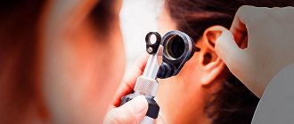
Diagnostics
The diagnosis of perichondritis of the costal cartilages is made on the basis of the patient’s complaints, anamnesis data (history of the disease), and the results of additional research methods - physical, instrumental, laboratory.
Physical examination findings will be as follows:
during examination, the patient subconsciously leans towards the affected area (to relieve tension in the inflamed tissues), often holding on to the affected area. With the development of a purulent form of the described pathology, the skin over the site of the lesion will be reddened and swollen. With purulent perichondritis, in the event of a breakthrough of the abscess, a defect in the skin is revealed, through which purulent contents are released;- upon palpation (palpation) - palpation pain in the affected area is determined; with purulent perichondritis of the costal cartilages it is especially pronounced, the skin over the purulent focus is hot to the touch. When a large abscess forms, a symptom of fluctuation (“pumping” of peculiar “waves” of liquid under the fingers) is revealed.
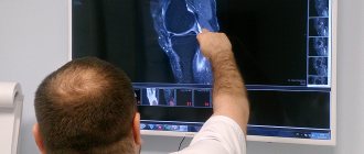
Instrumental research methods that are used in the diagnosis of perichondritis of the costal cartilages are as follows:
X-ray examination - X-ray images are taken, the blurred contours of the cartilage are noted at the site of the lesion. After 2-3 months from the onset of the pathology, radiographs reveal changes such as calcification of the cartilage, narrowing of the intercostal space and thickening of the anterior fragment of the bone rib;- computed tomography (CT) - computer sections help to identify changes in the costal cartilage as a whole, as well as deeper disorders in the tissues of the perichondrium and costal cartilage;
- magnetic resonance imaging (MRI) - its capabilities are equal to those of CT, but magnetic resonance examination is more informative for identifying changes in the soft tissues surrounding the costal cartilages;
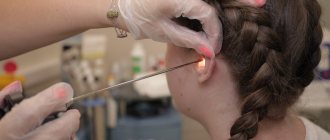
- puncture - if the formation of an abscess is suspected, it is punctured and the contents are collected, followed by examination under a microscope;
- biopsy - tissue is taken from the affected periosteum and then examined in the laboratory under a microscope;
- fistulography – performed when a fistula forms. A contrast agent is injected into the fistula tract, an X-ray is taken, and the characteristics of the fistula are assessed from it.
Laboratory diagnostic methods used for perichondritis of the costal cartilages are as follows:
- general blood test - an increase in the number of leukocytes and ESR is determined;
- microscopic examination of a biopsy - allows you to confirm the inflammatory nature of the pathological process and identify pathogens;
- bacteriological examination - if there is pus in the punctate, it is inoculated on nutrient media, and the infectious agent that provoked the development of the purulent process is determined from the grown colonies.
Rehabilitation period
Ear plastic surgery is performed on an outpatient basis, so it is necessary for the patient to be accompanied by relatives or friends.
After the operation, as a rule, the patient does not stay in the clinic, but goes home.
The doctor gives detailed recommendations for further rehabilitation and prescribes analgesics to eliminate possible pain. Postoperative swelling of the affected tissue may be observed during the first days.
Every 1-2 days for 10-14 days after otoplasty, the patient visits the surgeon for dressings, monitoring the tissue healing process and relieving possible complications. A bandage to help secure the ears should be worn for about a week after surgery. The results of surgery last a lifetime.
In case of reconstruction, 1-2 days of hospitalization are required to monitor the engraftment of the transplanted tissue.
Differential diagnosis
Differential (distinctive) diagnosis of perichondritis of the costal cartilages is primarily carried out with such pathologies as:
chondritis – inflammatory lesion of the costal cartilage;- intercostal myositis - an inflammatory process in the intercostal muscles;
- intercostal neuralgia is a pain syndrome that develops when intercostal nerves are damaged of various types;
- mediastinitis;
- tumor of the chest wall.
Treatment of rib perichondritis
Treatment methods depend on the type of pathological process. In all cases they are general and local.
With the development of aseptic perichondritis of the costal cartilages, general prescriptions are as follows:
- functional rest of the chest;
- non-steroidal anti-inflammatory drugs (NSAIDs);
- antibacterial drugs - for the prevention of septic complications.
The following are used as local treatments:
- physiotherapeutic methods;
- blockade with novocaine - with severe pain.
The most effective methods of physiotherapeutic treatment are:
- UHF;
- Microwave;
- dry heat.
If signs of suppuration appear, surgical treatment is performed:
- the purulent focus is opened;
- the abscess cavity is washed with antiseptics;
- carry out drainage.
In the postoperative period, antibacterial therapy is prescribed, drains are washed, and bandages are changed.
Important
If the perichondrium is critically damaged and cartilage is involved in the pathological process, it must be removed. If pus spreads to the bone fragment of the rib, 2-3 cm of bone tissue is removed.
Types of otoplasty
Otoplasty can be aesthetic or reconstructive.
Aesthetic, as a rule, implies a minor intervention: during the operation, the surgeon eliminates the external defect by changing the location, size or shape of the auricle. The operation takes from 30 minutes to 2 hours.
Reconstructive otoplasty is aimed at complete or partial restoration of the lost/absent auricle (a graft (cartilage material) from the patient’s rib is used as a reconstructive material). The period of reconstructive correction is divided into several stages - the formation of ear cartilage as the basis of the future ear, the formation of the auricle itself and the final aesthetic correction. The total duration of all manipulations is about a year.
The operation is performed both under general anesthesia (for example, for children) and under local anesthesia (for adults). The surgical technique is chosen by the surgeon based on the task. Auricular plastic surgery as an aesthetic operation does not affect hearing function in any way.
Forecast
The outcome of perichondritis of the costal cartilages is generally favorable. The aseptic type of pathology is a typical inflammatory lesion, the general principles of treatment of which have been successfully developed.
The prognosis worsens with the purulent type of the disease. In this case, timely diagnosis and adequate surgical treatment will help to cope with the disease.
Patients should remember that self-medication is fraught - especially the notorious overheating, which in the case of a purulent process can lead to critical consequences and even the death of the patient.
Kovtonyuk Oksana Vladimirovna, medical observer, surgeon, consultant doctor
18,272 total views, 5 views today
( 42 votes, average: 4.50 out of 5)
Schlatter's disease: symptoms and treatment
Tuberculosis of bones and joints
Related Posts
Advantages of the EMC Clinic
EMC Aesthetic Clinic is a leading Moscow clinic in the field of cosmetology and plastic surgery. EMC has been providing medical services for more than 27 years, strictly observing international protocols and representing the highest level of medicine.
The high professional skills of EMC specialists allow us to deal with the most complex cases and show amazing results in our work. The appearance of the ears and hearing acuity do not always radically change life, but they always affect its quality. Correction, reconstructive restoration of the auricles, restoration of hearing function, EMC’s task is to help its patients in any situation at the highest level of medical capabilities.