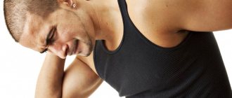
Protrusion of intervertebral discs is a common disease that occurs today in many people, both elderly and young. This is due to the fact that people rarely pay due attention to their health and are in no hurry to consult a doctor when the first signs of health problems appear, much less prevent the development of diseases. Therefore, back pain, especially in the lower back, is familiar to almost every second adult. In the vast majority of cases, they are caused by the occurrence of osteochondrosis, which over time leads to the formation of protrusion of the intervertebral disc.
Most often, it is the L5-S1 disc that suffers from such pathological changes, i.e., located between the last 5th vertebra of the lumbar spine and the 1st vertebra of the sacral spine. This is often accompanied not only by severe pain, but also by neurological complications, which, in the absence of timely treatment, can even lead to disability. Therefore, a negligent attitude towards one’s own health and attributing lower back pain to simple fatigue can have quite serious consequences.
What is L5-S1 disc protrusion and why does it occur?
Protrusion is the deformation of a disc with the formation of a protrusion in one direction or another. As a rule, this is a direct consequence of the lack of treatment for osteochondrosis, i.e., the initial stage of degenerative changes in the intervertebral disc. Previously, osteochondrosis was considered an age-related disease, but in recent years its significant “rejuvenation” has been observed. In most cases, this is due to a sedentary lifestyle and sedentary work.
Today, osteochondrosis and L5-S1 disc protrusion occur even in adolescents.
The reasons for the early occurrence of degenerative-dystrophic changes in the intervertebral discs are the increased loads placed on them. Since it is the lumbosacral spine that bears the greatest load in everyday activities, the discs located between its vertebrae are much more likely than others to suffer from osteochondrosis and protrusions. Moreover, the leader in the frequency of occurrence of deformities is the L5-S1 disc.
The vertebrae, and therefore the discs of the lumbar spine, are distinguished by their greatest width, the value of which exceeds even their height. This is directly related to the need to compensate for high loads.
Normally, the intervertebral discs are slightly abraded with every human movement, but then quickly restored due to the continuous diffuse supply of chondroitin, glucosamine and other biologically active compounds into them. But under the influence of excessive loads, the balance between these processes is disrupted for the worse, which leads to the early onset of degenerative-dystrophic changes in cartilage tissue.
This usually results from:
- the presence of excess weight, even a slight one, since each extra kilogram greatly increases the load on the lumbosacral spine and the L5-S1 disc in particular;
- sedentary work, sedentary lifestyle, since static loads can be even more destructive to cartilage tissue than active physical work;
- excessive physical activity, especially related to heavy lifting;
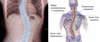
- poor posture, the habit of slouching or curvature of the spine, which leads to uneven distribution of the load.
Various metabolic disorders, especially diabetes mellitus, can aggravate and accelerate the process of destruction of intervertebral discs.
As a result of these factors, the nutritional process of cartilage tissue is disrupted, which is why it cannot be restored correctly and in a timely manner. Against this background, the level of hydration of the intervertebral disc decreases, but it consists of 80% water. Therefore, the slightest changes lead to a decrease in the elasticity of the numerous fibers of the fibrous ring surrounding its internal contents.
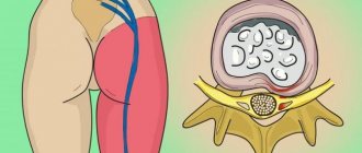
As a result, they can no longer withstand the high loads placed on them and evenly stretch and then shrink. Therefore, over time, microscopic cracks form in the fibers of the annulus fibrosus. They gradually increase, which is aggravated by increasing pressure inside the nucleus pulposus (the internal contents of the disc), since the cartilage is excessively compressed by the L5 and S1 vertebrae.
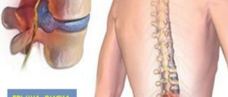
Sooner or later, the fibrous ring cannot stand it and becomes deformed, that is, it protrudes in the most vulnerable place. Thus, a protrusion of L5-S1 is formed. All this is accompanied by an inflammatory process and chronic pain of varying degrees of intensity. In this case, the outer shell of the disc still retains its integrity, but the bulging part can compress the surrounding anatomical structures, which will certainly make itself felt.
If the pathological process is not intervened at this stage, the L5-S1 protrusion will increase in size, increasing the risk of compression of surrounding tissues, blood vessels and nerve structures. Ultimately, the weakened fibers of the fibrous ring will not be able to withstand it and will finally rupture, as a result of which the nucleus pulposus, equalizing the pressure inside the disc, will leak out. Thus, the protrusion is transformed into an intervertebral hernia of L5-S1.
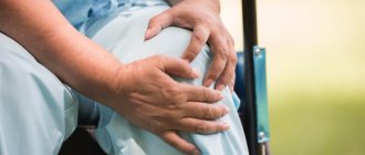
Due to the fact that during pregnancy the load on a woman’s body doubles, it is very important to put the spine in order. Our clinic has developed and prepared a special program of comprehensive examination and treatment for pregnant women and those planning pregnancy.
Types and symptoms of L5-S1 protrusion
The intervertebral disc has a close to ellipsoidal shape. On one side (posterior), it, together with the vertebral bodies, forms the spinal canal, where the spinal roots that form the cauda equina pass at the level of L5-S1. On the other side (front) they adjoin the soft tissues bordering the pelvic organs.
Therefore, anterior and building protrusions L5-S1 are distinguished. The former do not pose a serious danger, since the pressure they exert on surrounding organs is not enough to cause severe pathological changes to develop. The latter, on the contrary, are fraught with serious complications and require the earliest possible diagnosis and initiation of treatment.
Posterior or dorsal protrusions can compress the nerve fibers of the cauda equina, which will lead not only to acute, shooting pain, but also to the development of radicular syndrome. Therefore, if initially L5-S1 protrusion can only manifest itself as frequent or constant aching pain in the lower back and sacrum, which tends to intensify after prolonged walking, sitting or doing physical work and goes away after rest, then over time its manifestations can completely deprive a person of his ability to work.
Signs of advanced protrusion requiring immediate treatment are:
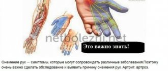
- pain comparable to an electric shock, originating in the lumbar region and radiating to the middle part of the buttock, the back and outer lateral surface of the thigh and lower leg, as well as the foot, especially the little toe;
- disturbances in the sensitivity of these areas of the body, which is manifested by numbness of the skin, the appearance of goosebumps, changes in the perception of hot and cold, pain even with light touches, etc.;
- changes in leg mobility, muscle weakness with gradual subsequent hypotrophy, i.e., a decrease in volume, which leads to changes in gait, lameness, tucking of the foot and other disorders.
If such signs are present, it is said that the L5-S1 protrusion provoked the development of radicular syndrome, that is, it pinched the spinal root passing at the level of this spinal motion segment. If measures are not taken when radicular syndrome appears, the progressive disease can lead to paralysis and disability.
In this case, the symptoms of radicular syndrome can appear simultaneously on both legs and buttocks, or only on one side of the body, or in an intermittent manner. This directly depends on in which segment of the posterior surface of the L5-S1 intervertebral disc the protrusion has formed.
If the protrusion is located clearly in the center of the spinal canal (median protrusion L5-S1), it can simultaneously infringe on both spinal roots of this area. Therefore, violations will be observed on both sides simultaneously or alternately. If the protrusion is shifted to the left or right, we speak of its paramedian location. Then the symptoms will be observed from the corresponding half of the body.
If the radicular syndrome manifests itself very clearly and little time has passed from the appearance of the first discomfort to its development, this indicates the presence of foraminal protrusion. In this case, it is located in a narrow hole formed by the vertebral arches, through which the spinal root passes. Therefore, such a protrusion, even at a very small size, can provoke severe impairments of mobility, sensitivity and pain on one side of the body.
Sometimes diffuse protrusions occur. This term refers to protrusion of the fibrous ring over the entire area, which is fraught with the most severe symptoms.
Symptoms
The first symptoms of this disease appear after compression of the nerve endings begins. In the initial stage, pain of relatively low intensity occurs, occurring after certain loads with irradiation into the buttock and leg. As a rule, they are preceded by a crunching sensation in the damaged area of the spinal column. Then the pain syndrome becomes more and more pronounced, accompanied by sensory disturbances, weakness and cramps in the calf muscles, foot, big toe, and stiffness in the lumbar region.
Diagnosis of the disease
If signs of protrusion of the L5-S1 intervertebral disc occur, you must immediately make an appointment with a chiropractor, vertebrologist or neurologist. Only a doctor can give a correct assessment of the patient’s condition and determine the cause of pain and other symptoms, since some other diseases can manifest themselves in a similar way.
Initially, the doctor will conduct a thorough survey of the patient, find out the nature of the complaints made about the state of health, the characteristics of the appearance of pain and their relationship with various physical activities. He will also be interested in the characteristics of the patient’s lifestyle, his work activity and the presence of other chronic diseases. After collecting an anamnesis, the chiropractor proceeds to an examination, during which he not only identifies the most painful areas and determines the level of damage to the spine, but also conducts a series of neurological tests. Their results make it possible to detect signs of radicular syndrome and assess the degree of neurological deficit.
To confirm the presence of L5-S1 protrusion, to clarify the features of its location and size, instrumental diagnostic methods are prescribed. It can be:
- MRI;
- CT;
- X-ray of the spine in two projections.
Priority is always given to MRI, since only this diagnostic method can provide comprehensive information about the condition of the intervertebral discs and at the same time has a high level of safety. Thanks to MRI, a series of layer-by-layer images of the lumbosacral spine is obtained, with a slice thickness of 2 mm. This allows you to detect the slightest deviations from the norm and accurately determine the type and size of L5-S1 disc protrusion. The data obtained make it possible to develop the most effective treatment tactics and determine whether L5-S1 protrusion can be treated conservatively or whether the patient should be immediately referred for surgery.
In our clinic you can also learn in more detail about the composition of your body and the state of the vascular system, which is involved in the blood supply to internal organs, musculoskeletal muscles, and the brain. Our experienced doctors will explain the data obtained to you in detail. Bioimpendansometry calculates the ratio of fat, muscle, bone and skeletal mass, total fluid in the body, and basal metabolic rate. The intensity of recommended physical activity depends on the state of muscle mass. Metabolic processes, in turn, affect the body's ability to recover. Based on the indicators of active cell mass, one can judge the level of physical activity and nutritional balance. This simple and quick test helps us identify disturbances in the endocrine system and take the necessary measures. In addition, it is also very important for us to know the condition of blood vessels for the prevention of diseases such as heart attacks, hypertension, heart failure, diabetes and much more. Angioscan allows you to determine such important indicators as the biological age of blood vessels, their stiffness, stress index (which indicates heart rate), and blood oxygen saturation. Such screening will be useful for men and women over 30, athletes, those undergoing long-term and severe treatment, as well as everyone who monitors their health.
In this case, body composition analysis gives us information that adipose tissue predominates in the body, and the bone-muscle component is in relative deficiency. These data will help the rehabilitation doctor competently draw up a physical activity plan, taking into account the individual characteristics of the patient.
Treatment of L5-S1 protrusion
If the size of the pathological disc protrusion does not exceed 8 mm (sometimes 10 mm), patients are initially prescribed conservative therapy. The earlier the protrusion is detected, the easier and more effective it will be. But in practice, it turns out that people usually come to a neurologist with already advanced degenerative-dystrophic processes in the intervertebral discs.
Conservative treatment is selected individually, but it is always comprehensive and includes:
- drug therapy;
- osteopathy;
- traction therapy;
- manual therapy;
- physiotherapy (phonophoresis, carboxytherapy, ozone therapy);
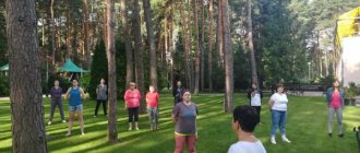
- individual sessions with a rehabilitation specialist.
Additionally, each patient is recommended to increase the level of physical activity within acceptable limits, take breaks from work at least every hour if it is sedentary, and purchase an orthopedic mattress. It is important not to be lazy and strictly follow the recommendations received from the neurologist, since the effectiveness of treatment largely depends on this.
Drug therapy
It is impossible to completely cure protrusion with the help of medications, but it is possible to reduce the severity of pain and inflammation. Therefore, patients are prescribed a complex of medications, each of which is necessary to solve individual problems:
- NSAIDs – have analgesic and anti-inflammatory effects;
- corticosteroids – have pronounced anti-inflammatory properties, making them effective even in severe inflammatory processes;
- muscle relaxants – help eliminate muscle spasms, which are often the body’s reflex response to pain and increase it;
- B vitamins – improve the conduction of nerve impulses along compressed spinal roots;
- Vitamin D is a remedy responsible for the condition of bone tissue, as well as for higher brain functions, such as memory, memory, attention, and speech.
- chondroprotectors are drugs aimed at activating the restoration of cartilage tissue, but giving positive results only when used at the very first stage of the development of osteochondrosis. To prevent diseases of the musculoskeletal system, we recommend to our patients the most effective drug Mermaids Marine Collagen;
Often, patients are additionally prescribed chondroprotectors, but their use is only advisable to prevent the formation of protrusions of other discs if signs of osteochondrosis are detected in them during an MRI.
Traction therapy
Spinal traction or traction therapy is often used to increase the space between the vertebrae of the lumbar and sacral spine. Thanks to this, optimal conditions are created for the restoration of the pathologically altered L5-S1 disc. Additionally, this helps to reduce compression of the spinal roots and manifestations of radicular syndrome or completely eliminate it.
Thus, traction therapy sessions can reduce pain, improve the patient’s motor abilities and reduce the manifestations of sensory impairment. They are carried out using special equipment and are completely safe.
Manual therapy
Manual therapy can be called the basis for the treatment of L5-S1 protrusion, since a specialist can directly mechanically influence the affected spinal motion segment and solve a whole range of problems. With the help of manual therapy it is possible to:
- restore the normal position of the vertebrae of the spinal column and thereby create favorable conditions for the regeneration of the L5-S1 disc;
- activate the body’s natural reserve capabilities and direct them to fight the existing disease;
- eliminate functional blocks and spasms of the lower back muscles, which reduces pain, improves spinal support and allows you to restore normal range of motion;
- release compressed nerve roots and significantly improve the patient’s well-being;
- activate blood circulation in soft tissues and restore normal nutrition of the L5-S1 intervertebral disc;
- increase the body's adaptive capabilities and strengthen the immune system.
But all this becomes possible only when manual therapy is performed by a true specialist in this field. To ensure that manual pressure on the spine brings only benefit and does not cause harm, it is important to take the choice of a clinic and a specific specialist seriously. Otherwise, incorrect calculation of the impact force or direction can only worsen the situation.
One of the most well-proven methods of manual therapy is the Gritsenko method, which has been practiced for decades. It involves a subtle and even jewel-like influence on the position of the vertebrae and intervertebral discs, which allows one to achieve excellent, and most importantly, lasting positive results in the treatment of L5-S1 protrusion.
Manual therapy for this disease involves a course of procedures consisting of 10-15 sessions. But patients notice the first noticeable positive changes in their well-being after the first procedures.
Physiotherapy
Physiotherapeutic methods of influence are used as a complement to other methods of treatment. But their use is possible only outside the acute period of the disease. Patients are often advised to undergo treatment courses:
- electrophoresis;
- laser therapy;
- ultrasound therapy;
- UHF.
Between procedures, a break must be taken, the duration of which is selected individually. It is possible to carry out two procedures on the same day, but only those with the same mechanism of action.
Thanks to physiotherapy sessions, there is a faster resolution of the inflammatory process, elimination of residual pain, an increase in the body's ability to regenerate, as well as other positive changes.
Causes of the disease
Most often, the trigger for protrusion is osteochondrosis. But it can also occur with an insufficiently active lifestyle and metabolic disorders. This is due to the fact that the intervertebral disc is nourished by the diffusion of substances into it from surrounding tissues due to the lack of blood vessels. When this mechanism is disrupted, “starvation” and destruction of disc tissue occurs.
The risk group includes people involved in heavy physical labor and athletes. Hereditary features of the development of the spine and frequent infectious diseases also cause protrusion.