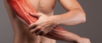
Nature and duration of pain
Short-term pain (usually localized in the navel area on the right and left), which passes without taking painkillers, is often provoked by neurosis and is accompanied by diarrhea (diarrhea).
Urgent medical intervention is not required. However, if episodes occur repeatedly, it is worth making an appointment with a therapist. You may need to consult a neurologist. Acute pain in the abdomen on the right for the first time is a serious reason to seek medical help. Pain in the upper abdomen on the right may indicate biliary colic and peptic ulcer.
Violent (most intense) pain in the abdomen on the right side occurs when a stone moves along the ureter from the right kidney. An attack of renal colic is accompanied by vomiting, flatulence, frequent urge to urinate and the passage of a small amount of urine. In severe cases, blood pressure decreases and the patient loses consciousness. At the slightest suspicion of an attack of kidney stones, you should call an ambulance.
Severe tenderness in the lower abdomen on the right side is often associated with appendicitis. However, in the initial stage of inflammation of the appendix, the pain is aching (less intense when lying on the right side with bent legs), intensifies over time and radiates to the stomach and to the right of the navel. Subsequently, the pain increases, the abdomen becomes hard.
Cramping abdominal pain occurs with intestinal inflammation (including intestinal infections, such as salmonellosis) and poisoning. Pain is localized in the navel area, accompanied by nausea/vomiting, diarrhea, fever and intoxication (weakness, muscle pain, headache). In diseases of the colon (its ascending part, located on the right side), pain occurs during the movement of feces and is often accompanied by constipation/diarrhea and bloating.
Sharp pain in the right hypochondrium , intensifying with inspiration, is characteristic of right-sided pleurisy, which complicates the course of pneumonia. In this case, all the symptoms of lung damage are observed: fever, shortness of breath, cough.
People who lead a sedentary lifestyle often complain of throbbing pain at the navel on the right Unpleasant sensations are associated with blood stagnation and quickly pass after warming up. However, with constant pulsation in the abdomen, an abdominal aortic aneurysm cannot be ruled out.
If you experience acute abdominal pain, call emergency services!
A nagging, aching pain in the abdominal region on the right indicates chronic inflammation. Unpleasant sensations occur periodically or are permanent. In Crohn's disease, pain is often localized in the right iliac fossa (right, below) and mimics appendicitis.
Pyelonephritis is characterized by dull pain in the back, radiating down the abdomen, groin area and leg on the side of the affected kidney. In this case, the patient suffers from severe intoxication. With chronic inflammation, swelling appears in the legs; high a/d is difficult to reduce with conventional antihypertensive drugs.
Aching pain in the right hypochondrium after eating , nausea, heaviness and bursting sensations in the abdomen (in the upper part on the right side), lack of appetite are characteristic of chronic cholecystitis and liver inflammation. With hepatitis and cirrhosis, jaundice, dark urine, and discolored feces appear.
Pain may indicate cancer . However, in most cases, the initial stage of cancer is painless. The patient notes weight loss for no apparent reason, lack of appetite, perversion of taste, unstable stools (usually diarrhea), fragments of undigested food in the stool, jaundice (with liver cancer). Intense pain indicates the spread of the malignant process.
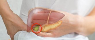
Causes of pain in the right hypochondrium
Biliary dyskinesia
With the hyperkinetic variant of gastric dyskinesia, colicky pain in the right hypochondrium predominates, developing 20-30 minutes after finishing a meal.
A painful attack can be provoked by physical activity and stress. Cramping pain is accompanied by nausea and then vomiting. Unpleasant sensations go away on their own or are relieved with antispasmodics. Between attacks, general health is good, there is no pain under the ribs on the right. Hypokinetic dyskinesia is characterized by constant aching pain, a feeling of heaviness and fullness in the hypochondrium. Eating fatty foods increases pain. When palpating the right upper abdomen, a person feels a dull pain. With this form of dyskinesia, pronounced dyspeptic disorders occur. Patients complain of bitterness in the mouth, belching, and loss of appetite.
Hepatitis
Pain in the right hypochondrium is a typical symptom found in all types of hepatitis. There is a dull soreness and a feeling of pressure in the specified area, usually not associated with food intake or stress. Acute hepatitis is characterized by sharp pain, which is aggravated by palpation of the abdomen in the hypochondrium area. The pain syndrome is supplemented by dyspeptic disorders, signs of intoxication, and jaundice.
Chronic cholecystitis
When the gallbladder is inflamed, moderate pain is felt in the hypochondrium, which persists for several days and even weeks. Irradiation of the pain syndrome to the scapula, lower back, right shoulder girdle or shoulder is typical. Eating fatty and spicy foods and carbonated drinks contributes to increased pain. The disease occurs with periods of remission, during which the pain disappears, and patients are only bothered by discomfort or heaviness under the right costal arch.
With acalculous cholecystitis, the symptoms are less pronounced, the general condition remains satisfactory. As a rule, dyspeptic disorders come to the fore. In chronic calculous cholecystitis, in addition to dull pain, severe painful attacks may occur. In this case, the pain becomes cramping and sharp. In addition, severe nausea, vomiting with bile, and low-grade fever are noted.
Hepatic colic
Symptoms are due to exacerbation of cholelithiasis. A person experiences excruciating pain, which can be cramping, cutting, tearing or burning in nature. At the time of an attack, the patient lies on his side, tucking his legs to his stomach, or rushes around the bed to find a comfortable position. Pathognomonic irradiation of painful sensations into the right shoulder and scapula. The clinical picture is complemented by nausea, vomiting bile, and flatulence.
Pain in the right hypochondrium
Biliary peritonitis
Initially, sharp pain appears in the right hypochondrium with irradiation to the corresponding scapula and shoulder. The patient lies motionless on his side, with his knees pulled towards the abdominal wall. When palpating the right abdomen, the pain intensifies. At the second stage of bile peritonitis, the pain syndrome persists, repeated vomiting occurs, and intoxication increases. If a person does not seek medical help, a terminal phase begins when the pain subsides due to the death of nerve endings.
Parasitic infestations
With echinococcosis of the liver, the pain syndrome gradually increases. At first, the patient is worried about heaviness and discomfort, especially after eating large amounts of food. An increase in the size of the hydatid cyst is accompanied by increased pain. The development of symptoms after physical activity is typical. Pain in the right hypochondrium also occurs with other infections: amebiasis, opisthorchiasis, ascariasis.
Cirrhosis of the liver
It is asymptomatic for a long time. Patients experience periodic dull pain in the right abdomen, provoked by diet disorders or alcohol consumption. There is rapid satiety and heaviness under the ribs after finishing a meal. When cirrhosis is complicated by portal hypertension, pain spreads to the epigastric and periumbilical areas.
Liver cancer
Painful sensations in the right hypochondrium are caused by an increase in the size of the tumor and stretching of the liver capsule. They bother a person constantly and are not associated with external provoking factors. Typical for oncological pathology is a dull pain, a feeling of fullness and heaviness in the right side. With malignant degeneration of the liver tissue, progressive weight loss, symptoms of dyspepsia, anemia and hemorrhagic syndrome are observed.
Hemolytic anemia
Constant dull pain on the right under the ribs is characteristic of toxic anemia caused by the action of toxic substances or medications. Less commonly, abdominal pain under the costal arch on the right is a sign of paroxysmal nocturnal hemoglobinuria, autoimmune damage to red blood cells. Severe pain in the liver area is possible with exacerbations of hereditary anemia - membranopathies, fermentopathy, hemoglobinopathies.
Heart failure
Discomfort and pain in the right hypochondrium are typical for dysfunction of the right ventricle of the heart, when blood stagnates in the venous bed. Patients complain of distension in the abdomen, which worsens with active movements, after eating. In addition to pain, swelling of the lower extremities, shortness of breath and fatigue during physical activity, and swelling of the neck veins are determined.
Referred pain
Often, when the pathological process is localized in the right iliac region, irradiation of pain in the hypochondrium is observed, which is due to the peculiarities of innervation. The pain is sharp, stabbing, dull or squeezing in nature. If the symptom is caused by surgical pathology, unbearable painful sensations develop, sharply intensifying when pressing on the abdominal wall. The most common causes of radiating pain:
- Appendicitis.
This localization of pain is typical for the retrocecal location of the appendix. Intense pain is felt in the right upper quadrant of the abdomen, nausea with vomiting, and stool disturbances. - Gynecological diseases.
In women, pain on the right side of the hypochondrium can be a sign of exacerbation of endometritis or right-sided adnexitis. Often the pain syndrome is caused by surgical pathologies: disrupted ectopic pregnancy, apoplexy of the right ovary. - Colon damage.
Pain in the right hypochondrium sometimes occurs with colitis and Crohn's disease. They indicate an exacerbation. Pain may radiate to the right into the hypochondrium in patients with polyposis and colon cancer.
Rare causes
- Gallbladder diseases
: cholesterosis, polyps, malignant tumor. - Vascular pathologies
: Budd-Chiari syndrome, veno-occlusive disease, liver infarction (ischemic hepatitis). - Rare liver diseases
: hepatoptosis, perihepatitis. - Damage to the gastroduodenal zone
: acute and chronic gastritis, gastroduodenitis, peptic ulcer of the stomach and duodenum. - Complications of pregnancy
: gestosis, cholestasis of pregnant women.
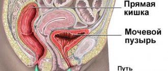
Causes of abdominal pain on the right in women
Pain in the lower abdomen in women occurs before menstruation and is caused by contractions of the uterus. The discomfort goes away after 2-3 days. Often, on the 14th day from the start of menstruation during the period of ovulation (maturation and release of the egg into the uterine cavity), women complain of nagging pain or cramps in the lower abdomen on one side. In this case, there is slight bloating and an increase in vaginal discharge. The condition does not require medical intervention. If pain on one side (right or left) is not associated with menstrual bleeding, gynecological pathology should be excluded. In this case, the nature of the pain will help make a preliminary diagnosis.
- Acute pain - ectopic pregnancy, rupture of an ovarian cyst, adnexitis (inflammation of the appendages). The pain syndrome is often accompanied by bloody discharge, and the patient's condition rapidly deteriorates.
- Aching pain - fibroids, uterine flexion, endometriosis, corpus luteum cyst. The pain intensifies during sexual intercourse and is accompanied by pathological discharge from the vagina. If the intensity of the pain increases sharply and blood appears, you should urgently seek medical help.
Important! All women who complain of pain in the lower abdomen are referred to a gynecologist for consultation.
Pain in the right side may be associated with pregnancy - the growing uterus puts pressure on the abdominal organs. There is an unpleasant, throbbing pain, frequent urination, and heartburn. Fetal movements are often mistaken for pulsation. The first conscious movement is recorded at 18-20 weeks; before this period, fetal movements are usually not felt. Pain in the abdomen on the right and in the groin area during pregnancy is caused by the divergence of the pelvic bones. However, with severe abdominal pain on the right side in a pregnant woman, acute surgical pathology cannot be ruled out: cyst torsion, intestinal obstruction, abdominal aortic aneurysm.
Important! If there is pain in the lower abdomen on the right or left, warming procedures (using a heating pad) are strictly prohibited. This will only increase bleeding during menstruation, and in pregnant women it can cause a miscarriage. Expectant mothers should not take medications (painkillers, antispasmodics) without a doctor’s prescription. Many modern medications negatively affect the fetus and uterine tone.
Cramping pain in the abdomen after childbirth, which intensifies during breastfeeding, is a normal phenomenon caused by contractions of the uterus. However, due to overstretching of the ligamentous apparatus in the postpartum period, an umbilical hernia often forms. Protrusion of the abdominal wall is accompanied by painful sensations in the navel area on the right or left, bloating and rumbling in the abdomen.
Constipation can cause abdominal pain in women giving birth. Therefore, it is important to do squats after childbirth, in consultation with your doctor. Abdominal exercises will speed up uterine contractions, reduce blood loss and strengthen the abdominal wall.
Important! Taking laxatives is contraindicated for nursing mothers. This will lead to diarrhea in the baby. Prunes, oatmeal, fermented milk products and consuming 1 tbsp. will help improve defecation after childbirth. l. sunflower oil in the morning. Of the medications, the safest are glycerin suppositories.
The development of postpartum endometritis cannot be ruled out. After cesarean section, inflammation of the uterus occurs in 43% of cases. The disease is manifested by fever, purulent discharge and increased bleeding, pain in the lower abdomen and groin. The risk of disease is reduced by a prophylactic course of antibiotics.
Syndrome after cholecystectomy in the practice of a general practitioner and gastroenterologist
The problem of recurrent pain that develops or recurs after cholecystectomy has appeared recently in historical terms, just as the first cholecystectomy was recently performed - in 1882 by the German surgeon C. Langebuch. At the same time, he formed the concept that made it possible to carry out this operation: “The gallbladder should be removed not because it contains stones, but because it produces them.” The attractiveness of this concept is that it creates a false impression of the therapeutic effect of removing the gallbladder, and not of the costs that the disease leads to and which force the removal of a very important functional organ. Already 40 years after the start of surgical removal of the gallbladder, it became clear that not all patients experience improvement (in some patients, pain persists, in others new sensations appear that worsen the quality of life). In 1926 [1], the French surgeon Mally-Guy proposed the term “postcholecystectomy syndrome” (PCES), which included purely surgical aspects (surgical injury to the ductal system of the liver; leaving a long stump of the cystic duct, operations in the “acute period” of the disease, etc. .). The very experience of that time (6000 cholecystectomies), when 497 reoperations were required (which amounted to 8.1%), led to the understanding that even in a purely surgical concept, PCES is heterogeneous. In addition to surgical heterogeneity, functional disorders began to be recorded, based not on surgical errors, but on the operation itself [1]. It became clear that some of the reasons were associated with insufficient preoperative examination of patients and after surgery the clinical manifestations returned again, some of the manifestations worsened, and new ones appeared. Recent statistics (Zvyagintseva T.D., Shargorod I..I., 2011 [7]) show that only 46.7% of operated patients experience improvement, in 25% the operation does not bring relief, and in 28% a return is recorded attacks [7]. PCES began to increasingly take on general medical significance, and not only surgeons, but also gastroenterologists and therapists began to be involved in its study [2–4]. From today’s point of view, “postcholecystectomy syndrome” is a collective concept that unites a variety of pathological conditions that arise directly or indirectly after cholecystectomy and sometimes have no causal connection with the absence of a gallbladder in the patient, but are accompanied by chronic recurring pain without a clear source [5, 6] . At the same time, the Rome Consensus II (1999) proposes to consider postcholecystectomy syndrome as a purely functional disorder, characterized by dysfunction of the sphincter of Oddi (SDO), caused by disturbances in its contractile function, complicating the normal outflow of bile into the duodenum in the absence of organic obstacles.
From this time on, a new (therapeutic) period begins in the study of PCES and successes in its treatment begin to largely depend on pharmacotherapeutic effects.
What makes up and what explains the physiology of bile movement:
If you have a gallbladder
- hepatic secretion;
- rhythmic activity of the sphincters of the terminal part of the common bile duct;
- normal functioning of the gallbladder sphincter;
- normal operation of the cystic duct valve;
- absorption function of the mucous membrane of the gallbladder and ducts.
All these anatomical and functional formations participate in the formation (form) of the pressure gradient, which is the engine of bile. The movement of bile through the duct system also depends on the phase of digestion:
- outside the digestion phase, bile enters the gallbladder at the moment of closure of the sphincter of Oddi (SO), while in small portions it also enters the duodenum, so the SO is not permanently closed;
- during the digestion phase, the CO opens, the gallbladder contracts and bile enters the duodenum, participating in the emulsification of fats and the activation of digestive enzymes, triggering intraluminal digestion [8, 9].
With the removal of the gallbladder from the system that forms the pressure gradient, a very important link in this important (to a large extent) self-regulating system is excluded and the entire system begins to function according to the laws of pathophysiology.
The main pathogenetic links are represented by the following provisions:
- A necessary condition for ensuring local self-regulation is the presence of CO and the gallbladder.
- In the absence of the gallbladder (cholecystectomy), the process of regulation and self-regulation is disrupted, the main self-regulating formation becomes the SO, which, in the absence of the gallbladder, shows constant readiness for dysfunction.
- The work of the CO begins to largely depend on the pressure gradient (liver, ducts, duodenum).
- With low CO tone, bile is constantly discharged into the duodenum and in a short time chronic biliary insufficiency develops with indigestion of fats.
- In some patients, RS tends to spasm, which forms pain symptoms (paroxysmal, constant).
- At different periods of time, SO dysfunction can be of a different nature and form different clinical symptoms.
- In some patients, at different times after cholecystectomy, bacterial overgrowth syndrome (BOS) develops, leading to duodenal hypertension, changing the pressure gradient and passage of bile (with stagnation in the ducts).
- In some patients, duodenal hypertension develops due to chronic biliary insufficiency and intestinal digestion disorder, leading to intestinal hypertonicity.
- In some patients, impaired intestinal tone and abdominal hypertension lead to duodenogastric reflux and reflux gastritis.
Based on the above and the recommendations of the Rome Consensus II and III, which recommended considering PCES as a consequence of dysfunction of the sphincter of Oddi, the following options for PCES can be distinguished:
- spastic variant;
- variant with CO deficiency, chronic biliary insufficiency and fat digestion disorders;
- option with SIBO, duodenal hypertension, DSO, stagnation of bile in the ducts;
- option with CO dysfunction, with chronic biliary insufficiency, fat digestion disorders, intestinal hypertension, CO spasm;
- the same with duodenogastric reflux, reflux gastritis [9].
Based on the above and relying on the recommendations of the Rome Consensus II and III, the recommendations of the Society of Gastroenterologists, we proposed our own definition of PCES in 2012.
PCES should be interpreted as a functional disorder that develops after cholecystectomy and is largely associated with dysfunction of the sphincter of Oddi, not only of the biliary type, but also of the pancreatic type. PCES also includes other functional disorders (duodenostasis, chronic biliary insufficiency, bacterial overgrowth syndrome, functional intestinal disorders, duodenogastric reflux).
From our point of view, this definition has greater practical significance, since it clearly defines the nature of treatment and allows all factors influencing the formation of PCES to be divided into two main groups:
- Contributing (sphincter of Oddi dysfunction).
- Allowing:
— bacterial overgrowth syndrome; - duodenal hypertension; — chronic biliary insufficiency; - intestinal and abdominal hypertension; - duodenogastric reflux.
Identification of these factors allows you to most adequately create an examination program, formulate a diagnosis and prescribe treatment.
Statistics
- Every year, about 2.5 million planned and emergency operations are performed on the biliary tract around the world.
- There are 600,000 cholecystectomies performed annually in the United States.
- In Russia, until 2012, 340,000 cholecystectomies were performed. In the last 2 years, 500,000 cholecystectomies have been performed.
- Postcholecystectomy syndrome develops in 5–40% of operated patients (both are correct and depend on the time of group formation and the nature of the operations) [10].
The statistics presented are very important, because they, unfortunately, indicate an increase in operations for cholelithiasis, which, in turn, leads to an increase in the number of patients suffering from PCES.
Since the main factor contributing to the formation of PCES is SO dysfunction, we would like to present in this report the recommendations of the Rome Consensus II and III on this disorder. The Rome Consensus II (2002) proposes to distinguish two clinical types of DSO:
- biliary;
- pancreatic.
The biliary type has three subtypes.
The first is characterized by an attack of pain in the right hypochondrium (with or without irradiation) in combination with the following three signs:
a) an increase in AST and (or) alkaline phosphatase by 2 or more times with double measurements (during an attack); b) delayed release of contrast agent during endoscopic retrograde pancreatocholecystography (ERCP) (more than 45 minutes); c) expansion of the common bile duct more than 12 mm.
The second is an attack of pain with one or two signs.
The third is characterized only by attacks of pain.
The pancreatic type of DSO can be represented by classic pancreatitis with epigastric pain and increased serum amylase and lipase. In less severe forms of pain there is, but no increase in enzymes.
In cases where ERCP excludes the absence of stricture pathology, manometry of the biliary and pancreatic sphincters is indicated.
The Rome Consensus III (2006) retained the main types of dysfunction, but recommended excluding instrumental studies as more dangerous than the pathology itself (ERCP, manometry).
In this regard, the diagnostic range of studies should include:
- transabdominal ultrasonography;
- esogastroduodenoscopy with targeted examination of the FS;
- ultrasound examination (ultrasound) with assessment of the functional state of the mucus (nutritional and pharmacological tests);
- endoscopic ultrasonography;
- dynamic echography;
- dynamic cholecystography with T 99;
- hydrogen test;
- X-ray examination (if gastroduodenostasis syndrome is suspected, the clinical manifestations of which are heaviness and pain in the epigastric region with possible irradiation to both the right and left hypochondrium, a feeling of nausea, rapid satiety, vomiting, which brings relief).
X-rays show an increase in the volume of the stomach and duodenal bulb, and impaired evacuation from the stomach, mainly due to low muscle tone. The lumen of the duodenal bulb is expanded, the progress of contrast is slow, the backflow of contents from the duodenum into the stomach is recorded, peristalsis is sluggish, propulsive ability is sharply reduced [11].
Establishment of biliary insufficiency. Biliary insufficiency (BI) is a symptom complex of digestive disorders that develops either as a result of a decrease in the production of bile and bile acids, or as a result of irreplaceable losses of bile acids. BN is recorded by measuring the amount of bile and the decrease in bile acids entering the intestine 1 hour after the introduction of the irritant.
For patients after cholecystectomy, we proposed dynamic ultrasonography with food load. The technique for doing it is as follows.
- The diameter of the common bile duct (CBD) is searched and determined on an empty stomach.
- A food load is given: 20 grams of butter, cheese, sweet tea - 6.5 g/sugar, white bread.
- 30 minutes after the food load, the CBD diameter is searched and determined.
- 1 hour after the food load, search and determine the CBD diameter again.
- Clinical symptoms, their appearance, increase, duration or absence are also recorded. All these data are entered into the study protocol [8].
Interpretation of the research results:
30 minutes after the food load.
1. Dilation of the common bile duct after a food load may indicate either a spasm of the CO or organic stenosis. 2. A reduction in the diameter of the common bile duct indicates the normal functioning of the CO (an increase in bile synthesis after a food load changes the pressure gradient, and the CO opens - a physiological reaction). 3. The absence of fluctuations in the diameter of the common bile duct after a food load may indicate either CO hypotension or CO due to the adhesive process.
60 minutes after the food load:
4. The dilation of the duct increases, pain appears and intensifies. This is most likely evidence of SO stenosis. 5. The original diameter of the common bile duct is preserved, or the existing expansion is preserved without pairs (confirms point 2 - normally functioning CO). 6. Preserved original diameter of the common bile duct.
This study fully meets the requirements of the Rome Consensus III (so that the study itself is no more dangerous than the pathology that is being diagnosed) and provides information sufficient to make a treatment decision.
After establishing (suspicion) the presence of chronic biliary insufficiency, its degree is determined. The characteristics are presented in table. 1.
The importance of this definition is related to the physiological role of bile in digestion.
- Neutralization of acidic food gruel coming from the stomach into the duodenum.
- Activation of intestinal and pancreatic enzymes.
- Emulsification of fats, due to which the action of lipase is carried out over a larger surface.
- Activation of lipase, which promotes hydrolysis and absorption of fat digestion components.
- Facilitates the dissolution in water and absorption of fat-soluble vitamins A, D, E, K.
- Bilirubin, cholesterol, metabolic products of sex hormones, the thyroid gland and adrenal glands are removed from the blood with bile.
- Activation of intestinal motility.
- Excretes salts of heavy metals, poisons, medicinal and other substances.
Since duodenal intubation after cholecystectomy is associated with technical difficulties, this interpretation can be based on the clinical equivalents presented in Table. 2.
Our experience in diagnosing and treating patients with PCES
We studied 140 patients in whom pain in the right hypochondrium, epigastric region or left hypochondrium persisted, appeared, intensified, or changed at different times after cholecystectomy (from 6 months to 19 years, mostly women 110/30, age from 19 to 74 years). The nature of the survey is presented above.
As a result of the study, it was established:
- 60 patients had either SO spasm or its insufficiency (according to the prevailing clinical and instrumental symptom complex);
- 50 patients had a predominant failure of the mucous membrane with chronic biliary insufficiency;
- in 20 patients, in addition to DSO, gastroduodenostasis and SIBO were recorded;
- 40 patients - DSO, biliary insufficiency, indigestion (mainly fats), intestinal dysfunction with increased intra-abdominal pressure, gastroduodenostasis, reflux gastritis.
The research results and analysis of clinical manifestations allowed us to make the following diagnoses (within the framework of one nosology - PCES).
- Postcholecystectomy syndrome: spastic dysfunction of the sphincter of Oddi.
- Postcholecystectomy syndrome: hypokinesia of the sphincter of Oddi, chronic biliary insufficiency I, II, III degrees.
- Postcholecystectomy syndrome: dysfunction of the sphincter of Oddi (biliary 1, 2, 3; duodenogastrostasis, bacterial overgrowth syndrome 1st, 2nd, 3rd stage).
- Postcholecystectomy syndrome: DSO (biliary type), chronic biliary insufficiency, digestive disorder (mainly fats), intestinal functional disorder with a predominance of hypertension, duodenostasis.
- Postcholecystectomy syndrome: dysfunction of the sphincter of Oddi with a predominance of hyperkinesia, duodenogastrostasis, reflux gastritis.
After the diagnosis has been made and its pathogenesis has been deciphered, treatment is prescribed, the general principles of which can be reduced to the following provisions.
- Dietary therapy is considered an important component of treatment. In terms of timing - diet of the early postoperative period: split 6 meals a day with the last meal before bed, limiting fat, adding foods containing fiber to the diet, vegetables and fruits must be thermally processed. The diet of the period of functional adaptation preserves the fragmentation of nutrition with the restoration of existing restrictions. Diet for the period of uncompensated biliary insufficiency and impaired digestion of fats. It is necessary to restore fats in the diet in combination with replenishment of bile acids and enzymes.
- Pharmacotherapy depends on the form and severity of PCES (both main and associated symptoms are taken into account):
- anticholinergics;
- nitrates;
- calcium channel blockers;
- myotropic antispasmodics;
- “motor regulators”;
- anti-inflammatory (mainly antibiotics, antibacterial drugs are non-absorbable when SIBO is established);
- ursodeoxycholic acid preparations (if chronic biliary insufficiency (CBF) is established, the dose of which depends on the degree of CBI);
- enzyme preparations (for indigestion, mainly fats).
- Therapy should be permanent (duration and intensity are developed individually).
Own data
The first 2 variants of PCES - 60 patients (30 men, 30 women), age from 20 to 77 years. The duration of cholecystectomy ranges from 6 months to 19 years. All patients had pain in the right hypochondrium or epigastric region associated with eating. Pain occurred at different times after cholecystectomy, in 3 patients it was progressive in nature (in some patients the pain was paroxysmal, in some it was constant, aching, worsening after eating).
The patients underwent an ultrasound of the abdominal organs (with food load), and it was found that in 23 patients the common bile duct was 1.1–1.2 cm; in 37 patients the common bile duct was 0.5–0.6 cm.
Results of the study of the CBD after a food load:
| A) after 30 minutes 1 - 25 patients 2 - 21 patients 3 - 14 patients | B) after 60 minutes 4 - 4 patients 5 - 34 patients 6 - 22 patients |
Thus, spasm of the sphincter of Oddi or its organic stenosis was detected in 38 patients, which amounted to 60.38%; in the remaining 22 patients, insufficiency of the sphincter of Oddi was detected. This group of patients had no signs of gastroduodenal stasis and SIBO. It should be noted that the tendency of the CO to spasm is typical for patients in the early stages after cholecystectomy (the early period after surgery is the 1st year and the initial period of adaptation is 1–3 years; in subsequent years, CO deficiency was more often recorded).
Treatment of patients with the 1st variant of PCES - spastic
Choice of myotropic antispasmodic
The characteristics of drugs that have an antispasmodic effect are presented in Table. 3. It also shows the zone of distribution of the effect, which is important for treatment, since the drug should be characterized by the most selective effect.
As can be seen from the table presented, hymecromone (Odeston) has this effect.
Odeston (hymecromone) is a synthetic analogue of umbealopherin found in anise and fennel fruits, which are used as antispasmodics (papaverine-like effect). The second effect is related to its relationship with cholecystokinin (it affects the gallbladder and ducts as an antagonist, and has a synergistic effect on the sphincter of Oddi).
The initial dose of Odeston was 200 mg (1 tablet) × 3 times with a possible dose increase to 1200 mg/day, the duration of treatment was 3 weeks (as monotherapy). In 34 patients, pain decreased significantly during the 1st week of treatment; 11 patients subsequently required an increase in the dose of the drug to 800–1000 mg/day. In 4 patients, despite treatment, pain persisted. The lack of effect was regarded as a consequence of organic stenosis. They conducted a contrast study of the biliary ducts, established stenosis of the common bile duct (or the terminal part of the common bile duct), and performed papillosphincterotomy with effect.
These data allowed us to conclude that:
- Ultrasound with food load in patients after cholecystectomy makes it possible to establish the type of DSO and select an adequate antispasmodic.
- Odeston is a selective antispasmodic in relation to SO and is most effective for the treatment of type 1 PCES (spastic).
- The lack of effect of treatment with Odeston is the basis for a contrast study of the biliary ducts in order to establish organic stenosis and its degree and subsequent surgical treatment.
Treatment of patients with the 2nd variant of PCES (hypokinetic dysfunction of CO and chronic biliary insufficiency)
This group included 22 patients who, in addition to hypokinesia SO and CBN, had no signs of indigestion, SIBO and gastroduodenostasis. The treatment used was the motility regulator Itomed (Czech Republic), the mechanism of action of which is the blockade of D2 receptors and blockade of cholinesterase (thereby potentiating the effect of acetylcholine). Acting throughout the gastrointestinal tract, it affects the pressure gradient, restoring the passage of bile and increasing the tone of CO. Ursosan, a preparation of ursodeoxycholic acid (URDC) (Czech Republic, Pro-Med), was used to replenish bile acids. The daily dose of Itomed was 50 mg × 3 times, the dose of Ursosan depended on the degree of biliary insufficiency (7 patients had a mild degree - they received 500 mg/day, 15 patients had a moderate degree of biliary insufficiency - the dose of Ursosan was 750 mg/day, which they received in one reception at night). The duration of treatment is 3 weeks.
As a result of the treatment, pain (discomfort in the epigastric region and right hypochondrium) was relieved by the 10th day of treatment. Patients receiving 750 mg of Ursosan had an effect within the same time frame, but it was characterized by greater stability.
Discussion . Considering the impossibility of changing the anatomical and functional situation and the failure of the SO, therapy with “motor regulators” should be resorted to when pain appears. The duration of their use should be determined by the stability of pain relief. Biliary insufficiency requires constant replenishment with bile acid preparations. The present period of time is characterized by the development of doses and duration of administration. We are studying similar options (10 days of each month, a month of each quarter, constant use of small doses of 250–500 mg/day). Each of these regimens may be sufficient for a particular patient. The criterion for the effectiveness of treatment is the absence of progression of biliary insufficiency and the absence of signs of impaired digestion of fats.
Treatment of patients with the 3rd variant of PCES
CO dysfunction (according to the predominant component), duodenogastrostasis, SIBO. The observation group consisted of 40 patients (mostly women 30/10), age from 40 to 70 years. Alpha Normix was used as a basic therapy in a daily dose of 200–400 mg (every 8–12 hours), the dose of the drug and duration of treatment depended on the severity of SIBO (1st, 2nd, 3rd degree), duration of treatment 7 -10 days. The effectiveness of treatment was monitored using a hydrogen test. The convenience of the test is the speed of obtaining an answer. Since SO dysfunction was multidirectional, Trimedat (trimebutine), which belongs to the group of opiate peptides and is an agonist of peripheral μ-, δ- and κ-opiate receptors, was used as a motility regulator. Since these receptors form multidirectional effects, the overall effect is regulatory in nature. In this version of PCES, the objects of influence are DSO, duodenogastrostasis, formed and maintained by SIBO. In addition, the drug changes visceral sensitivity, which is important in the treatment of functional disorders. The daily dose of Trimedat was 600 mg (3 tablets). The total duration of treatment is 3 weeks.
Efficacy: pain, discomfort, rapid satiety, bloating of the upper abdomen and bowel disorders were relieved within 10 days. The speed of onset of the effect depended on the relief of SIBO, which, in all likelihood, maintained gastroduodenostasis and provoked DSO.
Thus, the basic drug in the treatment of the 3rd option is a non-absorbing antibiotic (Alpha Normix), which stops microbial contamination in the stomach and duodenum and helps relieve gastroduodenostasis. “Motility regulators” can be used as a means of restoring motility, and the drug of choice is Trimedat, which restores the tone of the CO and upper gastrointestinal tract.
The hydrogen test was carried out by department employees V. A. Loginov and M. A. Kruchinina.
Treatment of patients with the 4th variant of PCES
DSO, chronic biliary insufficiency, digestive disorder (mainly fats), duodenogastrostasis, intestinal functional disorder with a predominance of hyperkinesia.
The observed group consisted of 20 patients. The complex of drugs used: Duspatalin 600 mg/day (mebeverine hydrochloride, the advantage of which is a predominant effect on intestinal tone, without a significant effect on peristaltic activity); Ursosan - 500–750 mg/day; Pangrol is an enzyme preparation containing 10,000 and 25,000 IU of lipase (we used 25,000 × 3 times per meal). Normalization of intestinal microflora, sufficient enzyme and “bile acid replacement” led to the restoration of pressure in the stomach and duodenum and the restoration of CO tone. Subsequently, maintenance treatment with UDCA and enzyme preparations should be permanent. This eliminates risk factors for DSO and prevents recurrence of pain in the epigastrium and right hypochondrium. A sufficient maintenance dose of Pangrol is 10,000 units, and Ursosan is 500 mg/day with the development of the rhythm of administration.
Treatment of patients with the 5th variant of PCES
DSO, duodenogastrostasis, reflux gastritis.
There were 20 patients under our supervision. This option, in addition to the accepted examinations, requires studying the acid-producing function of the stomach (endoscopic pH-metry is sufficient) and, if hypersecretion is established, introducing it into a treatment complex that includes motility regulators Trimedat, Itomed, Ursosan, and also gastric secretion blockers. The basic drug in this situation is Ursosan, which, on the one hand, compensates for the lack of bile acids, and on the other, converts the synthesis of bile acids to increase the production of non-toxic ones, since the main damaging factors are bile acids, lysolecithin and pancreatic enzymes thrown into the stomach. The effect of treatment (elimination of pain and gastric dyspepsia is recorded by the end of the 1st week of treatment). UDCA drugs are used as maintenance therapy.
General conclusion
We must agree with the conclusion of the Rome Consensus II and III that the development of PCES is based on dysfunction of the sphincter of Oddi. The basis of this dysfunction is the removal of the gallbladder, an organ that is very important functionally. Surgical errors in the operation can hardly be the basis of PCES, since they are not directly related to dysfunction of the CO, which maintains the pressure gradient and normal passage of bile through the ductal system. Also, diseases that developed before surgery are not directly related to this.
Our materials show that there are several variants of PCES (we have identified 5), and they are formed as a result of various functional disorders that directly or indirectly lead to disruption of bile passage (gastroduodenostasis, indigestion, dyskinesia, intestinal dysfunction, SIBO, reflux gastritis). These disorders are risk factors for DSO and its maintenance. The therapeutic approach and the choice of pharmacotherapy that most adequately relieves the exacerbation of PCES depends on their presence.
Essentially, work on studying the therapeutic approaches of PCES and treatment has just begun, it needs to be continued and intensified with the hope of obtaining the desired effect.
Literature
- Malle-Guy P., Kestels P. J. Syndrome after cholecystectomy. M., 1973.
- Lazebnik L. B., Konanev M. I., Ezhova T. B. Comparative study of the quality of life in patients with cholelithiasis and postcholecystectomy syndrome. Materials of the 5th Slavic-Baltic Scientific Forum, St. Petersburg, Gastro-2003, p. 93.
- Kovalev A.I., Sokolov A.A., Akkuratova A.Yu. Postcholecystectomy syndrome: causes. Tactics of surgical treatment // News of surgery. 2011, vol. 19, no. 1, p. 20–21.
- Ilchenko A. A. Why does cholecystectomy not always improve the quality of life? // Pharmateka. 2012, 17, p. 23–29.
- Zimmerman Ya. S., Kuisman T. G. Postcholecystectomy syndrome: a modern view of the problem // Clinical medicine. 2006, no. 8, pp. 4–11.
- Sheptulin A. A. Roman criteria for functional disorders of the gallbladder and sphincter of Oddi: controversial and unresolved issues // Russian Journal of Gastroenterology, Hepatology, Coloproctology. 2005, No. 4, p. 70–74.
- Zvintseva T. D., Shargorod I. I. Postcholecystectomy syndrome: dysfunction of the sphincter of Oddi // Faces of Ukraine. 2011, no. 2, p. 100–106.
- Minushkin O. N., Maksimov V. A. Biliary-hepatic dysfunction. Toolkit. M., 2008.
- Minushkin O. N. Use of the drug “Odeston” in clinical practice, M., 2014.
- Vetshev P. S., Shkrob O. S., Beltsevich D. G. Cholelithiasis. M., 1998.
- Sokolov L.K., Minushkin O.N., Savrastov V.M., Ternovoy S.K. Clinical and instrumental diagnosis of diseases of the hepatopancreatoduodenal zone. M., 1987.
- Maksimov V. A. et al. Biliary insufficiency. M., 2008. P. 232.
O. N. Minushkin, Doctor of Medical Sciences, Professor
FSBI UMTS UDP RF, Moscow
Contact Information
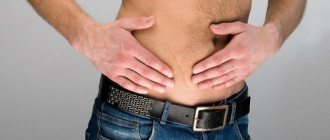
Abdominal pain on the right side in men
Dull abdominal pain in men most often occurs due to an inguinal or umbilical hernia. The reason is excessive physical activity. A bulge in the groin often develops in boys under 1 year of age due to weakness of the ligamentous apparatus, so babies should not be allowed to cry angrily for a long time. Umbilical and inguinal hernias are eliminated with plastic surgery (suturing the hernia opening or strengthening it with a special mesh). Without timely surgical intervention, strangulation of the hernia is highly likely. The pain increases, spreads to the entire abdomen, patients note frequent vomiting, stool retention and bloating. The surgery is performed on an emergency basis.
Pain in the lower back, radiating to the groin and leg, is characteristic of protrusion and herniation of the intervertebral disc. Patients often complain of weakness and numbness in the legs. Once the diagnosis is confirmed, the doctor will prescribe an effective course of anti-inflammatory drugs. If necessary, a therapeutic blockade or surgery is performed.
Diseases of the genitourinary system in men (cystitis, prostatitis, prostate adenoma) are manifested by pain in the lower abdomen, which subsides after defecation and urination. However, sometimes the pain is worse on the right or left.
With orchitis (inflammation of the testicle) and vesiculitis (an inflammatory process that develops in the seminal vesicles), the temperature suddenly rises, a sharp or dull pain radiates to the groin and perineum, and intensifies when walking down the stairs and physical activity. The damaged testicle swells. Inflammation of the testicles often complicates the course of sexually transmitted diseases and is fraught with infertility.
Varicocele (varicose veins of the spermatic cord) is diagnosed in every fourth man under the age of 30. The disease is often asymptomatic and is diagnosed during clinical examination of adolescents. Only in severe cases does aching pain occur on one side of the scrotum and in the groin. Pain increases with overheating, physical exertion and during sexual intercourse.
When testicular torsion occurs, a sharp pain suddenly occurs in the scrotum on one side, radiating to the groin, perineum and lower back. In young boys, the disease is accompanied by nausea and vomiting. Palpation reveals a higher position of the testicle in the scrotum. The pathology is eliminated urgently.
Important! If you suspect prostatitis, prostate adenoma or testicular disease, you should contact a urologist-andrologist.
Diagnostics
To make a diagnosis of abdominal pain on the right, the doctor prescribes the following tests.
Blood tests
A clinical blood test reveals signs of inflammation (increased leukocytes, accelerated ESR). Appendicitis is characterized by a sharp increase in the level of leukocytes to 15x109/l. Liver tests evaluate liver function.
Analysis of urine
A general analysis and, if necessary, urine culture exclude/confirm kidney inflammation and the presence of stones.
Stool tests
The coprogram diagnoses gastrointestinal diseases. If an intestinal infection is suspected, a stool culture is performed. A test for occult blood is prescribed when bleeding is suspected (for example, in Crohn's disease). Children who complain of abdominal pain are tested for dysbiosis and worm eggs.
Instrumental methods
Ultrasound (including Doppler mode) is the safest method for examining the abdominal cavity, pelvic organs and kidneys. The study is carried out for abdominal pain of any location in adults, pregnant women and children of all ages. If you suspect a disease of the stomach and gallbladder, it is advisable to undergo an FGDS. Pathology of the small intestine is diagnosed using x-rays. For colitis, colonoscopy is prescribed. However, the doctor receives the most accurate information about organic changes in the gastrointestinal tract using CT/MRI.
A child has a stomach ache: when to sound the alarm
Diagnosing abdominal pain in a child is sometimes a difficult task even for an experienced clinician. After all, children, especially preschool age, are not able to indicate a specific area of pain and note bowel movements. And parents cannot always remember the connection between a symptom and food intake or other provoking factors. So, what do you need to remember if your child complains of abdominal pain? And when should you not postpone a visit to the doctor?
How pain occurs
Most of the digestive system does not have pain receptors as such, and pain “in the abdomen” is caused by irritation of the membrane that covers the organs and separates them from each other (peritoneum).
This irritation may occur due to:
- spasm (in children, more often intestines)
- inflammation (edema),
- or overstretching of the organ, and accordingly, the peritoneum (for example, due to gas formation or obstruction).
And, depending on the cause, it is accompanied by a number of characteristic symptoms.
Qualitative differences
Thus, a catastrophe in the abdomen also has the name “acute abdomen”, is characterized by high intensity and infertility, has a diffuse (with appendicitis, cholecystitis, pancreatitis) or cramping character (with intestinal obstruction, volvulus, strangulated hernia), is accompanied by vomiting (usually multiple), temperature reaction and, unlike food poisoning, retention of stool and gases, and not diarrhea.
Treatment of such pathologies requires the mandatory participation of a pediatric surgeon and urgent research:
- clinical blood test with leukocyte formula (increased leukocytes) (0.D2.202),
- general urine test (differential diagnosis with kidney pathology, the pain in which can “radiate” to the abdomen),
- Ultrasound of the abdominal organs
- and, if necessary, radiographic examination with contrast.
And to clarify the degree of the inflammatory process, depending on the results of ultrasound and x-ray examination, a blood test may be prescribed for:
- pancreatic amylase and lipase (if acute pancreatitis is suspected),
- total bilirubin, Gamma GT and alkaline phosphatase (for signs of acute cholecystitis).
- By the way, these same tests are also indicated for less pronounced pain, but it occurs regularly and is associated with food intake.
And the main analysis, outside of acute pathology, in this case is a coprogram (general analysis of stool), where you should pay attention to:
- neutral fat (appears with a lack of bile or pancreatic enzymes)
- fatty acids (in case of impaired intestinal absorption function),
- intracellular (a sign of gastric pathology),
- and extracellular starch (lack of pancreatic amylase or too rapid movement of food through the intestines),
- as well as an increase in leukocytes
- and the appearance of red blood cells (blood).
- Intestinal causes of “chronic” abdominal pain include celiac disease (gluten intolerance), lactase intolerance (milk intolerance), as well as ulcerative enterocolitis and Crohn’s disease.
Wherein,
- in the first case, the distinctive feature is the “special” stench of feces and antibodies to transglutaminase and endomysium in the blood;
- in the second, the child’s stool takes on a foamy appearance and a sour smell, and stool containing carbohydrates allows the diagnosis to be confirmed;
- and the last two pathologies are characterized by the appearance of leukocytes, erythrocytes and calprotectin (the most effective marker of “intestinal” inflammation) in the feces.
It should also be remembered that “digestive” pathologies are accompanied by signs of vitamin deficiency, protein deficiency, anemia, calcium metabolism disorders (rickets, enamel destruction, fractures), skin pathologies (especially with celiac disease) and other deficiency conditions. Therefore, timely diagnosis of regular abdominal pain is necessary not only to maintain the health of the digestive system itself, but also the entire body as a whole.
How to relieve pain at home
- If you experience abdominal pain on the right side for the first time, you should not take analgesics on your own. Anesthesia may distort the clinical picture and make diagnosis difficult.
- If there are signs of an “acute abdomen” (pain, board-shaped abdomen, vomiting, stool retention, fever), cold (a heating pad with ice on the epicenter of pain), hunger, rest (the patient should be placed in a comfortable position) and calling emergency help are recommended.
- If the patient cannot wait until the ambulance arrives, it is permissible to give an antispasmodic tablet.
Treatment
Treatment tactics depend on the established diagnosis. Self-medication is fraught with deterioration in health and is sometimes associated with a risk to life. It is necessary to call an ambulance in several cases.
- Acute abdominal pain that does not go away within several hours.
- Within 1 hour after taking the antispasmodic, the pain does not stop or intensifies.
- Painful sensations are accompanied by fever, repeated/bloody vomiting, black feces or the presence of scarlet blood in the stool, and bleeding from the vagina.
- Pain in the abdomen develops against the background of a sharp deterioration in the patient’s condition.
- The abdomen has become hard (abdominal wall tension).
- Abdominal pain in a pregnant woman.
Make an appointment at the First Family Clinic of St. Petersburg if you are bothered by constant or recurrent abdominal pain of moderate intensity, in cases of deterioration or lack of therapeutic effect for a previously diagnosed disease.
Treatment tactics for abdominal pain on the right
- Gastrointestinal diseases - antispasmodics, antacids and antiulcer drugs, laxatives (for constipation), anti-inflammatory drugs and antibiotics (for inflammation, intestinal infections).
- Urogenital pathology and kidney diseases - antibacterial therapy, often requiring IV infusions to eliminate intoxication.
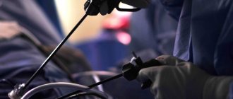
- Acute surgical pathology (appendicitis, ectopic pregnancy, intestinal obstruction) is an urgent operation.
The best solution, even if you have minor abdominal pain and no other symptoms, is to make an appointment with your doctor. Timely medical care will prevent the pathological process from worsening and often allows surgery to be avoided. Therapists, gastroenterologists and other specialized specialists at the First Family Clinic of St. Petersburg will prescribe only the necessary tests, quickly determine the exact cause of the pain and prescribe effective treatment. For a quick recovery, follow your doctor’s recommendations on the dosage and duration of taking medications, diet, and adhere to a healthy work and rest schedule.