Closed and open fracture of the humerus
Closed fracture
A closed fracture can occur in the upper part of the shoulder. There, the head of the bone, the lesser and greater tuberosity, the surgical and anatomical neck are susceptible to injury.
The symptoms that concern a patient who has received a closed injury to this part are the following:
- Pain in the joint area.
- If the injury is impacted, then the swelling is not too pronounced and grows slowly. The pain intensifies when trying to actively move the limb. Passive movements are not too limited.
- If displacement is observed during a closed fracture, then deformation of the arm is more often visible, and other symptoms, including pain, appear more clearly.
If a closed fracture of the shoulder occurs in the middle section, then most often the cause is a fall or a blow to the shoulder. The injury can be splintered, oblique, transverse or helical. A fracture of this part often entails damage to the nerve bundle, namely the radial nerve. In addition, the brachial arteries and veins are affected.
The main symptoms that indicate a closed fracture of the humeral body include:
- Strong pain.
- Deformation, in the presence of displacement.
- Decreased limb length.
- Crepitation of fragments.
- Swelling and hematoma, which can occupy a wide area, up to the hand.
- Movements are limited mainly in the elbow and shoulder joint.
- If the nerves have been damaged, there is a disturbance in finger movements and sensitivity.
- The hand cannot be kept in a raised state; it hangs limply.
Open fracture
The main features of an open fracture include:
- An open wound will be visible on the surface of the skin. Most often there is bone visible through it.
- There is severe bleeding, which must be stopped by applying a tourniquet. Its location is the upper third of the shoulder.
- The wound site is treated with any available antiseptic, after which a sterile bandage is applied.
- Only after treatment and stopping the bleeding should the hand be immobilized.
Department of Traumatology and Orthopedics
article in PDF format
QUOTE LINK:
1A. V. SKOROGLYADOV, 1A. P. RATIEV, 1K. A. YEGHIAZARYAN, 2E. A. KURUCH, 1A. V. GRIGORIEV
1Russian National Research Medical University named after. N.I. Pirogov, Moscow
2GBUZ MO Podolsk City Clinical Hospital, Podolsk
Information about the authors:
Skoroglyadov Alexander Vasilievich – Doctor of Medical Sciences, Professor, Head of the Department of Traumatology, Orthopedics and Military Field Surgery of the Russian National Research Medical University named after N.I. Pirogov
Ratyev Andrey Petrovich – Doctor of Medical Sciences, Associate Professor of the Department of Traumatology, Orthopedics and Military Field Surgery of the Russian National Research Medical University named after N.I. Pirogov
Egiazaryan Karen Albertovich – Candidate of Medical Sciences, Associate Professor of the Department of Traumatology, Orthopedics and Military Surgery of the Russian National Research Medical University named after N.I. Pirogov
Kuruch Evgeniy Aleksandrovich – traumatologist-orthopedist of the 1st traumatology department of the Podolsk City Clinical Hospital
Grigoriev Alexey Vladimirovich – postgraduate student of the Department of Traumatology, Orthopedics and Military Surgery of the Russian National Research Medical University named after N.I. Pirogov
Fracture-dislocations of the proximal humerus are the most complex injuries and are among the most severe pathologies of this location. These injuries still remain the most important problem of modern traumatology and require careful analysis in each specific case. The article provides an analysis of foreign literature devoted to diagnosis, research methods, complications and treatment of various types of fracture-dislocations of the proximal humerus.
Key words: fracture-dislocation of the shoulder, rotator cuff, aseptic necrosis.
Introduction
The shoulder joint, due to its anatomical architecture and functional purpose, is considered one of the most complex from a biomechanical point of view [1–3] and is most often susceptible to fracture-dislocations. The large number of associated injuries is due to the complex anatomy of the shoulder joint, which is surrounded by a large number of muscles, tendons, vessels and nerves that are of fundamental importance in the functioning of the joint.
Fracture-dislocation of the shoulder is a complex injury to the joint, characterized by the associated displacement of two or more fragments of the proximal epiphysis of the shoulder relative to the joint anteriorly, posteriorly and downward. The incidence of fracture-dislocations among all injuries of the proximal shoulder ranges from 2.6 to 8% [4]. According to foreign literature, the prevalence of shoulder fracture-dislocations in Europe is 1/100,000 of the population per year [5, 6].
During a fracture-dislocation of the shoulder, soft tissue injuries and osteochondral fractures of the glenoid often occur (the most common fracture is the anteroinferior angle). Contracture of the shoulder joint in fracture-dislocations is explained by the involvement of soft tissues (tendons, capsules, ligaments) and heterotopic ossifications in patients after treatment [7, 10, 11]. You need to remember these injuries and avoid unnecessary manipulation of the joint. It is known that incorrect and rough manipulations can transform a two-part fracture dislocation into a four-part one with worsening damage to soft tissues, the rotator cuff, brachial artery or brachial plexus [6, 12]. Neer [7] classified these fractures according to the number of fragments and the direction of displacement (anterior, posterior, or inferior).
Clinical assessment
Shoulder fractures and dislocations most often occur during high-energy trauma. The same can happen during a banal domestic injury, more often in women. In most cases, this occurs while running on a hard surface and falling on the shoulder joint or on the arm extended at the elbow joint [6, 8, 12]. In rare cases, fracture-dislocation of the shoulder can occur during electrical trauma or during an epileptic seizure [6, 12]. In the emergency department, a patient with a fractured shoulder dislocation looks exhausted and supports the injured limb with his healthy arm. There is soft tissue swelling in the shoulder joint area. While trying to perform minor movements, the patient feels a sharp pain in the shoulder joint and may scream loudly. Subcutaneous hematoma is rarely seen in the acute period. It occurs in older people or in people receiving anticoagulant therapy. In young people, a subcutaneous hematoma appears 24–48 hours after the injury and is located on the arm and chest.
In the case of a two-part anterior fracture-dislocation of the shoulder, the acromial protrusion is more noticeable, at the same time, soft tissue swelling appears along the anterior surface of the shoulder girdle and the coracoid process is not palpable. The acromial protrusion is more difficult to visualize in three- and four-part anterior fracture-dislocations.
With posterior fracture-dislocations of the shoulder, the clinical picture is completely different. The coracoid process and the posterior part of the deltoid muscle are clearly visualized. The position of the upper limb is also assessed in these patients. In the case of a two-part anterior fracture-dislocation, the passive mobility of the upper limb is blocked in the position of adduction and external rotation. An attempt to rotate the upper limb medially causes severe pain. And, conversely, with a posterior fracture-dislocation, the upper limb is internally rotated and outward rotation is impossible. When the fracture involves the surgical neck, the axis of the upper limb is in a neutral position [8].
1. X-ray examination
To make a diagnosis and select treatment tactics, the primary method of examination is to perform radiographs of the shoulder joint in standard projections. If the X-ray images are insufficiently informative, computed tomography is performed, which allows one to reliably assess the fracture lines, the direction of head dislocation, damage to the anatomical structures of the shoulder joint, including the involvement of the lesser and greater tuberosities, as well as damage to the glenoid [16–18]. In patients over 40 years of age, it is advisable to perform an MRI to evaluate rotator cuff and joint capsule damage.
2. Associated damage
1. Rotator cuff injuries
The rotator cuff is often injured in a two-part fracture-dislocation with a fracture of the greater tuberosity. Robertson et al. [6] reported rotator cuff injury in 33.4% of 3633 fracture-dislocation cases. This injury may involve the interval between the supraspinatus and subscapularis muscles. Rotator cuff tears can be especially severe in three- and four-part fracture-dislocations. These lesions are usually identified and treated during osteosynthesis. However, this can be detected before surgery if an MRI is performed.
2. Neurological damage
Damage to the brachial plexus or peripheral nerves as a result of fracture dislocation or during its repair accounts for 2–30% of cases. Robertson et al. [6] reported nerve injury in 13.5% of cases after 3633 anterior fracture-dislocations (2250 men and 1383 women; mean age 47.6 years). The nature of the injury is determined by the age of the patient, the energy of the injury, the type of injury and the time between dislocation and reduction. Electromyography is the most reliable diagnostic method in identifying the severity of nerve damage. The brachial nerve is most often damaged and recovers in approximately 4–5 months, in rare cases requiring surgical treatment.
3. Vascular damage
Fracture dislocations rarely occur with damage to large vessels. In such cases, the brachial artery or vein is usually affected in older people with vascular atherosclerosis. The mortality rate during surgery for such injuries is 50%.
4. Depressed fractures and glenoid fractures
Depressed fractures of the humeral head during dislocation were first described by Hill Sachs and McLaughlin [20, 21] in the 19th century and later classified by Neer [7]. It is known that with all dislocations there is a depressed fracture of the head of the humerus and the edge of the glenoid cavity. The severity of these injuries depends on the patient's age, the energy of the injury, and the time between dislocation and repair. Impression fractures of the humeral head can be in the anterior part, near the insertion of the subscapularis tendon in the case of a posterior dislocation, or in the posterosuperior part of the head in an anterior or inferior dislocation. These injuries often go unrecognized. CT and MRI studies can identify these fractures and adjust treatment. Detailed knowledge of pathological anatomy and the time between injury and primary treatment influence the further management of the patient. For deep and extensive injuries of the humeral head, remplissage or bone fragment transplantation is indicated, especially in young people and if the bone defect is less than 45% [24]. If the defect is more than 50%, prosthetics are indicated.
3. Treatment
1. Two-part fracture-dislocations
The shape of these dislocations depends on the pathological involvement of the greater tuberosity and the surgical neck of the humerus. Two-part fracture-dislocations involving the surgical neck of the humerus are extremely rare. If the first attempt to set the head into the glenoid cavity under local anesthesia was unsuccessful, then further treatment is carried out under intravenous anesthesia. After reduction, X-ray monitoring of a fracture of the surgical neck of the humerus is necessary, since in case of its occurrence and displacement, surgical treatment is indicated. In case of two-part fracture-dislocations, there is often a fracture of the greater tuberosity [10.33%]. Bahrs et al. [25] in a study of 100 patients with a fracture of the greater tuberosity found that in 50% of cases it was associated with anterior shoulder dislocation. In these forms, an attempt to reduce the dislocation should be as atraumatic as possible and performed after a complex of examinations, including CT and radiography. After repositioning the head, the displacement of the greater tubercle spontaneously corrects. Surgical treatment is indicated for persistent displacement of more than 5 mm.
Fractures of the lesser tubercle with two-part fracture dislocations are extremely rare. Posterior shoulder dislocation is assessed by clinical presentation and CT scan. Mostly, fractures of the small tubercle occur in posterior dislocations. The treatment tactics are the same as for other two-part fracture-dislocations; they involve extension and traction of the limb along its length. If the displacement persists more than 1 cm, surgical treatment is indicated. For two-part fracture-dislocations, the deltopectoral approach is used. After selecting v. cephalica, the fascia is cut, the subacromial bursa is exposed and excised. The deltoid muscle moves laterally, and the coracobrachialis moves medially. In this way, the surgeon can evaluate the damage to the superficial part of the rotator cuff and see the possible interposition of the biceps between the fragments. If the dislocation cannot be reduced even under anesthesia, the biceps tendon is most likely caught between two main fragments. In these cases, the rotator cuff must be opened and a tenotomy performed. Thus, open reduction of the head, reposition and fixation of fragments can be performed. In the postoperative period, the upper limb is fixed with an orthosis for 4-6 weeks, only passive and active movements in the elbow and wrist joints are allowed.
2. Three-part anterior fracture-dislocations
Most often, three-part fracture-dislocations occur with anterior dislocation of the shoulder and have two fracture lines. These are a fracture of the greater tuberosity and a fracture of the surgical neck of the humerus. In such cases, the small tubercle remains attached to the head of the humerus. This is a positive sign in terms of clinical outcome, since vascularization is preserved. During the operation, it is important not to damage the circumflex artery, which supplies the head. In three-part fracture-dislocations, a fracture of the surgical neck of the humerus and the greater or lesser tuberosity usually occurs. The delto-pectoral approach is used for the operation, the biceps is used as a guide for repositioning the head. Sometimes biceps tenotomy and tenodesis, open reduction and internal fixation of fragments are performed. In more complex cases, when there is a glenoid fracture requiring osteosynthesis, the m.subscapularis is separated and a vertical capsulotomy is performed. After this, osteosynthesis of the glenoid cavity is performed with cannulated screws.
3. Three-part posterior fracture-dislocations
Three-part posterior fracture-dislocations are characterized by two fracture lines, including the surgical neck of the humerus and the lesser tubercle. A CT scan and careful preoperative planning are necessary. The decision to osteosynthesis a fracture or replace the humeral head with an endoprosthesis depends on the patient’s age, bone quality, grinding of bone fragments, and damage to soft tissues. For these operations, deltopectoral access is also used.
4. Four-part anterior and posterior fracture-dislocations
Four-part fracture dislocations are a complex joint injury, the treatment of which is still under discussion [8, 9, 11, 12, 22, 26, 27]. The stumbling block to this problem is ischemia of the head, due to impaired blood supply [28, 29], which in most cases leads to necrosis of the humeral head [6, 7, 9, 12, 15, 17, 30]. Based on these studies, shoulder arthroplasty is generally preferred in middle-aged and elderly patients, while open reduction and internal fixation may be attempted in younger patients.
conclusions
Thus, fracture-dislocations of the shoulder remain one of the most important problems in modern traumatology. Before choosing treatment tactics, a thorough examination and preoperative planning (performing functional tests, radiographs, CT, MRI), assessment of the nature of the fracture, the presence of neurocirculatory complications and taking into account the patient’s age are necessary. A positive treatment outcome directly depends on the damage to the soft tissues, the nature of the fracture, the surgical technique and the experience of the surgeon. Incorrectly chosen surgical tactics without taking into account the classification of the fracture, possible damage to the rotator cuff, periarticular vessels and nerves can lead to serious complications, such as non-union of the fracture, aseptic necrosis of the humeral head, contracture of the shoulder joint with limited limb function and loss of ability to work with possible subsequent disability of the patient.
Bibliography
1. Goldstein, B. Shoulder anatomy and biomechanics. Phys. Med. Rehab. ClinicsNorth Am. 2004, 15; 313-349.
2. Halder AM, Itoi E., An Kn Anatomy and biomechanics of the shoulder. Orthop. Clin. North Am. 2000; 31:159-176.
3. Poppen nK, walker PS Normal and abnormal motion of the shoulder J. Bone JointSurg. 1976; 2: 195-201.
4. Grokholsky V.N. [2006] Journal Bulletin of Physiology and Pathology of Respiration. Issue NoS22/2006. Page 85.
5. Court-Brown CM, McQueen MM [2001] Epidemiology of proximal humeral fractures. Acta Orthop Scand 72:365–371.
6. Robinson CM, Akhtar A, Mitchel M, Beavis C [2007] Complex posterior fracture-dislocation of the shoulder. Epidemiology, injury patterns, and results of operative treatment. J Bone Joint Surg Am 89:1454–1466.
7. neer CS 2nd [1970] Displaced proximal humeral fractures: I. Classification and evolution. J Bone Joint Surg Am 52:1077–1089.
8. Bigliani LU [1998] Fractures of the proximal humerus. In: Rockwood CA, Matsen FA [eds] e Shouider 2ndedn. wB Saunders, Philadelphia, pp. 278-334.
9. Ackermann C, Lam Q, Linder P, Kull C, Regazzoni P [1986] Problems in classification of fractures of the proximal humerus. Z Unfallchir Versicherungsmed Berufskr 79:209–215.
10. Rouleau DM, Hebert-Davies J [2012] Incidence of associated injury in posterior shoulder dislocation: systematic review of the literature. J Orthop Trauma 26:246–251.
11. Shrader Mw, Sanchez – Sotelo J, Sperling Jw, Rowland CM, Coeld Rh [2005] Understanding proximal humeral fractures: image analysis, classification and treatment. J Shoulder Elbow Surg 14:497–505.
12. Duparc J, Largier A [1976] Fracture-dislocation of the upper end of the humerus. Rev Chir Orthop Reparatrice Appar Mot 62:91–110.
13. Kristiansen B, Andersen UL, Olsen CA, Varmarken JE [1988] Neer classification of fractures of proximal humerus. An assessment of interobserver variation. Skeletal Radiol 17:420–422.
14. Sidor ML, Zickerman JD, Lyon T, Cuomo F, Schoenberg n [1993] Neer classification system for proximal humerus fractures. An assessment of interobserver reliability and reproducibility. J Bone Joint Surg Am 75:1745–1750.
15. Brien H, no all F, MacMaster S, Cummings T, Lendells C, Rockwood P [1995] Neer's classification system: a critical appraisal. J Trauma 38:257–260.
16. Bernstein J, Adler LM, Blank JE, Dalsey RM, Williams GR, Iannotti JP [1996] Evaluation of the Neural system of classification for proximal humerus fractures with computerized tomogre with scans and plain radiographs. J Bone Joint Surg Am78:1371- 1375.
17. Au arth A. Mayer M. Koer B et al [2013] E interobserver reliability in diagnosing osseous lesions a er rst-time anterior shoulder dislocation comparing plain radiographs with computed tomography scans. J Shoulder Elbow Surg 22[11]:1507–1513. S1058-2746[13]00210-3.
18. Bono CM, Renard R, Levine RG, Levy AS [2001] E ect of displacement of fractures of the greater tuberosity on the mechanics of the shoulder. . J Bone Joint Surg Br 83:1056–1062.
19. Frank MA, Laratta JL, Tan V [2012] Irreducible luxation of erecta humeri caused by an aberrant position of the axillary nerve. J Shoulder Elbow Surg 21 e6-e9.
20. McLaughlin hL [1952] Posterior dislocation of the shoulder. J Bone Joint Surg Am 24-A-3:584-590.
21. hill SA, Sachs MD [1940] e grooved defect of the humeral head. A frequent unrecognized complication of dislocation of the shoulder joint. Radiology 35:690.
22. Shultz TJ, Jacobs B, Patterson LR [1969] Unrecognized dislocation of the shoulder. J Trauma 9:1009–1023.
23. Rowe CR Zarns B [1982] Chronic unrecognized dislocations of the shoulder. J Bone Joint Surg Am 64:494–505.
24. Assom M. Castoldi F, Rossi R, Blonna D, Rossi P [2006] Humeral head impression fracture in acute posterior shoulder dislocation: new surgical technique. Knee Surg Sports Traumatol Arthrosc 14:668–672.
25. Bahrs C, Lingenfelter E, Fischer F, Walters EM, Schnabel M [2006] Mechanism of injury and morphology of the greater tuberosity fracture. J Shoulder Elbow Surg 15:140–147.
26. Duparc F, Huten D [1998] Conservative treatment of fractures of the upper end of the humerus. Rev Chir Orthop Reparatrice Appar Mot 84:121–189.
27. hertel R, hemp ng A, Stiehler M, Leunig M [2004] Predictors of humeral head ischemia and intracapsular fracture of the proximal humerus. J Shoulder Elbow Surg 13:427–433.
28. Gerber C, Schneeberger AG, Vinh TS [1990] E arterial vascularization of the humeral head. An anatomical study. J Bone Joint Surg Am 72:1486–1494.
29. Meyer C, Alt V, Hassanin H et al [2005] E arteries of the humeral head and their relevance in fracture treatment Surg Radiol Anat 27:232–237.
30. Russo R, Cautiero F, Della Rotonda G [2012] E classification of complex 4-part fracture revisited: the missing fragment and indication for surgery. Musculoskelet Surg 96:S13-9.
TREATMENT OF THE DISLOCATION-FRACTURES OF PROXIMAL HUMERUS: LITERATURE REVIEW
2E. A. KURUCH, 1A. V. GRIGORIEV, 1A. P. RATEV, 1K. A. EGIAZARYAN, 1A. V. SKOROGLYADOV
1Russian National Medical Research University named after NI Pirogov, Moscow
2 Podolsky City Clinical Hospital, Podolsk
Information about the authors:
Kuruch Evgeny Alexandrovich – doctor traumatologist-orthopedist 1st trauma department of Podolsk City Clinical Hospital
Grigoriev Alexey Vladimirovich – of the chair of traumatology, orthopedics and military eld surgery of the Russian National Research Medical University named a er NIPirogov
Ratiev Andrey Petrovich – Doctor of Medical Sciences, associate Professor of the chair of traumatology, orthopedics and military eld surgery of the Russian National Research Medical University named a er NIPirogov
Yeghiazaryan Karen Albertovich – PhD, associate Professor of the chair of traumatology, orthopedics and military eld surgery of the Russian National Research Medical University named a er NIPirogov
Skoroglyadov Alexander Vasilyevich – Doctor of Medical Sciences, Professor, head of chair of traumatology, orthopedics and military surgery of the Russian National Research Medical University named a er NIPirogov
Dislocation-fractures of proximal humerus are the hardest injuries and appertain to the most severe pathology of this localization. ese injuries are still the major problem of modern traumatology and require careful analysis in each case. e article is considered the analysis of foreign literature dedicated to the diagnosis, research methods, complications and treatment of various types of dislocation-fracture proximal humerus.
Key words: shoulder`s dislocation-fracture, rotator cu, aseptic necrosis.
Displaced humerus fracture
The following symptoms are typical for a displaced fracture:
- The occurrence of sharp pain that appears when trying to move the arm or when palpating the body of the humerus.
- Swelling, often with severe hematoma.
- Inability to perform active actions to abduct and raise the limb.
- With active palpation, crepitations are heard.
- The deformation is visible even when the bones have not broken through the soft tissue. Especially if the displacement of the fragments is pronounced.
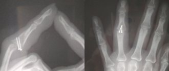
The final diagnosis can be made only after the doctor sees x-rays, which must be taken in two projections. If there is a pronounced displacement of the humeral head, then the prognosis is unfavorable. Since it will experience a nutritional deficiency, this will lead to its death or even resorption. Fractures, the line of which passes through the tuberosities, are also dangerous. Restoring full function of the limb is rare.
When the surgical neck is injured, the displacement occurs in an impacted manner with the formation of a chip. The fragment may move to the side (when the fracture occurs with the shoulder adducted) or to the middle (with the shoulder abducted). In this case, the decisive factor in where it will be directed is played not only by the position of the shoulder when injured, but also by the muscle contraction that occurs reflexively.
It is important not to confuse a displaced fracture with a dislocated shoulder. A distinctive feature of such fractures is the ability to freely move the shoulder (not at the joint), using passive force. There will be no springing effect. These signs are especially relevant for obese people, since a dense fat layer can make it difficult to conduct a full X-ray examination.
Operation Latarget
If the shoulder joint is unstable due to a deficiency of scapular bone tissue, transposition of the coracoid process of the scapula is recommended - the Latarget operation.
The operation is also used in cases of poor condition of the ligamentous apparatus of the shoulder joint, recurrence of dislocation after the Bankart operation, and the absence of a fibrous lip. During the operation, a fragment of the coracoid process (2x1 cm) is cut off. With the muscles attached to it, it is carried through the subscapularis muscle to the anterior surface of the glenoid cavity of the scapula (glenoid). After preparation and correct positioning, it is fixed with 2 screws.
The operation provides restoration of the bone defect of the glenoid due to the transfer of the coracoid process and a supporting effect due to the relocated muscles closer to the head of the humerus.
Recovery and return to previous loads is possible within 3 months after surgery.
Non-displaced humerus fracture
If there is no displacement during the fracture of the shoulder bone, then the signs of the fracture may be somewhat blurred:
- Depending on the location of the injury, the person will feel pain in the upper or lower arm. But its intensity is not as pronounced as in a fracture with displacement of fragments. It intensifies when trying to move.
- Swelling may not form immediately, but over several hours. This is due to the fact that the soft tissue around the bone will not be damaged by fragments.
- A hematoma is observed, but it also appears after some time; its size and severity depend on the cause of the fracture.
- The shortening of the limb is not noticeable without special measurements.
- There is no hand deformity.
It is important to provide first aid to the victim correctly so that the fragments do not shift and the injury does not become more serious. More often, non-displaced fractures of the humerus are observed in children, which is due to the structural features of their bone tissue.
Diagnostics
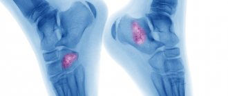
A preliminary diagnosis can be made based on the patient’s complaints, a history of a fall or blow, and examination data. To clarify the diagnosis, identify the nature of the fracture, and the position of bone fragments, an x-ray examination is performed. The picture is taken in several non-standard projections. In difficult situations, computed tomography is necessary.
This study makes it possible to accurately determine the nature of the damage in situations where the fragments on the radiograph are layered on top of each other, and it is technically impossible to perform the study in the desired projection due to limited mobility of the limb.
Fracture of the greater tuberosity of the humerus
Fractures of the greater tuberosity of the humerus are not uncommon. If it was damaged in isolation, then the injury occurs according to the avulsion type. Often the separation of the tubercle is accompanied by a dislocation of the shoulder. Due to the fact that the muscles responsible for shoulder abduction and rotation are attached to it, the torn fragment is always displaced due to their tension force.
Signs of avulsion of the greater tubercle are as follows:
- Swelling of the injury site.
- Pain that manifests itself locally above the shoulder joint.
- Impaired mobility of the shoulder joint.
- Due to muscle retraction, external rotation of the shoulder is impaired. This is one of the main symptoms indicating damage to the tubercle.
- If it is absolutely impossible to move your arm, then this is a sign of tendon damage.
After a fracture of the greater tuberosity, there is a danger that movement disorders in the shoulder joint may be severe. This is due to damage to the supraspinatus muscle. Sometimes there is no possibility of full recovery.
About the shoulder
Humerus, shoulder (lat. - Нumerus) - refers to the long bones. The humerus makes up the bone base of almost half of the free upper limb and is its initial section.
Like any long bone, the humerus consists of a body (diaphysis) and ends (epiphyses) - the proximal one, formed by the head of the humerus, connecting the shoulder to the scapula through the shoulder joint, and the distal one - forming the shoulder component of the elbow joint. Due to the large degree of freedom of movement of the shoulder in the shoulder joint (the ball and socket joint, the most mobile in the human body), the proximal part of the shoulder is the site of attachment of many muscles and ligaments, thanks to which an extremely complex and balanced process of movement of the shoulder is carried out in the shoulder joint, and with it and the entire free upper limb.
Moreover, during almost any movement, the head of the humerus is always strictly centered in the joint. The diaphysis of the humerus forms the lever of the shoulder and serves as the attachment point for part of the flexor and extensor muscles, providing the same movements of the shoulder in the shoulder joint, and the forearm in the elbow.
Due to the variety of functions of the shoulder and the complexity of the anatomical structure, damage to this area leads to significant dysfunction of the entire upper limb. Moreover, if we figuratively consider the free upper limb as a chain of successive links, and the shoulder is the initial link in the chain, then the higher the damage, the more significant the dysfunction of the entire chain.
The speedy restoration of the function of the upper limb plays a vital role in a person’s life: self-care, professional and creative activity, learning, tactile perception of the surrounding world.
Shoulder injuries are a fairly common pathology . There is a wide variety of injuries to both bones - bone fractures, fractures of hand bones, dislocation of the collarbone, and soft tissues (ruptures of muscles, tendons, ligaments, blood vessels and nerves), and various dislocations of the head of the humerus are also common.
Other types of humerus fracture
Special mention should be made of surgical neck fractures, transcondylar and comminuted fractures. Each of them has certain characteristics. In treating them you need to adhere to certain tactics.
Surgical fracture of the humerus
If the mechanism of injury is indirect, the surgical neck of the shoulder often suffers. Fractures are divided into adduction and abduction, depending on the position of the arm at the time of injury. The first occurs if the limb is adducted, and the second if it is abducted. When the hand is in the middle position, then the distal fragment is inserted into the proximal one more often. This is called a surgical impacted fracture.
If we consider the symptoms of this type of injury, they are as follows:
- The pain will be localized at the fracture site and becomes more intense when trying to make circular movements.
- It is uncomfortable for a person to hold a limb in weight; he tries to support it under the elbow.
- If attempts are made to move, the greater tubercle will move towards the head.
- Swelling occurs and a hematoma is observed.
- When displaced, crepitations will be heard.
- Pathological mobility is observed.
- The shoulder will become shorter compared to a healthy one.
A surgical fracture of the humeral neck is dangerous because at the time of injury, the integrity of the neurovascular bundle often occurs. The same damage can occur with improper reduction. This violation will lead to the fact that the function of the hand will not be fully restored.
Transcondylar fracture of the humerus
Such injuries are rare, due to the location of the bone. The damage is considered intra-articular, which means that the fracture line runs through the joint cavity. It runs transversely, from one condyle towards the other.
Symptoms of transcondylar fractures include:
- Painful sensations that radiate to the elbow and forearm area.
- Presence of swelling. Sometimes the swelling is pronounced.
- If there is a displacement, then deformation of this area will be visible.
- When you try to feel it, you hear a crunching sound.
- Movements of the elbow, if not completely blocked, are then significantly limited.
A characteristic feature of a transcondylar fracture is trauma to the brachial artery. This increases the risk of gangrene of the hand. If the artery is damaged, you will not be able to feel the pulse in the forearm.
In order for both condyles to be broken, an impressive force must be applied. This could be a fall on an elbow from a height, or industrial accidents, for example, a mine collapse. X-rays often show fractures that form a V or T shape.
The external condyle breaks more often in children. The displacement will be directed outward and upward. The internal and external epicondyles are rarely affected and are accompanied by elbow dislocation.
Diagnosis of shoulder fractures
Diagnosis of humerus fractures is a clinical examination of the patient (examination, comparative assessment, measurement, palpation, etc.), X-ray examination is standard radiography, special X-ray placement if necessary.
X-ray computed tomography to verify soft tissue damage is an MRI. To make an accurate and correct diagnosis, as well as to determine further treatment tactics, it is very important that the diagnosis is carried out by a qualified specialist doctor - an orthopedic traumatologist.
Comminuted fracture of the humerus
The most severe of all types of shoulder fractures is a comminuted, displaced one. The difficulty is that not only the nerves are damaged, but also the blood vessels. Therefore, treatment necessarily requires surgical intervention.
This type of injury is typical for the adult population.
Depending on the nature and location of the fracture, the following types of comminuted shoulder injuries are distinguished:
- A fracture of the upper part is accompanied by swelling and deformation of the joint. For recovery, surgical intervention is necessary, an obstacle to which can be either advanced age or the presence of a serious illness.
- A fracture of the shoulder in the middle part is dangerous because fragments can damage the radial nerve, veins and arteries. To fix them, it is necessary to use metal pins or plates, or an Ilizarov apparatus.
- If the injury occurred in the lower part of the humerus and there is no displacement of the fragments, then it is advisable to apply a plaster cast. If the fragments are significantly displaced, surgery is necessary.
First aid for a fractured humerus
First aid for injury is as follows:
- First, you need to calm the person down and offer him a pain reliever. The following can be used as an analgesic: analgin, nimesulide, ketorol. If a person is experiencing panic, then you can give him a tincture of valerian, 20 drops will be enough, or one tablet of tazepam or triocasin. Valocordin or cordiamine can be used as a cardiovascular drug.
- Then you need to limit your hand movements as much as possible. For this purpose, immobilization is carried out. Small planks can be used as improvised means. One of them should be bandaged tightly to the shoulder, and the other to the forearm. If there are not even planks, then it is advisable to place your hand on a scarf. She throws herself over her uninjured shoulder. In this case, the arm should be bent at a right angle at the elbow. To prevent displacement, it is best to tie such a bandage as tightly as possible to the body.
- During transportation, it is advisable for the person to sit.
Treatment of a humerus fracture
Three methods are used to treat a shoulder fracture: surgical, conservative, and skeletal traction. If the fracture is not complicated by displacement or it can be corrected by performing a one-stage reduction, then applying a plaster cast or other fixing agent is sufficient.
If we consider therapy at the fracture site, we can highlight the following features:
- Treatment of a large tubercle occurs by applying a plaster cast; sometimes it can be supplemented with an abduction splint. This is necessary in order to prevent the development of stiffness in the joint and ensure proper fusion of the supraspinatus muscle. If the tubercle fragment has moved out of place, it must be fixed in the correct position with knitting needles or a screw. After about 1.5 months, the structure will be removed.
- If the surgical neck has been injured, but no displacement has occurred, then you can get by with a plaster cast for a month. When reduction was required, and it was successful, the cast will have to be worn for two more weeks. If it is not possible to straighten the bone fragments, then surgical intervention is necessary. Fixation inside the bone is carried out using plates. If the fracture is of the impacted type, then it is advisable to use either an abductor pillow or a special scarf. The treatment period can be extended up to 3 months.
- When the fracture is localized on the body of the shoulder and displacement is observed, the most common method of treatment was skeletal traction. A person will have to spend up to a month in an immobilized position. Afterwards, plaster will be applied for the same period of time. Recently, the method of skeletal traction has faded into the background; it has been replaced by osteosynthesis, which does not confine a person to bed for such a long period of time.
- Transcondylar fractures are almost always accompanied by displacement of fragments. Their comparison is carried out under anesthesia, and then it is advisable to apply a plaster cast for up to two months.
If vessels or nerves are damaged as a result of fractures, then a special operation is necessary, which involves placing sutures on them. This increases the treatment period and it is not always possible to fully restore the functionality of the limb. As for medications, it is advisable to use calcium supplements, as well as analgesics and antibiotics.
On topic: 12 folk methods for home treatment
SLAP damage
Until recently, establishing a diagnosis of SLAP injury was very difficult. With the development of MRI diagnostics and improvement of arthroscopic technologies, this pathology has become mandatory in the practice of shoulder surgery.
SLAP (superior labrum anterior posterior) is characterized by separation of the fibrous lip from the glenoid in its upper segment with anterior and posterior distribution. In this localization, the tendon of the long head of the biceps begins from the fibrous lip, which is the main vector of traction during injury.
The cause of damage is most often trauma: a fall with support on the abducted arm, a blow to the shoulder area, often found in “throwing” athletes (handball, baseball, water polo), boxers.
Conservative treatment rarely leads to full recovery, because a return to specific loads provokes a recurrence of pain and progression of the rupture.
However, in patients without heavy physical and sports activities, complex therapy provides long-term relief from pain. First of all, rest is ensured for the shoulder joint by fixing the arm on a support bandage. Anti-inflammatory non-steroidal drugs are prescribed. To reduce the inflammatory reaction and reduce pain, physiotherapeutic procedures are necessarily used, such as phonophoresis with drugs, high-intensity laser (HILT), shock wave therapy (SWT), massage, and taping. Stimulation of regeneration is achieved by taking chondroprotectors and intra-articular administration of platelet-rich plasma (PRP). After inflammation has been relieved and the resting stage has been completed, proper rehabilitation under the supervision of a physical therapist will be an important factor in restoring function.
Arthroscopic fixation of the fibrous labrum, by analogy with the usual dislocation of the shoulder, is the most rational method of treatment, because ensures precise restoration of anatomical structures. Low-traumatic surgery reduces rehabilitation time. Under camera control, anchors are installed into the glenoid cavity of the scapula and the fibrous lip is returned to its place using non-absorbable sutures.
Immobilization for a humerus fracture
When complete immobilization of the limb is required, it is advisable to apply a thoraco-brochial bandage.
The technique for applying it is as follows:
- The victim should sit on a high stool or on a table. His limb must be flexed 80 °C for an injury to the upper part of the shoulder and 45 °C for a fracture of the lower bones.
- A layer of cotton wool should be applied to the body, which is secured with bandages.
- The joints of the hand, such as the wrist, elbow and shoulder, are also covered with cotton wool.
- Plaster splints are applied horizontally to the body, and they are attached vertically to the sides.
- One splint should be placed over the shoulder that was injured. It should be attached to the body with bandages.
- Then additional splints are applied over the body, shoulder girdle, forearm, up to the hand. All this is again fixed with bandages.
- A special spacer is inserted between the arm that will be in the cast and the body so that the limb cannot adhere to the body.
This way the limb will be immobilized and bone fusion will proceed correctly.
Rehabilitation after a humerus fracture
After the bandage is removed, it is necessary to proceed to rehabilitation measures. They are an integral part of bone restoration and play no less important role than adequate therapy.
Rehabilitation necessarily includes:
- Physiotherapeutic treatment - you will need to complete several courses, which consist of 10 procedures. Electrophoresis with novocaine and calcium chloride may be recommended. Ultrasound treatment has proven itself well.
- A massage that, if it is impossible to visit a specialized office, can be performed independently. To speed up healing and stimulate blood circulation, you can use specialized ointments and oils.
- Performing a set of special exercises.