Diagnostics
The disease can be identified during a neurological examination and based on specific patient complaints. To clarify the diagnosis, a specialist may additionally prescribe instrumental studies:
- Rheoencephalography – helps assess blood circulation. If necessary, it is supplemented with magnetic resonance imaging or vascular angiography.
- Echoencephalography – assesses the state of the liquor system. When mixed, a formation (hematoma, tumor or abscess) can be suspected, which can provoke vestibular ataxia.
- Electroencephalography – helps analyze the bioelectrical activity of the brain.
- Computed tomography and magnetic resonance imaging can detect demyelinating processes in the brain and the presence of neoplasms.
- X-ray of the skull and spine is performed if the possibility of a craniovertebral anomaly is suspected.
Patient's office
Request a call
Symptoms and treatment of vertebral artery syndrome
From this article you will learn: what is vertebral artery syndrome. What diseases lead to the occurrence of pathology. Manifestations of this syndrome and examination methods to establish a diagnosis. Methods for correcting any violations that have arisen.
Content:
Causes of occurrence
Classification Manifestations of the syndrome Diagnostics Treatment
Prognosis
Vertebral artery syndrome (abbreviated as VAS) is a combination of symptoms from the brain, vascular and autonomic systems that arise against the background of damage to the nerve plexus of the artery itself, deformation of its wall or narrowing of the lumen.
In the medical community, such pathology is usually associated with diseases of the cervical spine, but predisposing factors in some patients are the anatomical features of the artery itself or concomitant vascular diseases, leading to changes in the elasticity of their walls and (or) narrowing of the lumen.
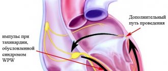
Regardless of the cause, the clinical manifestations of the syndrome are associated with two mechanisms that often combine to worsen the course of the disease:
1. Squeezing, or compression, of the artery leads to disruption of blood flow to part of the brain. 2. Irritation, or irritation, of the nerve fibers that surround the vessel leads to the activation of substances that cause narrowing of the arterial wall. This further disrupts the flow of blood to the structures of the central nervous system.
Negative manifestations of SPA include:
1. increased risk of developing an acute disruption of the blood supply to the brain of a temporary or permanent nature (transient ischemic attack, stroke); 2. decreased ability to work due to the need for a number of restrictions on movement and environmental conditions; 3. significant psychological discomfort against the background of clinical manifestations, especially in young patients.
Carrying out the necessary treatment significantly reduces the manifestations of the syndrome, but does not eliminate it completely.
Even after surgical correction methods, there are often residual effects of the disease, and the need for a restrictive regime of work and rest remains. Therapy significantly reduces the risk of acute vascular manifestations in the brain and the risk of fatal disruption of blood flow. Neurologists and neurosurgeons deal with the problem of diagnosis, choice of tactics for management and treatment of patients with SPA.
Factors that increase the risk of developing
| Group | Immediate factors |
| Features of artery anatomy | Deformation Tortuosity Kink Disturbance of the wall structure Incorrect movement |
| Diseases leading to a decrease in the lumen of the vessel | Atherosclerosis Hypertension Arteritis (inflammation of the wall) Thrombosis and embolism |
Vertebral artery syndrome occurs with the following diseases that cause compression of the vessel:
Osteochondrosis
– pathological destructive changes in the disc between two vertebrae, which begin in the central part (core), progressively spreading to the entire motor part of the vertebra.
Spondylosis deformans
– changes are associated with the aging process of the body, when, against the background of decreased blood flow in the capillaries, the nutrition of the intervertebral discs is disrupted, they lose their elasticity and decrease in size. This leads to the formation of bone spurs (osteophytes) on the front and side of the vertebra.
Spondyloarthrosis deformans
– pathology of the intervertebral joints, arising due to occupational hazards or peculiarities of the formation of the musculoskeletal system.
Deforming osteoarthritis of the spine
– one of the elements of systemic destructive damage to the joints of osteoarthritis. Affects two or more motor segments of the vertebrae.
Ligamentosis ossificans (Forestier disease)
– systemic damage to the ligamentous apparatus of the human skeleton, characterized by the deposition of calcium in the ligaments, which progressively reduces their extensibility and mobility.
Kimmerly Anomaly
– pathological structure of the first cervical vertebra.
Basilar impression
– violation of the location of the occipital bone, it is pressed into the cavity of the skull, compressing the spine.
Cervical spine injuries
associated with sudden hyperextension.
Compression of the artery by the neck muscles in certain head positions. Destructive changes in cervical localization of osteochondrosis lead to the development of RAS in 42.5–50% of patients.
Classification
Vertebral artery syndrome is classified according to the main causative mechanism, but in most cases the disease is of a mixed nature.
| Mechanism of occurrence | Its features |
| Compression | Compression of the lumen of the vessel from the outside |
| Irritative | Irritation of the nerve fibers of the perivascular plexus |
| Angiospastic | Reflex narrowing of the artery lumen in response to mechanical activation of receptors in the wall |
| Mixed | Any combination of the above options |
Manifestations of the syndrome
Patients with SPA experience constant difficulties in performing any tasks. Every wrong movement can cause clinical manifestations, forcing the patient to limit himself in work and everyday activities. The symptoms themselves are quite painful and disturb the psychological peace of the sick, forcing them to consult doctors to correct it.
In the clinical course of the syndrome, two phases of development are distinguished:
| Phase | Her characteristics |
| 1. Functional or dystonic | Symptoms are not persistent Good response to treatment Low risk of stroke |
| 2. Organic, or ischemic | The appearance of a focus of persistent disturbance of blood flow High risk of developing acute posterior cerebral stroke The effect of treatment is reduced, residual effects on the brain are possible |
In vertebral artery syndrome, symptoms are highly variable and affect many functions:
| Clinical variant of the symptom | Its manifestations |
| Posterior cervical sympathetic syndrome (Barré-Lieu) | Pain in the neck and back of the head with reflection (irradiation) to the forehead Constant, pulsating pain Intensifies in the morning after sleep, with movement and shaking, sudden turns of the head Noise in the ears Decreased hearing Flashes before the eyes |
| Basilar migraine | The attack begins with changes in vision (sparks, loss of part of the image) Severe dizziness and gait disturbance Incoherent speech Tinnitus Pain in the back of the head |
| Vestibulo-atactic syndrome | Impaired gait and balance Nausea and vomiting Dizziness Darkness in the eyes Rapid pulse Increased blood pressure |
| Cochleovestibular syndrome | Hearing loss Tinnitus that changes when you turn your head Unsteadiness when walking Swaying |
| Visual or ophthalmic syndrome | Flashes, flickering before the eyes Deterioration of vision Tearing Rapid eye fatigue Redness of the eyes Feeling like “sand has been poured into the eyes” Periodically loss of part of the visible image |
| Autonomic dysfunction syndrome | Feeling hot or cold Increased sweating Cold feet and hands Difficulty swallowing Voice problems Insomnia or drowsiness |
| Vertebral syncope (Unterharnscheidt) syndrome | A short attack of loss of consciousness during certain movements of the neck or prolonged stay in an uncomfortable position |
| Transient ischemic attack | Impaired active movement of the limbs Changes in skin sensitivity Loss of vision Loss of balance when walking Dizziness with nausea and vomiting Double vision Inability to speak and swallow |
| Drop attack attacks | Sudden paralysis of the whole body with a fall after throwing back the head. The duration of the attack is from several seconds to a minute |
| Mental changes | Unpleasant sensations of foreign material in the upper half of the body Refusal to eat Decreased all types of activity Thoughts about a fatal disease and the inability to recover from it |
The disease can manifest itself in only one of the listed options or a combination of them.
Diagnostics
Vertebral artery syndrome is a multifaceted disease that often imitates various pathologies of the organs of vision, hearing, neck, and brain.
Therefore, the main method of establishing the correct diagnosis is a thorough interview of the patient to identify the prevailing syndrome of the disease. To clarify the cause of the pathology, a number of additional procedures are required:
| Study | Target |
| X-ray of the cervical spine and skull bones with functional loads | Primary assessment of bone elements Identification of gross skeletal pathology |
| Ultrasound scanning of the vessels of the neck and head | Inspection of the artery itself for anatomical changes, pathology of the wall and lumen, identification of compression and kinks of the vessel |
| Multislice computed tomography | Assessment of all bone structures, identification of pathology associated with the destruction of elements of the spinal column |
| Magnetic tomography of the cervical spine and blood vessels of the head and neck | Assessment of “soft” skeletal elements (discs, ligaments), identification of pathology from the artery itself and its branches |
| Angiography | A clarifying method in complex diagnostic situations. Indicated for suspected blockage of an artery by a blood clot to select subsequent treatment. |
Treatment
Treatment of vertebral artery syndrome includes both medicinal effects on the vessels and surrounding tissues, and surgical methods for correcting compression by the bone structures of the vertebrae. There is no complete cure for the disease, since pathological processes in the elements of the spinal column are irreversible .
But a complex effect on all elements of the mechanism of occurrence makes it possible to slow down and (or) stop changes, as well as reduce the clinical manifestations of the syndrome.
Medicinal methods of correction
1. Drugs to relieve swelling and inflammation
When the lumen of the bone canal decreases, the venous vessels are the first to be compressed, and against the background of impaired blood outflow from the brain, increased compression of the vertebral artery occurs. Any prolonged compression leads to a local inflammatory reaction. Therefore, spa treatment begins with these medications:
— Troxerutin; — Diosmin; — Nimesulide; — Celecoxib; - Lornoxicam.2. Medicines that normalize blood flow in the arteries Against the background of disruption of the normal blood supply to brain structures, the risk of stroke and transient attacks increases. To prevent complications use: - Pentoxifylline; — Vinpocetine; — Cinnarizine; — Nicergoline; - Instenon. 3. Drugs to protect nerve cells Under conditions of lack of blood flow, neurons are exposed to free radicals and suffer from a lack of oxygen. Medicines from this group are indicated in the phase of organic disorders in SPA in order to prevent the development of persistent impairment of brain function. — Citicoline; — Gliatilin; — Actovegin; - Cerebrolysin; — Piracetam; — Mexidol; - Mildronate; - Sumatriptan; — B vitamins; - Antispasmodics. 4. Non-medicinal treatment is aimed at relieving spasm of the muscles surrounding the spine and improving blood flow in this area - massage; - acupuncture; - PRP therapy; - physiotherapy (UHF, shock wave, HILL laser, magnet, amplipulse); — therapeutic exercises and physical education; - sanatorium treatment.
Surgical correction
To relieve pressure on the artery, a number of techniques are used to normalize the supporting function of the spinal column:
1. Percutaneous spinal fusion
– fixation of two adjacent vertebrae relative to each other
2. Fenestration of discs between vertebrae
– creation of artificial defects in them for the growth of connective tissue
3. Autodermoplasty of discs
– replacement of the intervertebral disc with one’s own tissues
4. Replacement of the disc
with a titanium-nickel explant
Prognosis
Vertebral artery syndrome cannot be completely cured cure.
This is due to causal pathological factors - changes in the osteoarticular component of the spinal column do not undergo reverse development. Any surgical techniques provide a temporary or incomplete therapeutic effect. Drug correction is aimed at reducing the clinical manifestations of the disease, but has virtually no effect on the primary cause of the pathological process. Despite this, all treatment methods significantly alleviate the course of the disease and stop its progression.
More details
Treatment
It is recommended to take antihypertensive drugs, as well as drugs that help lower cholesterol. The person is given advice on changing his lifestyle and is prescribed a special diet. A prerequisite is to quit smoking.
To regulate the blood supply and functioning of the nervous system, as well as relieve symptoms, the use of antioxidants is indicated - Actovegin, Mildronate, Mexidol. Recommended drugs include Cavinton, Instenon and Trental.
For ischemia, symptomatic therapy includes antidepressants, usually of the benzodiazepine type. Among them, Grandaxin has the least number of side effects.
Neurologists who treat patients during rehabilitation procedures usually include physiotherapeutic procedures in their treatment.
In some cases, specialists resort to surgery for therapy. In this case, the operation is performed on the vertebral artery, at the intersection of the vasomotor fibers. This intervention helps to reduce spasms in the arteries and improve blood flow.
Physiatry requires daily monitoring of blood pressure and blood tests for cholesterol levels.
Vertebral artery syndrome (VAS)
The term SPA, to a certain extent, is a collective concept and unites a complex of cerebral, vascular, autonomic syndromes that arise as a result of damage to the sympathetic plexus of the vertebral artery, deformation of its wall or changes in the lumen.
According to various data, the frequency of dysgemia in the vertebrobasilar region ranges from 25 to 30% of all cerebrovascular accidents, including up to 70% of transient ischemic attacks. In the structure of the causes causing disruption of blood flow in the vertebrobasilar basin (VBB), a significant place is occupied by atherosclerotic lesions of the vertebral arteries (VA), hypoplasia, abnormalities of the bone bed, damage to the craniovertebral junction, pathological tortuosity and displacement of the mouth of the vertebral artery.
The most significant etiopathogenetic factor in the development of these disorders is the pathology of the cervical spine, which has become increasingly widespread in recent years, especially in young people. The leading place in the pathogenesis of these disorders is given to degenerative-dystrophic processes in the cervical spine and abnormal processes in the atlas, which disrupt blood flow in the vertebral arteries and cause cerebral circulatory disorders. These changes belong to the group of compression narrowings of the vertebral arteries, which occur under the influence of many extravascular factors, and are collectively termed vertebral artery syndrome (VAS). In ICD-10, vertebral artery syndrome is considered under the code G99.2 and includes the clinic of posterior cervical sympathetic syndrome, repeated episodes of vertebrobasilar insufficiency, episodes of drop attacks, and Unterharnscheidt syndrome.
The etiological factors of SPA can be divided into 3 main groups:
- Occlusive arterial diseases (atherosclerosis, thrombosis, embolism, arteritis of various origins);
- Deformations of arteries (pathological tortuosity, kinks, anomalies of structure and course);
- Extravasal compression of arteries (compression by bone anomalies, ribs, muscles, osteophytes and articular processes of the cervical vertebrae, scars, tumors, etc.).
The multiplicity of etiological factors in the development of this process somewhat “blurs” the interpretation of SPA, because This concept can mean almost any vascular lesion, including acute cerebrovascular accidents in the vertebrobasilar region. But in clinical neurological practice, as a rule, the diagnosis of SPA is made to patients who have a certain set of complaints and clinical syndromes that can be associated with degenerative lesions or anomalies of the cervical spine. Thus, despite the polyetiology of SPA, in clinical practice this term refers to the compression version of this syndrome. In our opinion, it is more correct to use the term “vertebrogenic vertebral artery syndrome” (VSPA). However, an analysis of the available literature data allows us to conclude that in scientific and clinical practice, SPA is a reflection of the vertebrogenic nature of dyshemia in a given area. And here we are faced with the other extreme - most of the authors we cited associated PAS exclusively with posterior cervical sympathetic syndrome (Barre-Lieu syndrome), while ignoring other possible clinical manifestations of compressive effects on the trunk or autonomic plexus of PAS. Based on this, we will further use the term “vertebral artery syndrome” to denote the vertebrogenic nature of the process.
Anatomical prerequisites for the development of vertebrogenic SPA
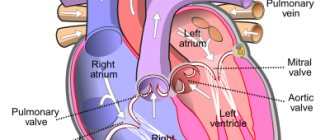
To understand the pathogenesis of the development of VA compression syndrome, it is necessary to have an idea of the anatomical features of this vessel. There are extra- (I–III segments) and intracranial (IV segment) sections of the VA. Segment I begins from the place where the VA exits the subclavian artery and ends at the level of the entrance to the bone canal. Segment II is located in the bone canal along the C II C VI vertebrae; segment III - from the place of exit from the bone canal at level C II to the entrance to the cranial cavity (the bends of the VA are located in this area); segment IV – intracranial - from the entrance of the artery into the skull to its confluence with the VA of the opposite side. One of the most important features of the structure of the cervical spine is the presence of holes in the transverse processes of the VI–VII cervical vertebrae. These openings form a canal through which the main branch of the subclavian artery passes - the vertebral artery with the sympathetic nerve of the same name (Frank's nerve).
The vertebral artery, when leaving the canal, goes to the foramen magnum, making bends. Then, at the lower edge of the pons, both vertebral arteries join to form the basilar artery. The vertebrobasilar basin is connected to the carotid basin through the circle of Willis. The vertebral artery vascularizes a wide area: segments of the spinal cord from C I to D III inclusive (superior medullary vascular basin), inner ear, stem structures of the brain with its reticular formation and vital centers, occipital lobes, mediobasal sections of the temporal lobes, cerebellum, posterior sections hypothalamic region. From the stellate ganglion, formed by the sympathetic centers of the C III DI segments of the spinal cord, the spinal nerve (posterior cervical sympathicus, or Frank’s nerve) arises. The latter enters the canal of the transverse processes, densely entwining the vertebral artery with its branches. In addition, branches that participate in the formation of the sinuvertebral nerve of Luschka depart from the vertebral nerve. The latter innervates the capsular-ligamentous apparatus of the cervical spinal motion segments, the periosteum of the vertebrae and intervertebral discs.
The possibility of damage to the VA in cervical osteochondrosis is determined by its topographic and anatomical position. A significant part of the extracranial segment of the VA passes in a mobile bone canal formed by the transverse processes of the cervical vertebrae and rudiments of the ribs. In this case, the lateral wall of the artery is adjacent to the uncovertebral joint, and the posterior wall is adjacent to the superior articular process. At level C I–C II, the artery is covered only by soft tissue, mainly by the inferior oblique muscle of the capitis. The condition of the perivascular plexuses and the lower cervical sympathetic ganglion, which determines the sympathetic innervation of the vertebral artery, is also of great pathogenetic importance in the development of SPA.
The main pathogenetic mechanisms of PA syndrome are compression of the artery trunk, autonomic plexus and narrowing of the lumen of the vessel due to reflex spasm, which contribute to a decrease in blood flow to the posterior parts of the brain with subsequent cerebral circulatory failure.
Osteophytes formed during osteochondrosis and spondylosis deformans in the area of uncovertebral joints have the greatest compressive effect on the vertebral artery. Displacement and compression of the vertebral arteries in cervical osteochondrosis can also be observed as a result of subluxation of the articular processes of the vertebrae. Due to pathological mobility between the individual segments (two vertebrae connected by a disk) of the cervical spinal column, the vertebral artery is injured by the tip of the superior articular process of the underlying vertebra. Most often, the vertebral artery is displaced and compressed at the level of the intervertebral cartilage between the V and VI cervical vertebrae, somewhat less often - between IV and V, VI and VII, and even less often - in other places.
A certain role is assigned to abnormal processes in the atlas region, which disrupt blood flow in the vertebral arteries. Also, pathogenetic variants of the development of PA syndrome in degenerative-dystrophic processes can be uncovertebral arthrosis, arthrosis of the facet joints, pathological mobility, posterior extensor subluxation of the articular processes according to Kovach, blockades and instability of the joints of the head, disc herniations, reflex muscle (inferior oblique muscle of the head, anterior scalene muscle) compression, the location of the vertebral arteries in the openings of the bone canal of the transverse processes of the cervical vertebrae, which easily shift relative to each other during movements of the head and neck. In addition, they are closely adjacent to the vertebral bodies. In this case, even under normal physiological conditions, compression and restriction of blood flow occurs in one or both arteries. Normally, blood circulation in them is usually not impaired due to sufficient compensatory capabilities. The situation changes with hypoplasia (anatomical narrowing) or atherosclerotic stenosis of the arteries. Then extravasal factors (compression by articular processes due to instability of the cervical spine or osteophytes in uncovertebral areas, etc.) become the decisive causes of circulatory failure in the VBB. Compression of the vertebral arteries is also possible by the neck muscles (scalenus, longus colli, inferior oblique capitis) when they contract in certain positions of the head. The most common cause of vertebral artery syndrome is uncovertebral arthrosis. Taking into account the extremely close functional and topographic-anatomical relationship of this articulation with the vertebral artery, it is clear that even small uncovertebral exostoses can have a mechanical effect on the vertebral artery. Initially, osteophytes cause dynamic irritation of its sympathetic plexus only during certain positions or movements in the cervical spine. Severe osteochondral growths of the uncovertebral joint can cause severe compression of the lumen of the vertebral artery canal.
Among the significant factors of SPA one can also highlight Kimmerly, Powers anomalies, and basilar impression. In addition to mechanical compression, vascular spasm may occur as a result of irritation of the periarterial nerve plexus. Most often there is a combination of these factors.
Clinical classification of vertebral artery syndrome (Kalashnikov V.I., 2009)
- Pathogenetic factors of SPA (by the nature of the compressive effect on PA).
- Subluxation of the articular processes of the vertebrae.
- Pathological mobility (instability, hypermobility) of the spinal motion segment.
- Compression by osteophytes.
- Vascular spasm as a result of irritation of the periarterial nerve plexus.
- Compression in the atlas region (Klippel-Feil anomaly, Kimmerly anomaly, atlas anomalies, platybasia).
- Uncovertebral arthrosis.
- Osteoarthritis of the facet joints.
- Blockades and joint instability.
- Herniated intervertebral discs.
- Reflex muscle compressions.
- Clinical stages of SPA.
- According to the degree of hemodynamic disturbances.
- Dystonic (functional).
- Ischemic (organic).
The functional stage of vertebral artery syndrome is characterized by three groups of symptoms: headache with accompanying autonomic disorders, cochleovestibular disorders, visual disorders. The headache is throbbing or aching, burning, constant and worsening in paroxysms, especially when moving the head, with its prolonged forced position, spreading from the back of the head forward to the forehead. Cochleovestibular disorders can also manifest themselves in the form of paroxysmal non-systemic dizziness (feeling of instability, swaying) or systemic dizziness. They can be combined with paracusis, mild hearing loss and give rise to confusion with Meniere's disease.
Visual disturbances are limited to the following: darkening in the eyes, a feeling of sand, sparks and other manifestations of photopsia, slight changes in the tone of the fundus vessels.
In conditions of prolonged and intense vascular spasms, the development of foci of persistent ischemia is possible - the organic stage of vertebral artery syndrome.
The organic stage of the vertebral artery is manifested by transient and persistent disturbances of cerebral circulation. Transient circulatory disorders in the vertebrobasilar system manifest themselves in the form of dizziness, ataxic disorders, nausea, vomiting, and articulation disorders. There are other forms of transient cerebral ischemia characteristic of vertebrogenic lesions of the vertebral arteries. As a rule, they occur when turning or tilting the head. This pathology includes attacks of sudden falling while maintaining consciousness lasting up to several minutes (drop attacks), as well as attacks with loss of consciousness lasting from two to three to ten to fifteen minutes (syncope). Regression of symptoms usually occurs in a horizontal position. After attacks, general weakness, headaches, tinnitus, photopsia, and autonomic lability are noted. The pathogenetic mechanism of these paroxysms is transient ischemia of the brain stem with localization in the area of the intersection of the pyramids (during drop attacks) and the reticular formation (during syncope).
According to the nature of hemodynamic disorders:
- Compression.
- Irritative.
- Angiospastic.
- Mixed.
In the compression version, the narrowing of the lumen of the vessel occurs through mechanical compression of the artery wall. The irritative version of the syndrome is formed as a result of vertebral irritation of the efferent sympathetic fibers of the spinal plexus, causing vascular spasm. As a rule, in clinical practice there are mixed (compression-irritative) variants of this syndrome. Angiospastic syndrome manifests itself in the form of a reflex spasm that occurs in response to irritation of receptors in the area of the affected SDS. In angiospastic syndrome, diffuse vegetative-vascular disorders predominate, less associated with head turns. The compression-irritative version of the syndrome is more often associated with pathology of the lower cervical spine, while the reflex version is more often associated with pathology of the upper cervical spine.
Clinical SPA options
- Barre Lieu syndrome (posterior cervical sympathetic syndrome).
Clinically characterized by headaches in the cervical-occipital region with irradiation to the anterior parts of the head (like “removing the helmet”). The headache is constant, especially in the morning after sleeping on an uncomfortable pillow, when walking, shaking, or when moving the neck. Headaches can be pulsating or shooting in nature, begin in the cervical-occipital region and spread to the parietal, temporal and frontal regions. The pain intensifies when turning the head, at night and after sleep. Headache is accompanied by autonomic disorders, cochleovestibular and visual disorders.
- Basilar migraine.
A migraine attack that begins with bilateral visual disturbances, accompanied by dizziness, ataxia, dysarthria, and tinnitus. At the height of the attack, a sharp headache develops in the occipital region, accompanied by vomiting and, in some cases, loss of consciousness. Basilar migraine is not a consequence of compression of the VA itself; it is based on a narrowing of the basilar artery (BA) and/or its branches, but taking into account the direct anatomical and physiological unity of OA and VA, as well as a certain commonality of clinical symptoms with other forms of BAS, this syndrome is necessary considered in the context of clinical manifestations of PA syndrome.
- Vestibulo-atactic syndrome.
Subjective symptoms predominate: dizziness, a feeling of body instability, darkening of the eyes, imbalance with nausea and vomiting, cardiovascular disorders. Symptoms intensify when you move your head or when you are forced to position it.
- Cochleo-vestibular syndrome.
Cochlear disorders are manifested by noise in the ear or back of the head, paresthesia, hearing loss, decreased perception of whispers, and changes in the audiogram. These disorders are usually combined with paroxysmal non-systemic dizziness (feeling of instability, swaying) or systemic dizziness. Tinnitus is characterized by persistence and duration of manifestations; its character can vary depending on the position of the head.
- Ophthalmic syndrome.
Visual impairment is characterized by transient photopsia, atrial scotoma, fatigue, and decreased vision during reading and other visual stress. Phenomena of conjunctivitis may be observed: pain and sensation of a foreign body in the eyes, redness of the conjunctiva, lacrimation. There are also episodes of paroxysmal loss of visual fields or parts thereof, most often associated with the position of the head.
- Syndrome of autonomic disorders.
The most common vegetative symptoms are the following: feeling of heat, chills, cold extremities, hyperhidrosis, changes in dermographism. Laryngopharyngeal disorders may be detected, as well as paroxysmal sleep and wakefulness disorders. These changes, as a rule, are not isolated; they almost always occur during an exacerbation of the PA syndrome and are combined with at least one of the syndromes described in this classification.
- Transient ischemic attacks.
The ischemic stage of PA syndrome can manifest itself in the form of transient circulatory disorders in the vertebrobasilar region. The most common clinical symptoms are: transient motor and sensory disturbances, complete or partial loss of vision, homonymous hemianopsia, ataxia not associated with dizziness, paroxysmal dizziness, which may be accompanied by nausea, vomiting, diplopia, dysphagia, dysarthria.
- Unterharnscheidt syndrome (vertebral syncopal syndrome).
Unterharnscheidt's syncopal attack is an acute circulatory disorder in the reticular formation of the brain stem, characterized by a short-term loss of consciousness during a sudden movement of the head or a prolonged forced position.
- Drop attack attacks.
An attack of sudden falling is associated with ischemia of the caudal parts of the brain stem and cerebellum and manifests itself in the form of pyramidal tetraplegia with a sharp throwback of the head with rapid subsequent restoration of motor function.
Diagnostics
Diagnosis of SPA is quite complex due to the polymorphism of complaints and clinical symptoms. In clinical practice, we often encounter both over- and underdiagnosis of SPA.
Overdiagnosis of SPA is most often associated with a basic underexamination of the patient. Most often, this occurs when patients have vestibulo-atactic and/or cochlear syndrome, when the clinician fails to recognize or suspect pathology of the labyrinth. Despite the variety of complaints presented by patients with SPA (headaches, dizziness, unsteadiness when walking, noise and ringing in the ears, photopsia, transient disturbances of vision and consciousness, etc.), the clinician needs to identify the main clinical syndrome and compare it with the description clinical manifestations of SPA (see classification). Next, it is necessary to establish the presence of extravasal compressions and/or deformations of the VA. However, the available radiological correlates may not always be directly related to clinical symptoms. Therefore, to clarify the nature of the process, it is necessary to establish the fact of the compression effect on the VA, which is achieved using duplex scanning or Doppler ultrasound.
In our opinion, to establish a diagnosis of vertebrogenic SPA, the presence of 3 clinical diagnostic criteria is necessary.
- Clinical symptoms (presence of 1 of the 9 clinical options described above or their combination).
- The presence of changes detected during magnetic resonance or spiral computed tomography in combination with functional radiography of the cervical spine (osteochondrosis, deforming spondylosis in the area of uncovertebral joints, subluxation of the articular processes of the vertebrae, instability and hypermobility, abnormalities of the bone bed of the vertebral column, craniovertebral junction, etc. ).
- The presence of changes detected during duplex scanning of the VA and/or during vertebral Dopplerography using functional loads with rotation, flexion and extension of the head (compression of the vertebral artery, asymmetry of the linear velocity of blood flow in the vertebral arteries, vasospastic reactions in the vertebral and basilar arteries, hyperreactivity to functional tests).
Make an appointment with a neurologist
Be sure to consult a qualified specialist in the field of neurological diseases at the Semeynaya clinic.
Single contact center
8
weekdays 8-21, weekends 9-21
To find out more about prices for seeing a neurologist or other questions, please follow the link below
Manual therapy
Neurology
Reflexology
Physiotherapy
Prevention
Preventive measures are aimed at reducing the risk of ischemia. A prerequisite is giving up bad habits.
For prevention, it is necessary to avoid stress and reduce the duration of exposure to the sun. Physical activity should be moderate, but constant. One of the necessary preventive measures to prevent this pathology is the regulation of the patient’s body weight, because obesity directly affects the development of vestibular ataxia.