Inflammatory processes, developing in various parts of the eye - this is a fairly common pathology. Inflammation can affect one or more parts of the eyeball, as well as surrounding tissues, and it can be of either an infectious or non-infectious nature. Any of the inflammatory diseases of the organs of vision is characterized by similar clinical signs and leads to impaired eye function.
An ophthalmologist (ophthalmologist) deals with the diagnosis and treatment of inflammatory eye diseases.
Blepharitis
The focus of inflammation in this disease is localized along the edge of the eyelids. The causes of blepharitis are usually prolonged exposure to caustic substances, smoke, volatile liquids, the presence of a chronic infection in the body, or infection due to injury to the eyelids.
It is customary to distinguish 3 forms of the disease: simple, ulcerative and scaly.
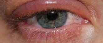
Simple blepharitis is accompanied by redness of the edges of the eyelids with slight swelling, without spreading the process to the surrounding tissues. The process is accompanied by a sensation of a foreign body in the eye, which causes the patient to blink frequently. Sometimes foamy discharge is observed in the inner corners of the eye.
Scaly blepharitis is accompanied by severe redness of the eyelid margins and noticeable swelling. Its characteristic feature is grayish or pale yellow scales at the edges of the eyelids. Their mechanical removal leads to mild bleeding. The patient feels itching in the eyelids, sometimes there is a feeling of a foreign body, pain when blinking. Advanced cases occur with pain and photophobia. Visual acuity often decreases.
Ulcerative blepharitis is the most severe form of the disease. Its characteristic sign is dried pus at the roots of the eyelashes, which causes them to stick together. After the crusts are eliminated, slowly healing ulcers form. Eyelashes fall out and often then grow incorrectly. Often accompanied by conjunctivitis caused by the spread of an infectious agent.
Risk factors
The causes of keratitis are known, and there are also risk factors that significantly increase the likelihood of developing the disease.
Immediate causes include:
- Infections, parasitic infestations.
- Mechanical injuries, chemical, thermal damage.
- Allergy.
These causes lead to the occurrence of infectious (for example, herpetic), traumatic or allergic keratitis, respectively.
Among the risk factors for developing the disease, the following are most important:
- Presence of autoimmune diseases.
- Long-term wearing of contact lenses.
- Dry eye syndrome.
- Lack of vitamins.
- Various metabolic disorders.
- The presence of some systemic diseases: diabetes, gout, rheumatism.
Based on the list of risk factors, it is clear that keratitis is a fairly common pathology.
Purulent eye infection
As a rule, its causative agent is streptococci or staphylococci, which penetrate inside when the eye is injured by a sharp object.
The course of the disease goes through 3 stages - iridocyclitis, endophthalmitis, panophthalmitis.
Iridocyclitis occurs a couple of days after an eye injury and is accompanied by severe pain of the eyeball upon palpation. The iris takes on a grayish or yellowish tint due to accumulated pus, and the pupil seems immersed in a kind of haze.
Endophthalmitis is a more severe form of purulent inflammation. The infection spreads to the retina, and pain is felt even with the eye closed. Visual acuity quickly drops to light perception. During the examination, characteristic signs are revealed - dilation of the conjunctival vessels, a greenish or yellowish tint of the fundus.
Panophthalmitis is a rare complication of endophthalmitis with timely treatment. Purulent inflammation spreads to all tissues of the eye. Unbearable pain occurs, the eyelids swell, the mucous membrane turns red and swells. Pus oozes through the cornea, the color of the white becomes greenish. The skin around the eye becomes red and swollen. An eye abscess may occur. Severe cases of the disease require surgery. Even with successful antibiotic therapy, visual acuity is noticeably reduced.
Retina
The retina is the thin inner layer of the eye structure and contains special photoreceptors. The diseases of this element are:
- Retinitis is an inflammation of the retina in question due to infections, traumatic, thermal or chemical damage. The clinical picture includes the appearance of spots, low visual acuity (especially in the dark), limited vision, and double vision.
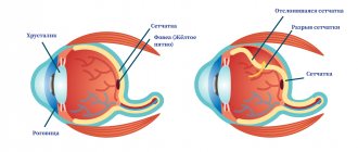
- Peeling. The retina is detached from the choroid. Signs: blurred vision or veil, seeing objects distorted, sparks with flashes, limitation of the visual field.
- Angiopathy is a disease with changes in the structure of the vessels providing blood supply. The problem occurs either due to high pressure inside the eye or due to traumatic injury. Any symptom will be obvious: flashes or glare, blurred or impaired vision, distorted picture.
- Dystrophy is a disease with death or depletion of retinal tissue.
Dacryocystitis
Inflammation of the lacrimal sac of an infectious nature. Predisposing factors to the disease are the characteristics of the lacrimal canal and fluid stagnation in the lacrimal gland.
The disease is manifested by liquid purulent discharge and lacrimation. A tumor develops in the inner corner of the eye - a swollen lacrimal gland. When pressed, pus flows out of it. Sometimes hydrocele of the lacrimal gland may develop.
Dacryocystitis can be easily cured with timely treatment. Complications often include keratitis and conjunctivitis with a slight decrease in vision.
Inflammation of the cornea
Keratitis is a fairly common eye disease, and it can be superficial (associated with external causes) or deep (associated with internal pathological processes). All these forms of the disease require immediate treatment, as complications may develop. Among the consequences, the most common are scleritis, adhesions on the pupil, decreased vision, and endophthalmitis.
Symptoms of keratitis are:
- lacrimation;
- Cutting pain;
- Narrowing of the palpebral fissure;
- Photophobia;
- Swelling of the eyelids;
- Itching.
Treatment of keratitis is carried out with local and systemic drugs. In this case, antibacterial, antifungal or antiviral drugs are prescribed. Oral vitamin complexes are also used. Local therapy is carried out using anti-inflammatory, antibacterial, disinfectant, and hormone-containing eye drops.
If the lacrimal canals are involved in the pathological process, then they are washed with a solution of chloramphenicol.
In case of herpetic nature of keratitis, laser coagulation or diathermocoagulation can be additionally performed. In addition to traditional drugs, phytotherapeutic treatment is sometimes prescribed.
Keratitis
This is an inflammatory process localized in the tissues of the cornea. There are exogenous and endogenous forms of the disease and its specific varieties (creeping ulcer of the cornea).
Exogenous keratitis develops after a chemical burn and infection with viruses, microbes and fungi. The endogenous form occurs against the background of a creeping corneal ulcer or infectious diseases of a fungal, bacterial and viral nature (syphilis, herpes, influenza). Sometimes the cause may be some metabolic abnormalities or hereditary predisposition.
The disease begins with tissue infiltration, continues with ulceration, and ends with regeneration.
The infiltrate is a fuzzy yellowish spot. The area of damage varies from microscopic to global, over the entire area of the cornea. Its formation leads to decreased visual acuity, the development of photophobia, lacrimation and spasms of the eyelid muscles (corneal syndrome).
After some time, an ulcer forms. If left untreated, it spreads to the cornea and internal structures of the eye. Its healing is possible only with antibacterial therapy.
If the area of the ulcer is small, visual acuity does not decrease. With extensive damage, blindness can occur.
Keratoconjunctivitis
A disease caused by an adenovirus that affects the conjunctiva and cornea.
Transmitted by contact.
From the moment of infection to the onset of the disease, 7-8 days pass. It begins with headache, chills, weakness and apathy. Then there is pain in the eyes and redness of the sclera, a feeling of a foreign body inside and profuse lacrimation with mucus discharge from the lacrimal canal.
The conjunctiva turns red, small bubbles with liquid inside appear on it - a characteristic manifestation of an adenoviral infection.
With timely treatment, after 5-7 days, signs of infection disappear, with the exception of photophobia. Cloudy lesions form in the cornea. Complete healing occurs after two months.
Viral conjunctivitis
The cause of the disease is the introduction of a viral agent. There are several forms of the disease, with a specific nature of the pathological process.
Herpetic conjunctivitis is typical for young children. The inflammatory process spreads beyond the mucous membrane into the surrounding tissues. It can be catarrhal, follicular and vesicular-ulcerative.
The most severe form of the disease is vesicular ulcerative. It is manifested by the appearance of small transparent bubbles with liquid on the mucous membrane of the eye. They are characterized by spontaneous opening with the formation of painful ulcers, which causes lacrimation and photophobia in patients.
After 5-7 days, the symptoms of the disease disappear spontaneously without additional therapy. Visual acuity does not change, no traces remain on the cornea.
Lens, vitreous body
What are the types of eye diseases in people that provoke dysfunctions or disorders of the structure of the lens - the natural lens?
- Cataract is a disease with clouding of the lens, in which the sick person’s vision decreases and the perception of images changes.
- Anomalies: biphakia (presence of two lenses in one eye) or aphakia (complete absence of a natural lens).
- Destruction of the vitreous body - a transparent substance with a gel-like structure that fills the space between the retina and the lens. This structural element is destroyed, which provokes hemorrhages, changes in visual functions and perception of visible objects.
Gonococcal conjunctivitis
Another name for the disease is gonoblennorrhea. This is an intense process of inflammation of the mucous membrane of the eye during the penetration of a gonococcal infection. The disease is transmitted exclusively through contact (sexual intercourse, from mother to fetus during childbirth, etc.).
The first symptoms of gonoblennorrhea in newborns appear 3-4 days after birth. The eyelids swell and become purplish-red or bluish in color. Bloody discharge appears from the lacrimal canal. Certain areas of the eye become ulcerated and cloudy. In advanced cases, panophthalmitis develops with loss of vision and atrophy of the eyeball. After therapy, rough scars may remain on the affected areas of the cornea.
Gonococcal conjunctivitis in adults is accompanied by general malaise, fever, and general pain.
Scleritis This is an acute inflammatory process in the sclera of the eye. More often it develops against the background of viral, bacterial or fungal general pathologies, as an ascending infection.
Episcleritis is a superficial scleritis affecting the upper layer of the sclera. The eye becomes red, its movements become painful. There is no lacrimation, which is considered a characteristic sign of the disease, photophobia rarely develops, and visual acuity does not change. An infected area of purple or red color appears on the sclera, slightly rising above the surface.
Deep scleritis spreads into the eye shells and in advanced cases spreads to the surrounding tissues, affecting the ciliary body and iris. Sometimes multiple foci of infection are possible, which often causes severe purulent complications.
Purulent episcleritis is caused by staphylococcus. The disease progresses rapidly, affecting both eyes. In the absence of therapy, it can continue for years, subside and recur. In areas of infection, the sclera becomes thinner and vision decreases noticeably. When the inflammatory process moves to the iris, glaucoma may develop.
Iris and choroid, pupils and ciliary body
The iris is a mobile, thin structure of the eye located behind the cornea and containing the pigment that determines the color of the eyes. The pupil is a hole in the iris through which all light rays emanating from the objects in question penetrate. The choroid is the middle element of the ocular structure, penetrated by blood vessels and supporting the blood supply. The ciliary body ensures the correct location of the lens and accommodation - a change in refraction depending on distance.
These structures are susceptible to eye diseases in humans from the list below:
- Uveitis with the spread of inflammation throughout the ocular choroid. Symptoms are redness and swelling of the eye, impaired vision.
- Iridocyclitis is an inflammatory disorder affecting the iris and ciliary body.
- Anisocria is a difference in pupil size caused by traumatic eye injuries. The main symptom is visual impairment. Photosensitivity also often occurs.
- Aniridia is an absent iris (its space is occupied by the pupil).
- Polycoria is the presence of several, usually asymmetrical pupils in one eye.
Choroiditis (posterior uveitis)
This is an inflammatory process behind the choroid. The reason for its occurrence is the introduction of microbes into the capillaries.
The disease is characterized by an initial absence of symptoms. As a rule, choroiditis is discovered incidentally during an ophthalmological examination.
If the focus of inflammation is localized in the center of the choroid, characteristic signs are sometimes observed: distortion of the contours of objects, flickering and light flashes before the eyes.
In the absence of timely antibacterial therapy, retinal edema develops with microscopic hemorrhages.
Inflammation of the tear ducts
When inflammation spreads to the lacrimal canals, we are talking about dacryocystitis. Since its patency is impaired, microbes accumulate in this area, which cause inflammation. The causes of the disease may be congenital characteristics, consequences of injury, or ophthalmological problems. Usually the inflammation is unilateral and is accompanied by redness, swelling and pain. There are also specific secretions. For treatment, adult patients are prescribed rinsing of the lacrimal canal with special disinfectant solutions. In childhood, massage of this area, antibacterial drops or tetracycline ointment are recommended. If conservative treatment is ineffective, then surgery is performed.
Barley
The disease is an inflammatory process in the sebaceous gland and ciliary bulbs, caused by staphylococcal and streptococcal infections against a background of weakened general immunity.
It is characterized by redness of the eyelid area, which then turns into infiltration and swelling. Gradually, redness spreads to nearby tissues, swelling of the conjunctiva increases. After 2-3 days, a cavity with pus forms inside the infiltration. On the 3-4th day from the beginning of the process, the purulent sac breaks through with the release of pus beyond the eyelid, pain and swelling subside. In severe cases, the process can spread to surrounding tissues.
By contacting the Moscow Eye Clinic, each patient can be sure that some of the best Russian specialists will be responsible for the results of treatment. The high reputation of the clinic and thousands of grateful patients will certainly add to your confidence in the right choice. The most modern equipment for the diagnosis and treatment of eye diseases and an individual approach to the problems of each patient are a guarantee of high treatment results at the Moscow Eye Clinic. We provide diagnostics and treatment for children over 4 years of age and adults.
Diagnostics
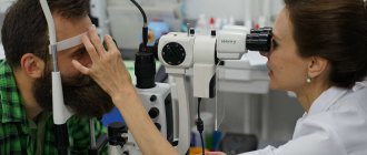
Eye diseases in humans, photos and descriptions of which this site contains, are identified by ophthalmologists. The diagnosis can only be made by a doctor after an initial examination and comprehensive diagnostics, including ophthalmoscopy, visometry, various tests, perimetry, refractometry, canal probing, ultrasound, CT, radiography, MRI, ophthalmometry, and sometimes blood tests. Only when the problem and its causes are identified can treatment begin.
Any disease requires a visit to the doctor and correct therapy. Contact an experienced specialist and follow his recommendations to preserve your vision.
Our doctors who will solve your vision problems:
Fomenko Natalia Ivanovna
Chief physician of the clinic, ophthalmologist of the highest category, ophthalmic surgeon. Surgical treatment of cataracts, glaucoma and other eye diseases.
Yakovleva Yulia Valerievna Refractive surgeon, specialist in laser vision correction (LASIK, Femto-LASIK) for myopia, farsightedness and astigmatism.
You can find out the cost of a particular procedure or make an appointment at the Moscow Eye Clinic by calling in Moscow 8 (499) 322-36-36 (daily from 9:00 to 21:00) or using the ONLINE REGISTRATION FORM.


